5A. 3: Cardiac + ECG cycle
0.0(0)
Studied by 0 people Call Kai
 Learn
Learn Practice Test
 Spaced Repetition
 Match
Match Flashcards
 Knowt Play
Card Sorting
1/20

Earn XP
Description and Tags
Last updated 12:10 PM on 10/11/26
Name | Mastery | Learn | Test | Matching | Spaced | Call with Kai | Chat |
|---|
No analytics yet
Send a link to your students to track their progress
21 Terms
1
New cards
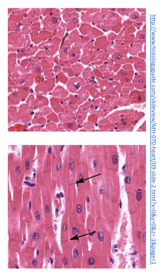
Cardiac muscle
* striated
* involuntary
\
Cardiomyocytes contain the same contractile filaments as skeletal muscle (sarcomere)
\
Cells are joined end-to-end and are connected via intercalated discs
\
Nuclei are centrally located, sometimes there are 2
* involuntary
\
Cardiomyocytes contain the same contractile filaments as skeletal muscle (sarcomere)
\
Cells are joined end-to-end and are connected via intercalated discs
\
Nuclei are centrally located, sometimes there are 2
2
New cards
2 types of cardiomyocytes
1. __**pacemaker**__
* Autorhythmic cells (“automaticity“)
* spontaneously contract
* SA node, some fibers in AV node, bundle of His, purkinje fibers
\
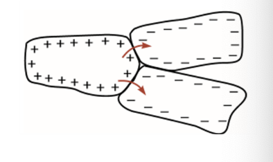
2. __**non-pacemaker cells**__
* bulk of heart
* basic contractile myocytes
* depolarization is induced by adjacent cells depolarizing
3
New cards
Syncytia
The heart can be divided into 2 syncytia:
* the atria syncytium
* ventricular syncytium
\
* allows for the atria to contract prior to ventricular contraction
* syncytium = network of cardiomyocytes connected via intercalated discs
* the atria syncytium
* ventricular syncytium
\
* allows for the atria to contract prior to ventricular contraction
* syncytium = network of cardiomyocytes connected via intercalated discs
4
New cards
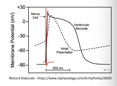
Action potentials
neuronal vs cardiomyocytes
\
__**neuronal**__ = rapid depolarization (1ms)
* depolarization caused by fast sodium channels
\
__**Cardiomyocytes**__ = 200-400 milliseconds
* pacemaker cells = slow response
* Myocytes = faster response
* depolarization caused by sodium + calcium
\
__**neuronal**__ = rapid depolarization (1ms)
* depolarization caused by fast sodium channels
\
__**Cardiomyocytes**__ = 200-400 milliseconds
* pacemaker cells = slow response
* Myocytes = faster response
* depolarization caused by sodium + calcium
5
New cards
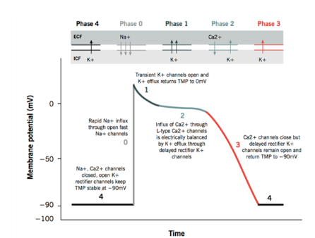
Refractory period
phases 0-3
* can’t be re-excited
* limits firing rate
\
* can’t be re-excited
* limits firing rate
\
6
New cards
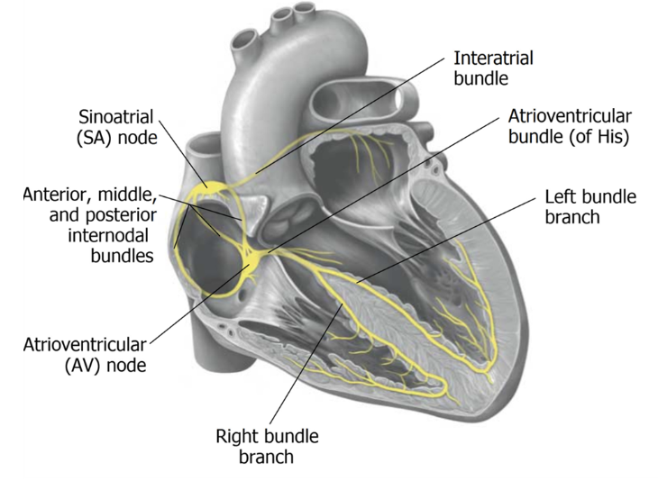
conduction system in the heart
__**SA node = pacemaker**__
* origin of cardiac impulse
* rate of depolarization is greatest here - which means it drives everything else
\
__**AV node**__
* located at the centre of the heart, in the floor of the right atrium, between the atria and ventricles
* electrically connects atria and ventricles via bundle of His
* slows the signal from the SA node
* allows for atrial blood to empty into ventricles
* origin of cardiac impulse
* rate of depolarization is greatest here - which means it drives everything else
\
__**AV node**__
* located at the centre of the heart, in the floor of the right atrium, between the atria and ventricles
* electrically connects atria and ventricles via bundle of His
* slows the signal from the SA node
* allows for atrial blood to empty into ventricles
7
New cards
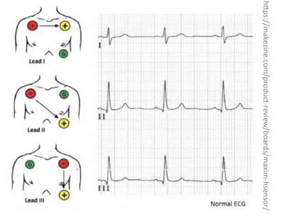
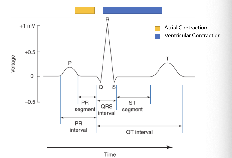
ECG
measures changes in electrical signal across cardiomyocytes
\
* as cardiomyocytes depolarize/ repolarize, electrical currents pass across the body
* electrical impulse picked up by electrodes
* Voltage measured as a difference between 2 electrodes
* toward +ve deflection, away from +ve deflection
multiple lead arrangements = many signals and characteristic patterns
\
* as cardiomyocytes depolarize/ repolarize, electrical currents pass across the body
* electrical impulse picked up by electrodes
* Voltage measured as a difference between 2 electrodes
* toward +ve deflection, away from +ve deflection
multiple lead arrangements = many signals and characteristic patterns
8
New cards
ECG configuration examples

9
New cards
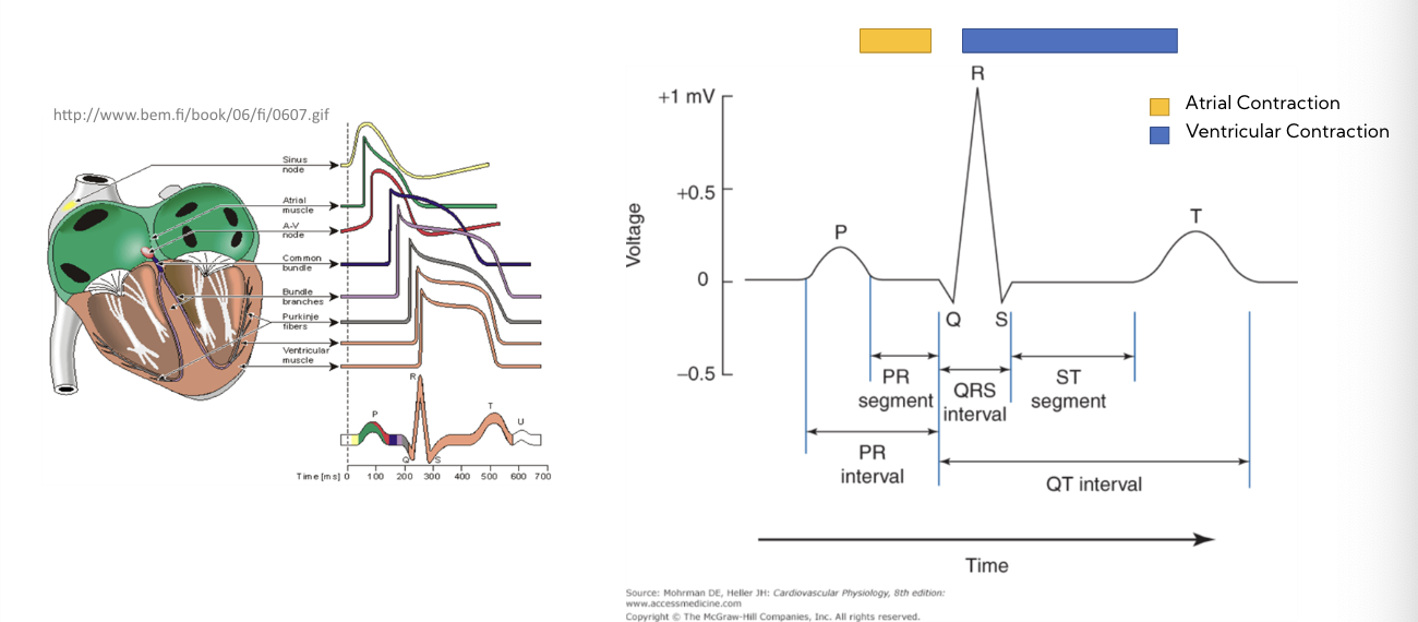
ECG recording
P wave = atrial depolarization
QRS = ventricular depolarization
T wave = ventricular repolarization
QRS = ventricular depolarization
T wave = ventricular repolarization
10
New cards
the different components of an ECG
* should be able to identify each component
11
New cards
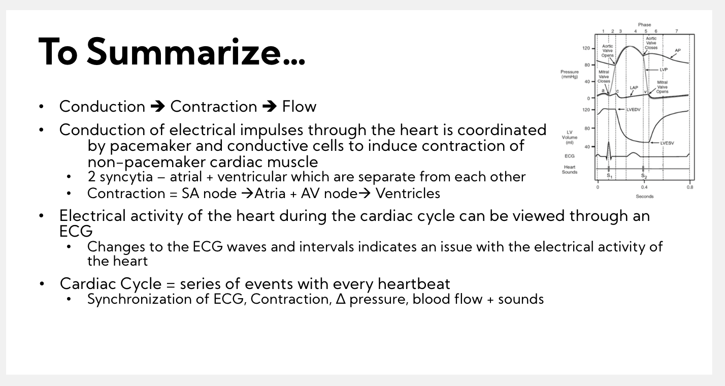
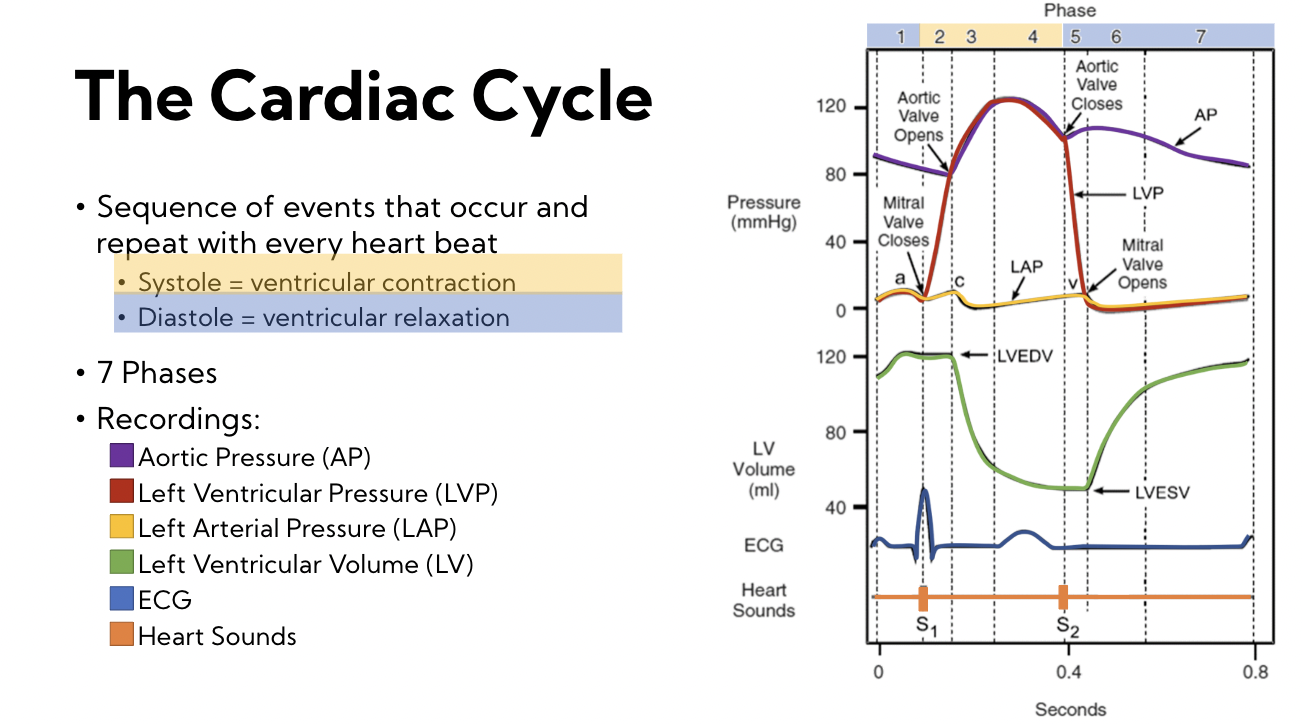
The cardiac cycle
The sequence of events that occur and repeat with every heart beat
* systole = ventricular contraction
* Diastole = ventricular relaxation
\
7 phases
**Recordings:**
* Aortic pressure (AP)
* Left ventricular pressure (LVP)
* Left Arterial pressure (LAP)
* Left ventricular volume (LV)
* ECG wave
* Heart sounds
* systole = ventricular contraction
* Diastole = ventricular relaxation
\
7 phases
**Recordings:**
* Aortic pressure (AP)
* Left ventricular pressure (LVP)
* Left Arterial pressure (LAP)
* Left ventricular volume (LV)
* ECG wave
* Heart sounds
12
New cards
basic principles
conduction → contraction → flow
\
blood flows from higher to lower pressure
* contraction increases pressure
* relaxation/emptying decreases pressure
\
valves open/ close based on pressure gradients
* atria are always filling
(no valves in vena cava or pulmonary veins)
\
\
\
blood flows from higher to lower pressure
* contraction increases pressure
* relaxation/emptying decreases pressure
\
valves open/ close based on pressure gradients
* atria are always filling
(no valves in vena cava or pulmonary veins)
\
\
13
New cards
heart sounds
S1 = mitral valve (left AV valve)
S2 = semilunar valve (aortic)
S2 = semilunar valve (aortic)
14
New cards
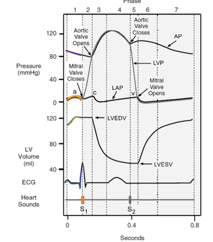
1. Atrial systole
* atria contract
* ventricles in diastole
* LVEDV = left ventricle and diastolic volume
* Pushes last bit of blood into ventricles
* increased pressure in ventricles closes AV-valve
* heart sound S1 (mitral valve)
* electrical activity precedes contraction
* QRS complex starts (ventricular depolarization)
* ventricles in diastole
* LVEDV = left ventricle and diastolic volume
* Pushes last bit of blood into ventricles
* increased pressure in ventricles closes AV-valve
* heart sound S1 (mitral valve)
* electrical activity precedes contraction
* QRS complex starts (ventricular depolarization)
15
New cards
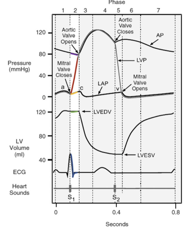
2. Isovolumetric contraction
* atria relax
* ventricles contract (systole)
* no blood is ejected = isovolumetric
* electrical activity precedes contraction - QRS complex starts
* ventricles contract (systole)
* no blood is ejected = isovolumetric
* electrical activity precedes contraction - QRS complex starts
16
New cards
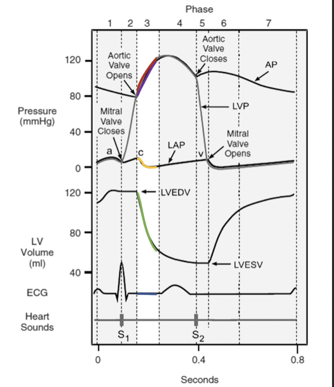
3. Rapid Ejection
* aortic + pulmonary valves open
* blood rushes into aorta + pulmonary trunk
* volume falls in ventricles rapidly
* pressure in ventricles continues to increase
* blood rushes into aorta + pulmonary trunk
* volume falls in ventricles rapidly
* pressure in ventricles continues to increase
17
New cards
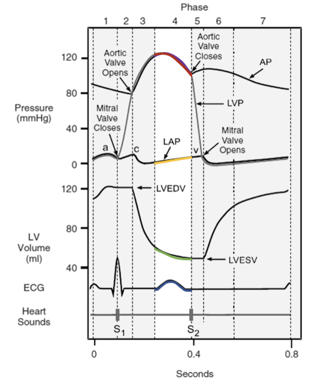
4. Reduced ejection
* pressure begins to decrease in aorta as the last bit of blood leaves the ventricles
* pressure in atria continues to rise as atria passively fill with blood
\- T-wave = ventricular
* repolarization
* pressure in atria continues to rise as atria passively fill with blood
\- T-wave = ventricular
* repolarization
18
New cards
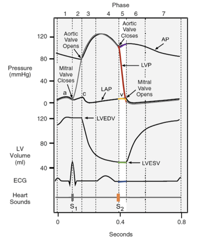
5. Isovolumetric relaxation
* semilunar valves close
* heart sound #2
* ventricles enter diastole
* AV valves are still closed thus volume of blood doesn’t change = isovolumetric relaxation
* LVESV = left ventricle end systolic volume
* heart sound #2
* ventricles enter diastole
* AV valves are still closed thus volume of blood doesn’t change = isovolumetric relaxation
* LVESV = left ventricle end systolic volume
19
New cards
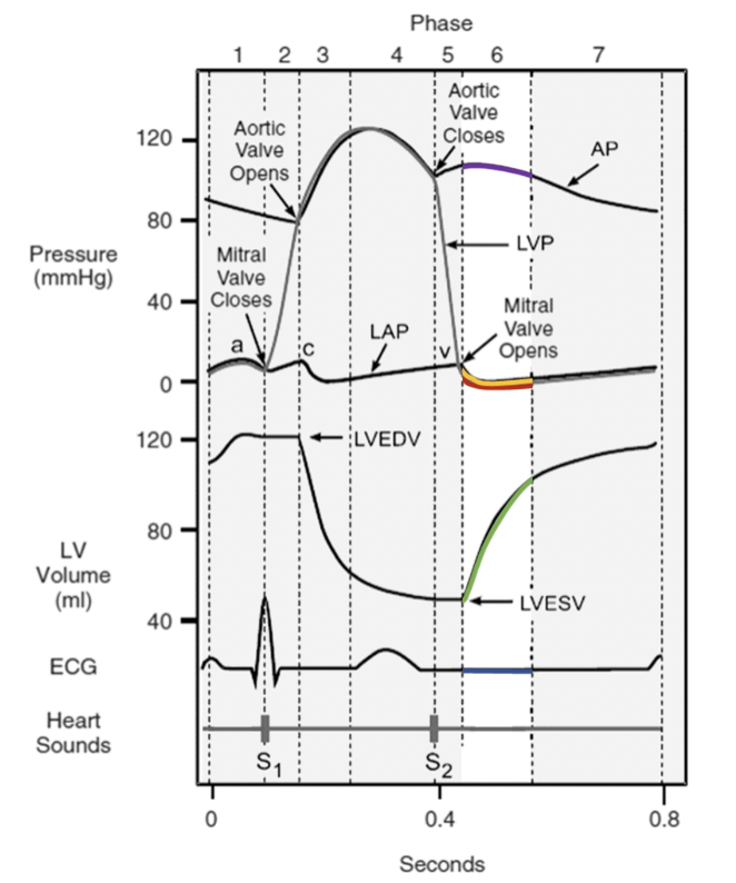
6. Rapid filling
* pressure in atria exceed pressure in ventricles and AV valves open
* Blood dumps into ventricles from atria “rapidly filling” them
* atrial volume + pressure drops
* ventricular volume + pressure rises
* Blood dumps into ventricles from atria “rapidly filling” them
* atrial volume + pressure drops
* ventricular volume + pressure rises
20
New cards
7. Reduced filing
* blood passively flows into heart from vena cava + pulmonary arteries
* AV valves are open, so it flows directly into ventricles
* ventricular volume (and pressure) slowly rises
* P-wave starts = atrial contraction triggered
* AV valves are open, so it flows directly into ventricles
* ventricular volume (and pressure) slowly rises
* P-wave starts = atrial contraction triggered
21
New cards
summary