Cell Bio E1 Ch 5
 Call Kai
 Learn
Learn Practice Test
 Spaced Repetition
 Match
Match Flashcards
 Knowt Play
1/106

Earn XP
Description and Tags
Cytoplasm, Water Homeostasis, and Ion Transport
Name | Mastery | Learn | Test | Matching | Spaced | Call with Kai | Chat |
|---|
No analytics yet
Send a link to your students to track their progress
107 Terms
A hallmark of sepsis is significant degradation of the glycocalyx on the vascular endothelium. How might this be problematic for vascular endothelial cells and contribute to disease progression?
The glycocalyx acts as a physical barrier, aids in cell adhesion, and concentrates circulating growth factors. Degradation weakens the physical barrier, promoting vascular permeability/leakage, decreasing proper cell-cell adhesion, disrupting growth factor signaling, and exposing endothelial cell receptors to inappropriate immune cell targeting
How might the organization of membrane proteins in lipid rafts be beneficial (or essential) for signaling?
they cluster receptors and relevant signaling targets togethering into signaling microdomains. By increasing the local concentration of interacting partners, lipid rafts drive the forward rate of reaction (A+B —>←-AB) even when the total cell concentration is low.
How would you redraw the low/high entropy diagrams if there were two blue dots already present in the bottom compartment? What if there were three?
if two dots are already in the bottom compartment, net movement down the concentration gradient continues until the concentration difference minimizes, balancing across both compartments.
of three dots are already in the bottom compartment (matching the top), the system is already at or near the equilibrium (high entropy), so net movement will be zero (deltaG = 0).

Which graph represent passive Transporter X in the presence of a non-competitive inhibitor?
Graph D. Non competitive inhibition lowers Vmax without changing Km (binding affinity)
What kind of transporters are Na+/H+ exchangers (NHE), and how do they help regulate intracellular pH?
secondary active antiporters. They utilize the favorable inward NA+ gradient to pump excess H+ out of the cell against its gradient, raising intracellular pH.
How would the transport rate of NHE and SGLT be affected by eating a salty meal?
Eating a salty meal increases extracellular Na+ levels, steepening the inward Na+ concentration gradient (detla Gion becomes more negative). this higher driving force increases the transport rate of both SGLT (symporter) and NHE (antiporter)
Does the Na+/H+ exchanger affect osmotic equilibrium across the membrane?
No. Because the NHE exchanges one Na+ for H+ (1:1 stoichiometric ion exchange), there is no net difference in the total number of solute particles crossing the membrane, preventing an osmotic gradient from forming
Draw/describe the transport kinetics of an aquaporin on a graph
Aquaporins are gated water channels, not carriers. Because channels do not bind solutes or undergo cyclic conformational changes during passage, they do not saturate. The graph is a straight linear line starting at (0,0) where transport rate increases proportionally with the osmotic/concentration gradient
phospholipids (spontaneously or non-spontaneously?) self-assemble into lipid bilayers in water.
spontaneously
cells membranes barriers are 3 things?
continuous, fluid, and semi-permeable barriers
the plasma membrane is 3 things? What does it contain (2 things)?
complex, asymmetrical, and heterogenous
contains diverse phospholipids and membrane proteins
glycosylation
addition of sugar side chains of membrane proteins occurs in the ER and Golgi lumen (extracellular side of cell)
glycocalyx
a thick coating from carbohydrate chains on the outside of cells (formed by glycosylated domains facing the extracellular surface) that serves as a physical barrier, helps with cell adhesion, concentrates circulating growth factors

membrane proteins are not randomly distributed or moving throughout the membrane. Then how do they get distributed?
immunogold labeled by a protein of interest to visualize the location after viewing with electron microscope (shown as clusters and not evenly distributed)
Why does clustering matter?
the proteins that work together can benefit from physically being close together (protein-protein interactions) important for cell signaling
3 ways to reduce lateral diffusion?
cell- cell junctions, cytoskeletal anchoring, lipid rafts
cell-cell junctions
some membrane proteins bind proteins on an adjacent cell, forming junctional complexes that divide the membrane into subdomains, such as apical vs. basolateral
cytoskeletal anchoring
some membrane proteins are connected to the cytoskeleton through spectrin-actin anchoring that restricts their movement
lipid rafts
function as signaling microdomains; membrane proteins can cluster into rigid lipid rafts, and the raft can move as a unit-or not move much at all
protein clustering
proteins stay near signaling proteins, easier protein-protein interactions, enhanced signaling
what happens if GPI-anchored proteins are cleaved from the membrane?
they can become free and bind to receptors, activating signaling
what happens when proteins cluster with signaling targets?
enhance signaling events
What can mediate signaling in cytoskeleton?
interactions between receptors and the cytoskeleton
clustering of membrane proteins in lipid rafts does what?
decrease fluidity of those membrane proteins since lipid rafts are large and bulky moving as a unit, restricting protein movement and decreasing fluidity
increases immobile fraction when lipid rafts interact with cytoskeleton, anchoring raft in place
more anchoring —> more immobile protein
What is the FRAP graph measuring?
how fluorescence intensity changes over time in a bleached area of a cell or tissue after high-intensity laser pulse
FRAP process
fluorescently label a protein
bleach a region
watch fluorescence recover as unbleached proteins move into the bleached area
more mobile protein shows faster/more recovery
less mobile protein shows less recovery/greater immobile fraction
After labeling Integral Membrane Protein A with fluorescence, perform a FRAP experiment. Now, add Drug X, which promotes more clustering of Integral Membrane Protein A into lipid rafts. Predict the effect of Drug X on the FRAP curve for Integral Membrane Protein A.
the Drug X should have a lower recovery plateau than control showing greater immobile fraction and less FRAP recovery.
plasma membrane are semi-permeable barriers. What does it create which is a major step towards producing a living cell?
compartment, the inside of which can now be specialized as different from the outside
Small nonpolar molecules
can diffuse easily (o2, CO2, N2)
small uncharged polar molecules
can cross, but less easily (H2O, NH3, glycerol)
large uncharged molecules
requires a transporter (glucose, sucrose)
ions
require transport proteins (Na+, K+, Cl-)
cytoplasm
contained within a plasma membrane, including cytosol, organelles
cytosol
liquid gel-like matrix contained within the plasma membrane and cotains water, small molecules (ions/small signaling molecules), and large molecules (non-membrane-bound cytoplasmic proteins) making up 30-50% of total cell volume
organelles
subcellular structures within the cytoplasm
free-floating ribosomes
site of soluble/cytosolic protein synthesis
FRAP simple diffusion graph
fast recovery

FRAP restricted diffusion graph
recovery occurs but doesn’t reach the same level.
difference between starting/ending levels relates to the mobile vs. immobile fraction

FRAP complex diffusion graph
recovery has different phases

FRAP graph with chart tells you what?
how freely molecules can move

Small molecules move about 4× _____ in cytoplasm compared to simple aqueous solution.
slower meaning cytosol is more viscous than regular water
large molecules (>100 kDa) movement is _______ restricted
more than smaller molecules moving in the cytoplasm
Immobilization and molecular crowding in the cytosol ________ the local concentration of a given macromolecule even if whole-cell concentration remains low.
increases

molecular crowding with small molecule
more available solvent around it

molecular crowding with large molecule
less available space bc many other molecules crowd around it, so even if the total amount of protein isn’t huge, the protein may experience a high local concentration
How can protein-protein interactions be modeled?
A+B ⇌ AB
the greater the concentration of A and/or B, the greater the reaction rate and more AB can form
Why might lipid raft organization help signaling?
molecular crowding increases local concentration so the likelihood of interaction (lipid rafts cluster protein together) therefore lipid rafts concentrations relevant proteins, increases local concentration, facilitates protein-protein interactions, and enhances signaling
concentration gradient
unequal concentration of a molecule within a space with low entropy (molecules are more ordered/non-randomly distributed)
In simple diffusion, molecules move spontaneously from what concentration to what concentration?
high to low inorder to increase entropy (more disorder (e.g. gas) and things become less concentrated)
Concentration gradients are associated with a _______ ΔG that serves as a driving force (move forward to products) for that molecule to equilibrate the concentrations.
negative
_________ is needed to establish a concentration gradient, and is released upon dispersal of a concentration gradient. what is it used for later?
energy; secondary active transport
creating a gradient cost what?
cost energy
going down a gradient releases what?
releases energy
ΔS > 0
more random/disordered (e.g. gas)
ΔS < 0
more non-random/ordered

How might you redraw the diagrams on the right if there were two blue dots already present in the bottom compartment? What about if there were three on the bottom?
with 2 blue dots already in the bottom and 3 on top, there is a net movement from top to bottom.
with 3 top and 3 bottom, there is no net diffusion/directional movement.
what are the 3 transport proteins?
channel, passive transporter, active transporter/pump
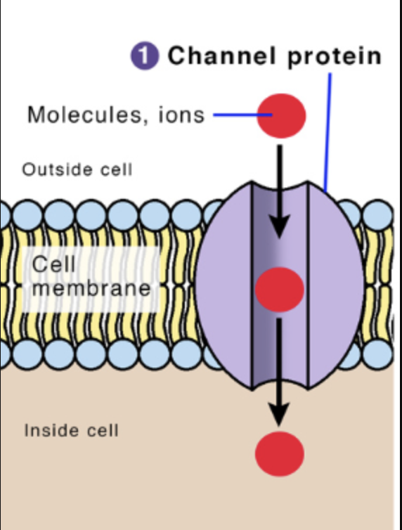
channel protein
A transmembrane protein/protein complex that produces a gated channel allowing solutes to move by simple diffusion down their concentration gradient. facilitated diffusion
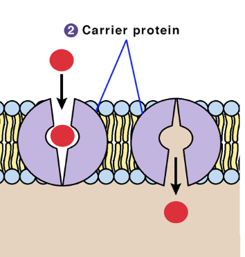
passive transporter/carrier protein complex
binds solutes, passively transports them, moves them down their concentration gradient via facilitated diffusion then saturates
active transporter/pump
transmembrane protein or protein complex that binds to and expends energy to actively transport solutes UP their concentration gradient via active transport (ATP)
ion channels
selective; Allow only specific permeants to pass through the channel, due to pore-lining residues that limit permeants based on size and charge; have open or closed conformations; DO NOT SATURATE
What are two mechanically gated ion channels?
ligand-gated (ligand binds and gate opens/closes)
voltage-gated (change in membrane potential causing channel to open/close)
what happens once channels are open?
ions move DOWN their concentration gradient by simple facilitated diffusion (no ATP needed)
permeant
molecules move through a specific transport protein/protein complex (e.g. Na+ channel —> Na+ is the permeant)

The ______ the concentration gradient, the ______ the driving orce and the _______ the movement of ions through an open channel.
larger/greater; greater; faster
Why do ion channels NOT saturate?
they con’t need to bind and undergo repeated conformational cycles like a carrier. as the concentration gradient increases, so does the transport rate.
channel = linear
transporter = saturating curve
saturation
the point where adding more permeant does not further increase transport rate; happens to transporters.
How are passive transporters like carriers?
both do not expend energy and can only move solutes DOWN their concentration gradient
How are passive transporters different from carriers?
carriers must bind to solutes to facilitate their transport across the plasma membrane, and therefore transporters can saturate and be described in terms of their kinetics
Michaelis-Menten kinetics
used to model enzyme-substrate reaction via v = (Vmax[S])/(Km+[S])
![<p>used to model enzyme-substrate reaction via v = (Vmax[S])/(Km+[S])</p>](https://assets.knowt.com/user-attachments/36fd3143-d1f9-48fb-8d99-6dd40ef8c8ac.png)
What is Vmax?
maximal rate of cellular uptake based on max number of binding sites available. Reflective of the expression level of the transporter

what is Km?
binding constant; the affinity of the transporter’s binding site for its permeant, which is the concentration at which half the binding sites are bound (1/2 Vmax)

higher binding affinity = low or high Km
low km
lower binding affinity = low or high Km
high Km
Model glucose transport through GLUT1 as a Michaelis-Menten curve. Read this curve to predict - what is the membrane transport rate if extracellular glucose increased or decreased?
GLUT1 - passive glucose transporter
if extracellular glucose increases then more glucose binds and transport rate increases, approaching Vmax.
if extracellular glucose decreases, then transport rate will decrease but once transporter is saturated, adding more won’t increase transport rate significantly.
What are 3 types of regulating transporter?
competitive, pure non-competitive, and uncompetitive

competitive inhibitor/activator
binds directly to permeant’s binding site and only changes binding affinity while Vmax stays the same
activators (agonists)
increase the function of trasporter
inhibitors (antagonists)
decrease the function of transporter
pure non-competitive inhibitor/activator
interacts with transporter or an upstream regulator and changes Vmax only while Km stays the same
Uncompetitive inhibitor/activator
binds at a site other than the permeant’s binding sites and produces a conformational change; affects Vmax and Km in same direction

You measure the kinetics of passive transporter X in some cultured cells.
Next, you apply a non-competitive inhibitor of Transporter X to the cells.
Draw the new kinetics curve for passive transporter X in the presence of your drug.
D; red "Drug" curve reaches a noticeably lower maximum height (Vmax) than the control curve, while keeping the same half-saturation point (Km)
graph of noncompetitive inhibitor
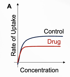
graph of competitive inhibitor

graph of Activator / Allosteric Enhancer

Active transporters
expend energy to move solutes against their concentration gradient; basis of membrane potential for ions
primary active transport
hydrolyze ARP and use that energy to move ions against their gradients
secondary active transport
one ion moves down its gradient and provides the energy to move another solute up its gradient
Predict the Michaelis-Menten curve for transport rate of the SGLT1 glucose secondary active transporter under control conditions vs. if treated with Phlorizin, a competitive inhibitor of SGLT1.
competitive inhibitor increases apparent Km while Vmax stays the same; SGLT1 curve with phlorizin should be shifted to the right to reach the same Vmax
Na+/K+ ATPase
contains an ATP hydrolysis domain that is able to hydrolyze ATP to ADP + Pi — an energetically favorable reaction; 25% of total ATP in cell
Na+/K+ ATPase process
3 Na+ ions bind along with ATP—> ATP hydrolysis switches conformation and releases Na+ outside the cell —> 2K+ binds to exposed sites —> K+ and Pi is released to reverse confirmation; cycle repeats (321 - NOKIA)

with 3 Na+ out and 2 K+ in is the concentration of what high inside and outside the cell?
high Na+ outside, high K+ inside
ion exchangers (uniporters, symporter, antiporter)
Ionic gradients store energy and can be used to power secondary active transporters, which leverage the highly negative ΔG of one gradient to compensate for the positive ΔG of concentrating other permeants against a gradient.

What equation represents ion exchangers?
ΔGtotal = ΔGion + ΔGsolute
when ΔGion is very negative, it outweights positive ΔGsolute
overall process can become energetically favorable
Secondary active transport, one substance moves DOWN its gradient which provides energy. But how?
one substance moving down, provides energy for another substance to move up its gradient
What happens if you reduce the concentration gradient of the coupled ion through symporters and antiporters?
weaken the energy source and transport rate decreases
the proximal tubule of the nephron (kidneys) are lined with what that allow filtering of blood and reabsorption of nutirnets?
SGLT which are Na+/glucose cotransporters that actively transport glucose from urine to the cell and then GLUT1 (passive transport) moves glucose from cell to blood
In the kidney, proximal tubule also express Na+ /H +
exchanger (NHE) proteins. What kind of transporters
are these and how might they help regulate
intracellular pH? How would transport rate of NHE and
SGLT be affected by eating a salty meal?
NHE is an antiporter moving Na+ and H+ in opposite directions which help regulate intracellular pH;
salty meal increased extracellular Na+ availability

how does Na+/K+ ATPase pump create an osmotic balance?
net efflux of osmotic solute which maintains cellular osmotic balance and cell volume
An imbalance in dissolved “stuff” in water produces _____ ______ that can drive movement of water to resolve an osmotic gradient.
osmotic pressure