Histology Identification Weeks 5-8
 Call Kai
 Learn
Learn Practice Test
 Spaced Repetition
 Match
Match Flashcards
 Knowt Play
1/144

There's no tags or description
Looks like no tags are added yet.
Name | Mastery | Learn | Test | Matching | Spaced | Call with Kai | Chat |
|---|
No analytics yet
Send a link to your students to track their progress
145 Terms
Hyaline Cartilage
What is the tissue present on the slide? (Monkey Trachea)

Chondrocytes
What are the yellow arrows pointing to in the slide? (Monkey Trachea)
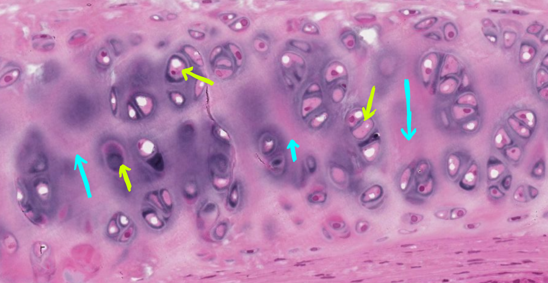
Lacunae
What is the space in which chondrocytes reside? (Monkey Trachea)

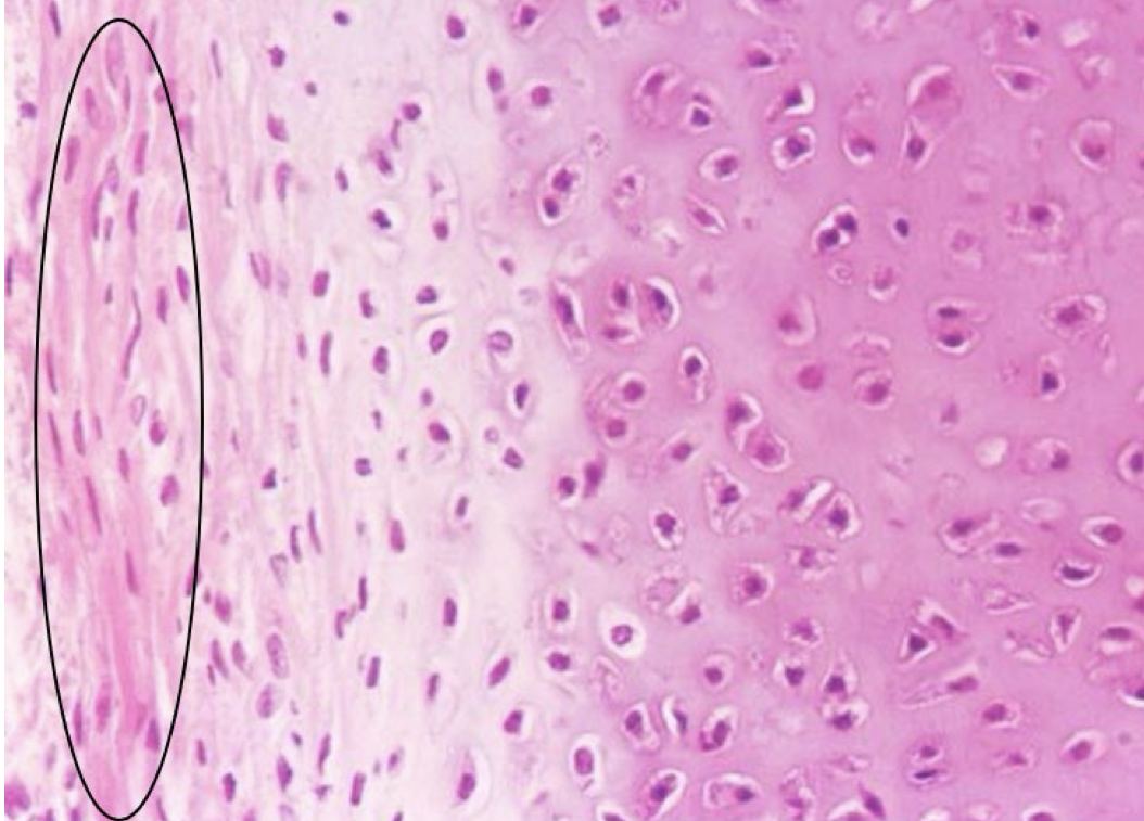
Perichondrium
What layer of cartilage is circled on the slide? (Monkey Trachea)
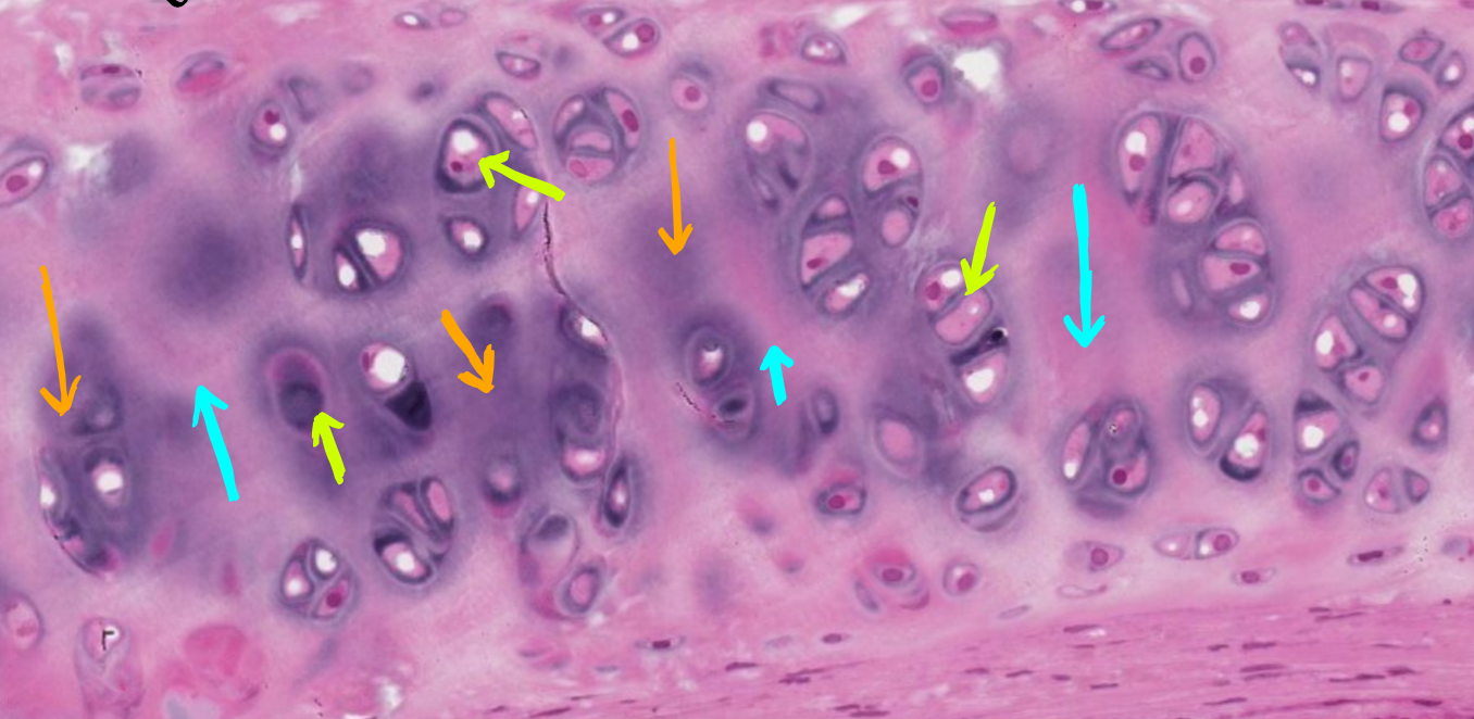
Territorial Matrix
What kind of matrix is being pointed at with the orange arrows in the slide? (Monkey Trachea)

Interterritorial Matrix
What kind of matrix is being pointed at with the blue arrows in the slide? (Monkey Trachea)
Epiphyseal Cartilage
What is the name for cartilage located on the end of a long bone? (Dog Humerus)

Growth Plate
What overall structure is pictured in this slide? (Dog Humerus)

Proliferation Zone
What is pictured by the red arrows in the slide? (Dog Humerus)

Hypertrophy Zone
What is pictured by the yellow arrows in the slide? (Dog Humerus)

Resorbing Zone
What is pictured by the green arrows in the slide? (Dog Humerus)

Osteogenic zone
What is pictured by the blue arrows in the slide? (Dog Humerus)

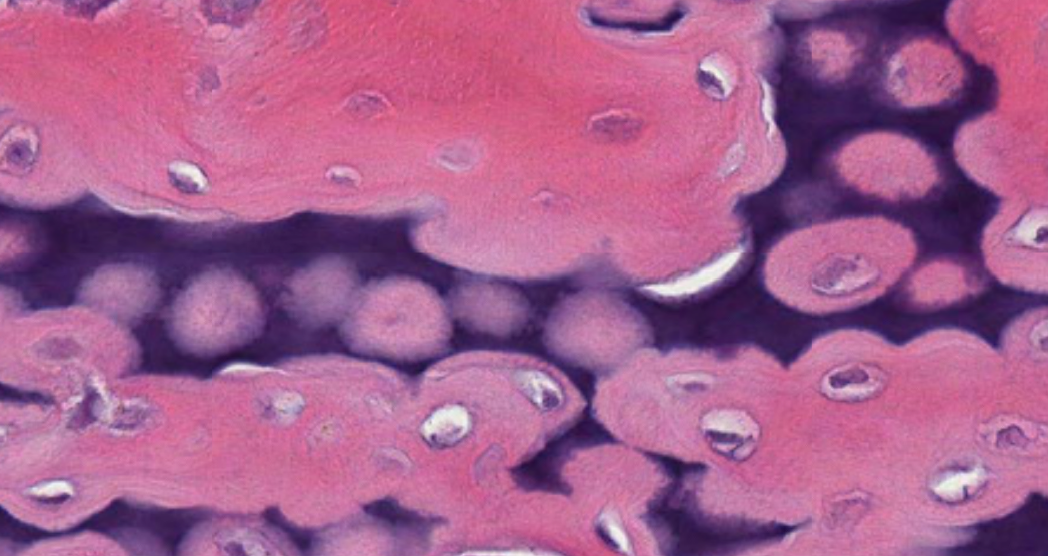
Bone, Cartilage
What stains pink in this slide? What stains blue in this slide? (Dog Humerus)
Articulating Surface
What overall structure is pictured in this slide? (Head of Humerus)

Elastic Cartilage
What type of cartilage is pictured in this slide?

Fibrous Cartilage
What type of cartilage is pictured in this slide?

Herringbone Weave, Fibrous Cartilage
What type of cartilage is pictured in this slide?

String of Pearls
This Dense Regular Connective Tissue is identified by what fibroblast nuclei pattern?

Trabeculae of Spongy Bone
What is the structure created by osteocytes pictured in the slide? (Dog Humerus)

Osteocytes
What is pictured by the black arrows on the slide? (Dog Vertebral Column)

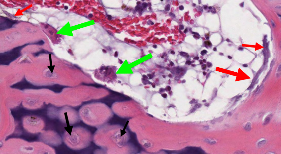
Osteocytes
What is pictured by the black arrows in the slide? (Dog Humerus)

Osteoid
What is the red material that makes up the bony spicule of spongy bone? (Dog Vertebral Column)
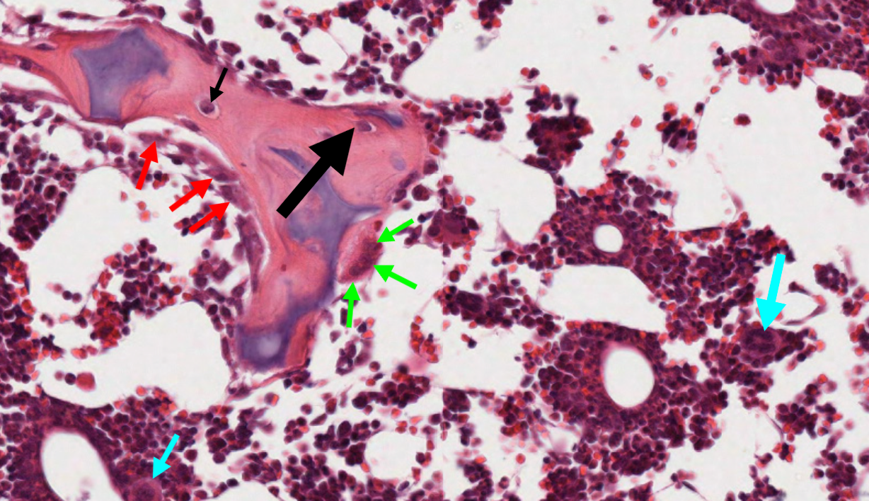
Osteoblasts
What is pictured by the red arrows on the slide? (Dog Vertebral Column)

Osteoblasts
What is pictured by the red arrows on the slide? (Dog Humerus)

Osteoclast, They are multinucleated
What is pictured by the green arrows on the slide? What separates them from other cells? (Dog Vertebral Column)
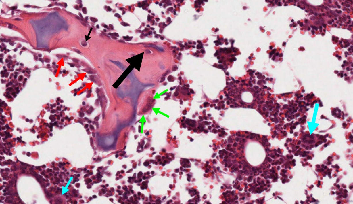
Osteoclasts
What is pictured by the green arrows on the slide?
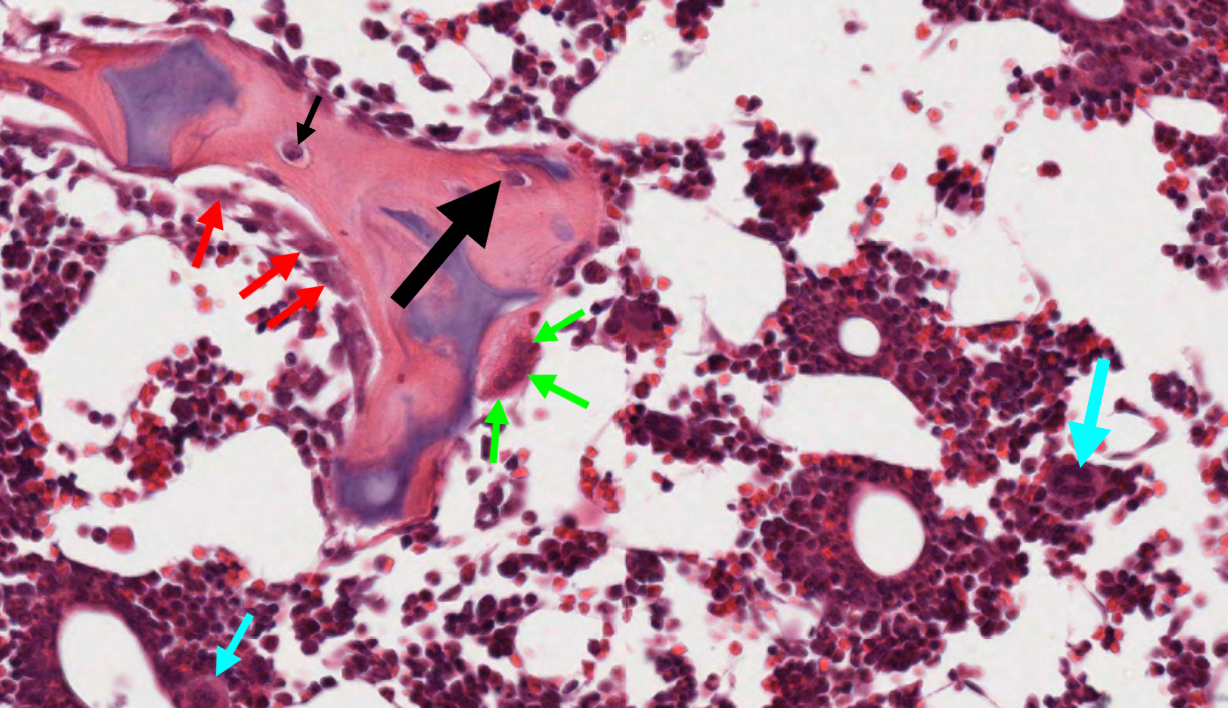
Megakaryocytes
What is pictured by the blue arrow on the slide? (Dog Vertebral Column)
Canaliculi
What is pictured by the black arrows in the slide? (Ground Bone)

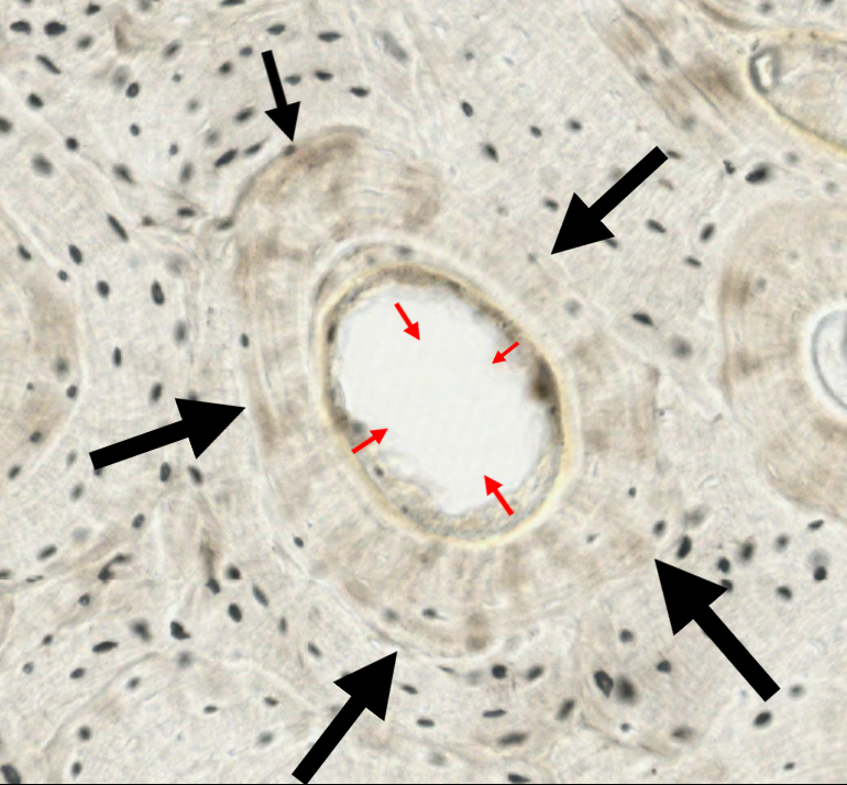
Osteon
What is the structure pictured in the slide by the black and red arrows? (Ground Bone)

Central Canal
What structure is pictured in the slide by the red arrows? (Ground Bone)
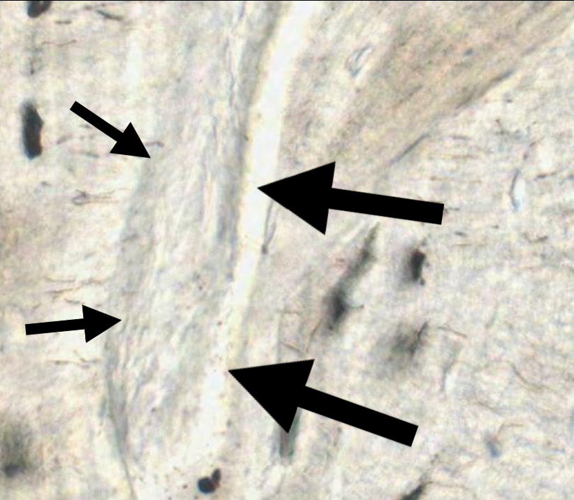
Perforating Canal
What structure is pictured in the slide by the black arrows? (Ground Bone)
Anisocytosis
What is the marked variation in red blood cell diameter (presence of macrocytes and microcytes)?

Macrocyte
What kind of RBC is >12 microns?
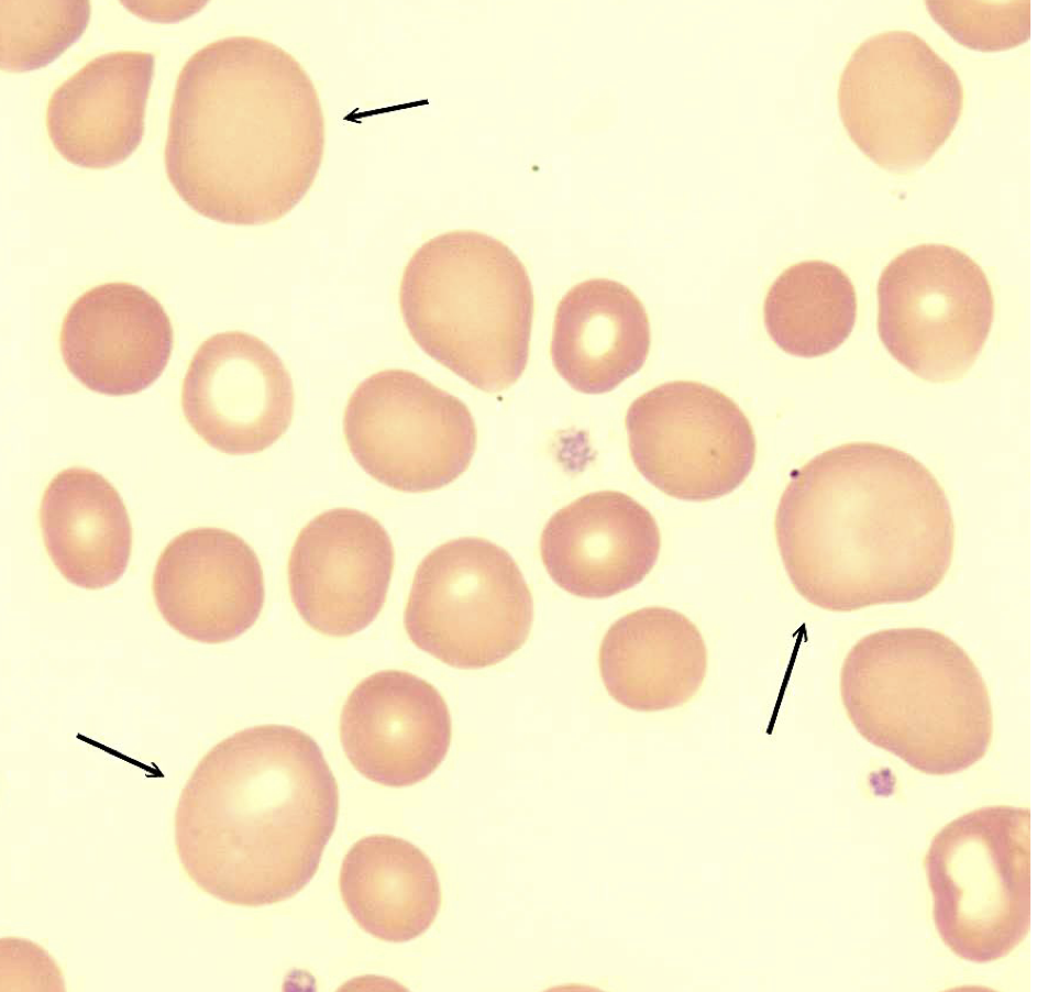
Microcyte
What kind of RBC is <6 microns?

Erthrocyte
What type of cell is pictured in the slide? (Canine Blood)

Reticulocyte
What cells are labeled by the orange arrows? (Canine Blood)

Poikilocytosis, Echinocytosis
What is the general term for a variation in shape seen in a red blood cell? What is the specific term for this variation? (Canine Blood)

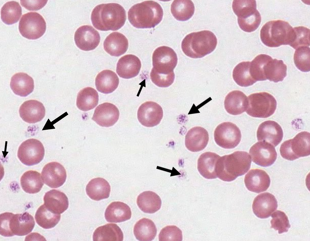
Platelet
What type of blood cell are the black arrows pointing to in the slide? (Canine Blood)
Mononuclear
What kind of nucleus do Lymphocytes and Monocytes possess?

Lymphocyte
What type of immune cell is pictured in the slide with the black arrow? (Canine Blood)

Lymphocytes
What type of immune cells are pictured in the slide with the black arrow? (Feline Blood)

Lymphocyte
What type of immune cell is pictured in the slide with the black arrow? (Equine Blood)

Lymphocyte
What type of immune cell is pictured in the slide? (Porcine Blood)

Monocyte
What type of immune cell is pictured in the slide? (Canine Blood)

Monocyte
What type of immune cell is pictured in the slide? (Feline Blood)

Monocyte
What type of immune cell is pictured in the slide? (Equine Blood)

Monocyte
What type of immune cell is pictured in the slide? (Porcine Blood)

Polymorphonuclear
What is the shape of nuclei within Neutrophils, Eosinophils, and Basophils?

Neutrophil
What type of immune cell is pictured in the slide? (Canine Blood)

Neutrophil
What type of immune cell is pictured in the slide? (Feline Blood)

Neutrophils
What type of immune cells are pictured in the slide? (Equine Blood)

Neutrophil
What type of immune cell is pictured in the slide? (Porcine Blood)

Eosinophils
What type of immune cells are pictured by the black arrows in the slide? (Canine Blood)

Eosinophil
What type of immune cell is pictured in the slide? (Feline Blood)

Eosinophil
What type of immune cell is pictured in the slide? (Equine Blood)

Eosinophil
What type of immune cell is pictured in the slide? (Porcine Blood)

Basophil
What type of immune cell is pictured in the slide with the black arrow? (Equine Blood)

Heterochromatic
In comparison to other immune cells, what nucleus coloration do lymphocytes have?

Band Cell
What kind of (immature) immune cell is pictured in the slide? (Canine Blood)

Rouleaux Formation
What is the organization of these RBCs called? (Equine Blood)

Thymus
What organ is pictured in this slide? (Canine)

Septae, Thymic Lobe
What is the unstained space dividing the structures of this slide? What structure is it dividing? (Canine Thymus)

T-cells
What are the red arrows pointing to in the slide? (Rabbit Duodenum)

Lobule
What is the entire structure pictured on the slide? (Canine Thymus)

Cortex
What is the black arrow pointing to in this lobule? (Canine Thymus)

Medulla
What is the red arrow pointing to in this lobule? (Canine Thymus)

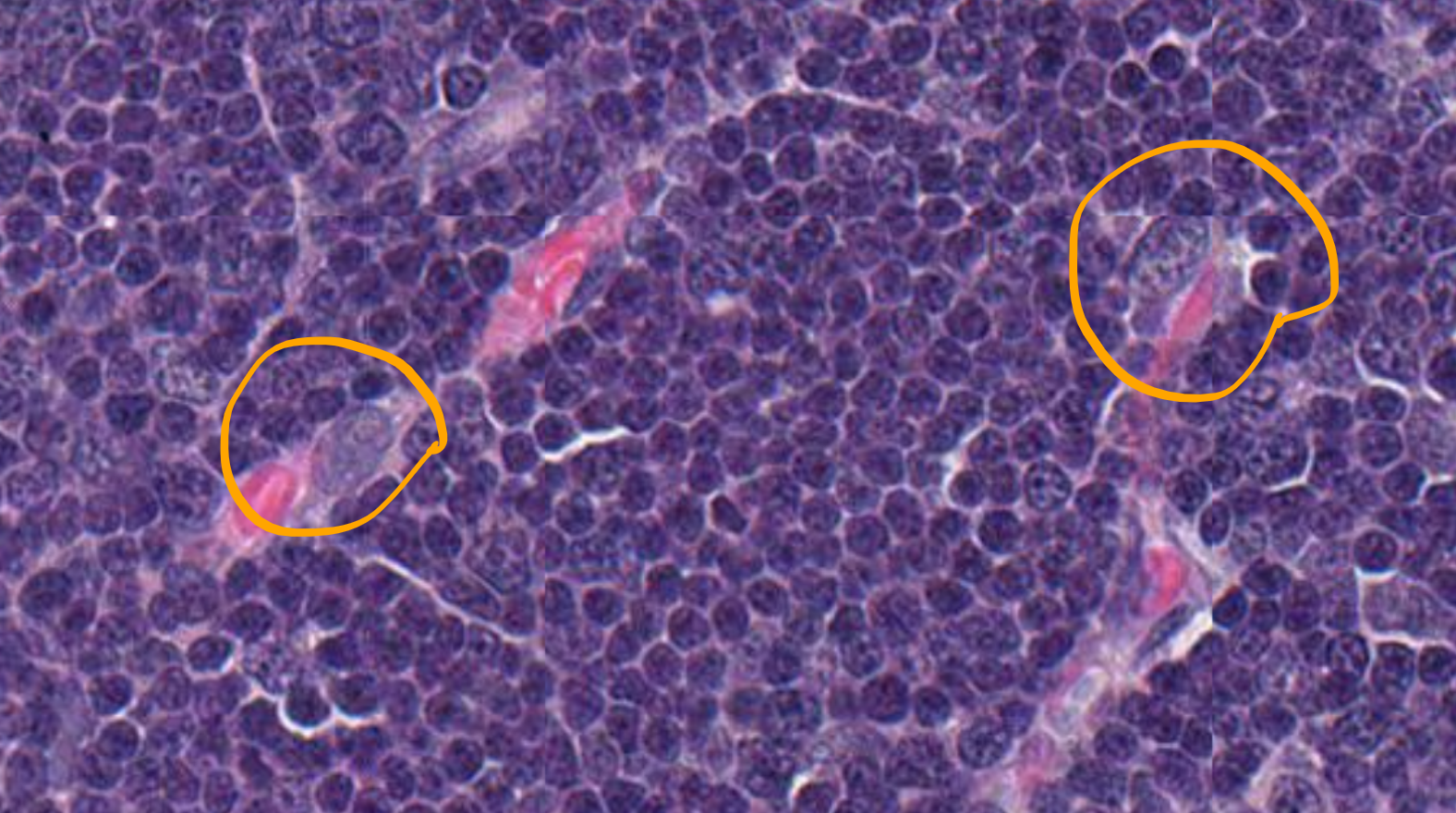
Epithelioreticular Cells
What is circled in orange on this slide? (Canine Thymus)
High Endothelial Cells, Blood-Thymic Barrier
What cells are labeled by the black arrows? Because these cells are fenestrated, what do they allow to be open for the transport of mature T-Cells? (Canine Thymus)

Continuous Endothelial Cells, Blood-Thymic Barrier
What cell is labeled by the black arrow? What structure is formed by these cells to prevent the passage of immature T-Cells? (Canine Thymus)

Thymic Corpuscles
What cellular structures are labeled by the orange arrows? (Canine Thymus)

High Endothelial Venules (HEVs), Post Capillary Venules
What structure is centered on the slide? What is a secondary name for this structure? (Canine Thymus)

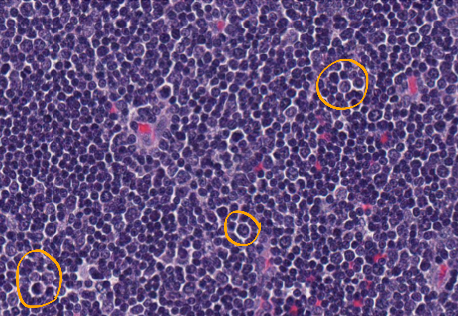
Macrophages
What cells are circled in orange? (Canine Thymus)
T-cells, Immature, Mature, Mitotic Figure
What are all of the arrows pointing to on the slide? What is the separation between red, green, and blue? (Canine Thymus)

Germinal Center
What are the black arrows pointing to on the slide? (Canine Tonsil)

Mantle Zone
What are the red arrows pointing to on the slide? (Canine Tonsil)

Tingible Body Macrophage
What cell is pointed at by the red arrows? (Canine Thymus)

Follicular Dentritic Cells
Antigen Presenting Cells
Interdigitating Dentritic Cell
Plasma cells
What cell is circled in orange on the slide? (Rabbit Duodenum)

T-Lymphocytes
What cells are outlined by the red arrows? (Rabbit Duodenum)

Large Reticular Cells
What structure is marked by the green arrow? (Canine Ileum)

Peyer’s Patch
What tissue is outlined by the arrows on the slide? (Canine Ileum)

Palatine Tonsil
What organ is pictured on the slide? (Canine)

Nasal-Associated Lymphatic Tissue (NALT)
What kind of lymphoid tissue is on the slide? (Canine Tonsil)

M-Cells
What cell type is labeled by the black arrows? (Rabbit Cecum)

Mucosa Associated Lymphoid Tissue (MALT)
What kind of lymphoid tissue is in the tonsils, esophagus, and ileum?

Afferent Lymphatic Vessels
What kind of lymphatic vessels (green arrow) are present in the lymph nodes? (Canine Lymph Node)

Efferent Lymphatic Vessels
What kind of lymphatic vessels (black arrows) are present in the MALT? (Canine Tonsil)

Gut Associated Lymphoid Tissue (GALT)
What kind of lymphoid tissue is pictured in the slide (orange arrows)? (Canine Ileum)

Diffuse Lymphoid Tissue
What kind of lymphoid tissue is outlined by the green arrows? (Canine Ileum)

Lymph Node
What organ is pictured on the slide? (Canine)

Fibrous Capsule
What structure is outlined by the black arrows? (Canine Lymph Node)

Subcapsular Sinus
What structure is outlined by the red arrows? (Canine Lymph Node)

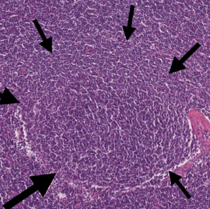
Primary Lymphoid Follicle
What is the structure outlined by black arrows in the slide? (Canine Lymph Node)
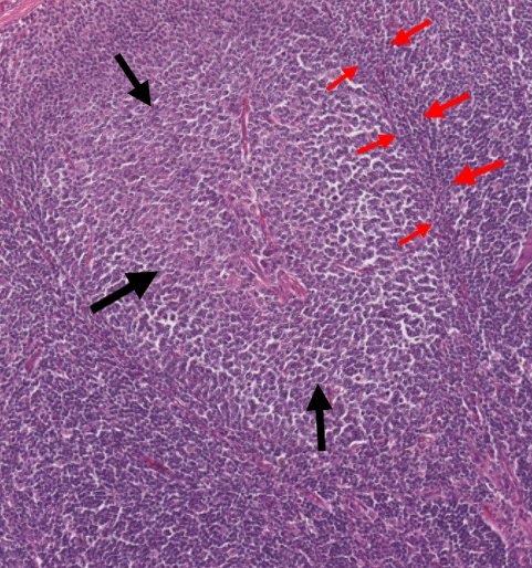
Secondary Lymphoid Follicle
What is the structure pictured in the slide (red & black arrows)? (Canine Lymph Node)
Germinal Center
What structure is outlined by the black arrows? (Canine Lymph Node)

Mantle Zone
What structure is outlined by the red arrows? (Canine Lymph Node)
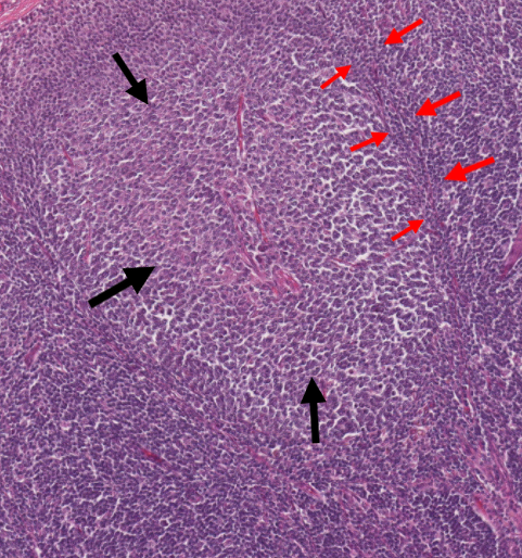
Medulla
What is the structure pictured in the slide (both red & black arrows)? (Canine Lymph Node)

Medullary cords
What structure is outlined by the black arrows? (Canine Lymph Node)
