BONES and BLOOD VESSELS: 9
 Call Kai
 Learn
 Practice Test
 Spaced Repetition
 Match
 Flashcards
 Knowt Play
1/29

There's no tags or description
Looks like no tags are added yet.
30 Terms
How is bone a living tissue?
Highly vascular (good blood supply)
Innervated (supplied by nerves)
Metabolically active capable of growth, remodelling and repair
Bone stores calcium and phosphorus and serves as a reservoir for these essential minerals
How is Bone formed?
The process (generally)
The two main ways bone is formed
In general:
Osteoblasts secrete osteoid, rich in collagen and proteoglycans
Alkaline phosphatase promotes deposition of calcium and phosphate forming hydroxyapatite crystals
Osteoblasts become trapped and differentiate into osteocytes
Osteoclasts resorb bone to maintain shape, strength and calcium balance
Ossification occurs in two main ways: Intramembranous and endochondral

Intramembranous:
Bone is going to directly form from the mesenchyme with osteoblasts forming osteoid. Examples of this is most bones in skull and facial bones form
Endochondral:
Where mesenchyme cells first form cartilage which serves as a template, this cartilage gets replaced by bone. KEY POINT: The cartilage does not turn into bone, it gets replaced by bone. Examples of this is most of the long bones: humerus, femur, vertebrae and ribs
What are the three important cells of the bone?
Osteoblasts: Bone-forming cells, that secrete osteoid (unmineralized matrix) and initiate mineralization
Osteocytes: Mature osteoblasts trapped in lacunea, maintain bone matrix and communicate via canaliculi
Osteoclasts: Large multinucleated cells that resorb bone, important in remodelling and calcium homeostasis

How does bone undergo remodelling?
Bone remodelling is a lifelong process, where old bone is replaced by new bone. Remodelling maintains bone strength, repairs microdamage and helps regulate calcium levels.
Osteoclast activation, which resorbs bone over a few weeks
Mononuclear cells then prepare the surface for osteoblasts, which lay down osteoid.
The osteoid mineralizes over several months.
What are the two main types of bone?
Compact (cortical) bone: Dense, strong and forms the outer layer of bones
Spongy (cancellous/ trabecular) bone: Porous, vascular (good blood supply) and found inside bones, contains bone marrow

The structure of Compact (cortical) bone:
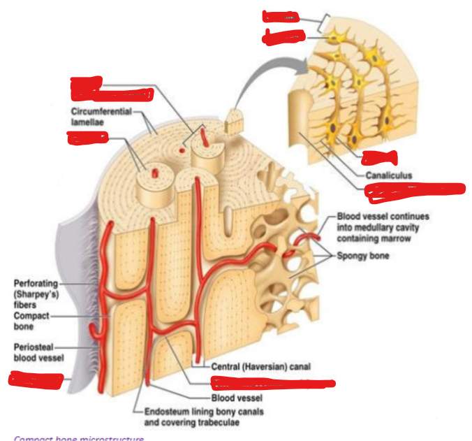

Organized into osteons (Haversian systems)
Central Haversian canals, which carry blood vessels, nerves and lymphatics
Lamellae are layers of bone matrix, that surround the canals
Osteocytes in lacunae communicate via canaliculi by extending their filopodia, link to each other via gap junctions
Canaliculi: Channels in the bone matrix
Filopodia: Cellular projections that run inside the channels
Interstitial lamellae: remnants of past osteons which remain after bone remodelling


The structure of spongy (cancellous/ trabecular) bone:

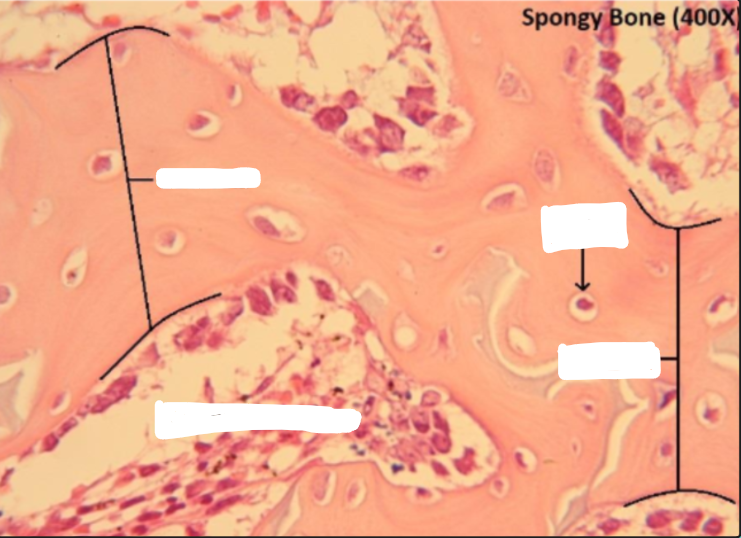

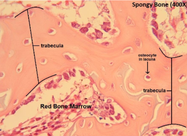
Composed of trabeculae, thin beams of bone with osteocytes
Spaces between trabeculae contain bone marrow (red or yellow)
Spongy bone lacks osteons, canaliculi connect directly to the surface or marrow for nutrient exchange
Outline Longitudinal (interstitial) growth in bones:
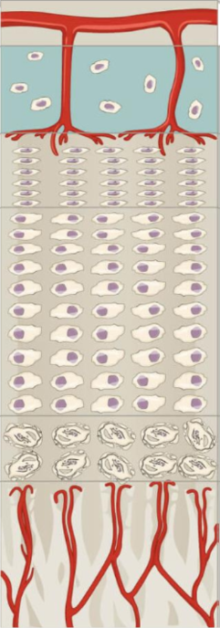
Occurs from birth through early adulthood, primarily through cartilage proliferation at the epiphyseal plates (growth plates) located at the ends of long bones.
The process slows and eventually stops when the growth plates fuse and are completely replaced by bone. [14-16 in females] and [16-18 in males]
Outline appositional growth in bones:
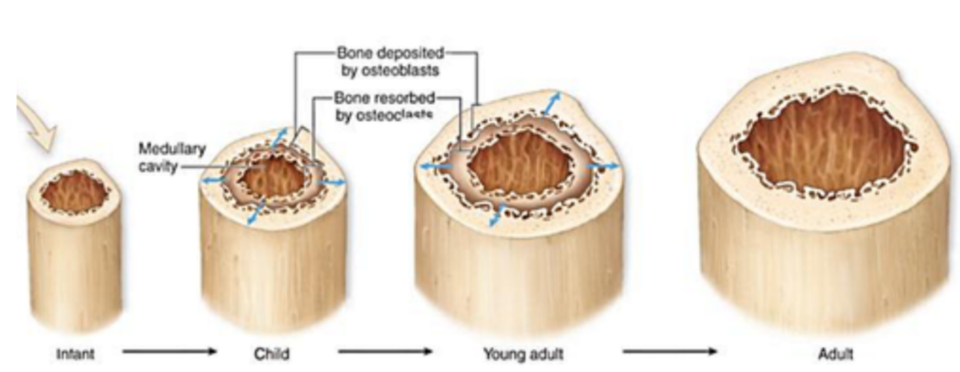
Increases in bone width
Primarily occurs during childhood, but appositional growth also occurs in adults as a response to stress from physical activity like weight-bearing exercise.
Osteoblasts deposit new bone on the outer surface, while osteoclasts resorb bone from the inner surface of medullary cavity
This allows bone to become thicker, without becoming overly heavy
Label the zones on the epiphyseal plates

Explain the importance of vitamin D in normal bone stability:
Vitamin D essential for calcium absorption and proper bone mineralization. Deficiency leads to soft, weak bones and leads to increased risk of fractures:
Children: Rickets - skeletal deformities such as bowed legs, shortened height and stature, painful to walk - older children
Adults: Osteomalacia - soft, pliable (easily bend without breaking) bones prone to fracture.
Sources: Sunlight (skin synthesis) and dietary intake (fortified foods, dairy)
Describe the genetic basis and phenotypical changes in Osteogenesis imperfecta
Most cases due to mutation in COL1A1 or COL1A2 genes, where mutated collagen fibres do not ‘knit together‘ or not enough produced or both.
Results in: weakened bones (fractures common), short stature, presence of blue sclera, hearing loss, hypermobility (loose joints), poor teeth development.
What is Osteoporosis? Discuss primary type 1, type 2 and secondary and how to overcome these risk factors?
Primary:
Type 1:
Occurs in postmenopausal women
due to an increase in osteoclast number, so increased bone reabsorption
due to loss of Oestrogen after menopause
Type 2:
Occurs in older men and women
due to loss of osteoblast function (senile osteoporosis)
Loss of both oestrogen and androgen
As we get older, osteoblasts (formation of new bone) functions less, and bone breakdown continues to happen, additionally as you get older there is reduced sex hormones (oestrogen and androgen) which contribute to loss of bone
Secondary:
Result from drug therapy (corticosteroids), which decreases osteoblast activity, less bone made, decreased calcium absorption from gut, increases bone loss
Process affects bone remodelling => reasons being malnutrition (not enough calcium, vitamin D to maintain bone) prolonged immobilisation → bone not experiencing their normal mechanical load, bone resorption increases
Weightlessness e.g. space travel, where bones aren’t loaded by gravity, so increased bone loss
Other disease e.g. hyperparathyroidism
Too much PTH → Increases bone resorption → increase calcium released from bone → prolonged excess PTH contribute to osteoporosis
To modify these risk factors:
Have a high sufficient calcium intake
Exercise: Physical activity required to maintain bone mass
What is cartilage?
Cartilage is an avascular connective tissue composed of chondrocytes embedded within an extracellular matrix.
Provides crucial support and flexibility to many body structures
Shock absorber
Forms smooth and low-friction surface in joints
Maintains soft tissues such as the ear and airways
Where in the body is cartilage found?
In foetal development, most skeleton is formed as cartilage, which is later replaced by bone through process of endochondral ossification which continues after birth.
In adults, cartilage is in specific areas such as joints, nose, respiratory passages and between certain bones. Where flexibility or smooth movement is required.
Describe the composition of cartilage in terms of its cells and extracellular components:
Cartilage consists of two main components:
Chondrocytes, which are mature cartilage cells derived from chondroblasts.
They occupy small spaces in the ECM called the lacunae and are responsible for producing and maintaining the matrix
Since the cartilage is avascular, nutrients and gases must diffuse through matrix from surrounding tissues or synovial fluid (viscous fluid that provides lubrication and nutrients to avascular cartilage)
Extracellular matrix:
Gives cartilage mechanical properties, firmness, elasticity and resilience.
Rich in water, type 2 collagen fibres, proteoglycans containing glycosaminoglycans (GAGs) such as chondroitin sulphate, these molecules attract and trap water, making matrix resistant to compression
Contains three types of cartilage: Hyaline cartilage (type 2 collagen), elastic cartilage (elastic fibres and type 2 collagen), fibrocartilage (type 1 and 2 collagens)
How cartilage, which is avascular obtains nutrients?
Cartilage is surrounded by perichondrium, a layer of dense connective tissue containing blood vessels and progenitor cells that form new cartilage
The perichondrium nourishes the cartilage via diffusion
Articular cartilage (a type of hyaline cartilage found at joint surfaces) however lacks perichondrium and is nourished by synovial fluid (articular surface) and the underlying bone/ bone marrow
Since no direct blood supply, healing and repair after injury is limited. Damage is usually replaced by fibrous scar tissue rather than new cartilage
Describe what Hyaline cartilage is, structure, locations, functions and clinical aspect:
Most common type of cartilage and serves as structural model for most developing bones.
Structure:
ECM contains fine type 2 collagen fibres, not visible under light microscope, so gives a smooth and uniform appearance.
Chondrocytes are located in lacunae often in small groups called isogenous clusters, showing cell division
Surrounding matrix rich in GAGs which are basophilic (appear blue/purple)
Locations:
Articular surfaces of moveable joints (the ends of bone that meet each other at a joint) for example knee and shoulder
Costal cartilages (connecting ribs to the sternum)
Nasal septum and cartilage of the nose
Trachea and bronchi
Epiphyseal (growth) plates of growing bones
Functions:
Provides smooth, low- friction surfaces for joint movement
Serves as a precursor for bone formation during growth
Maintains shape and structure in respiratory passages
Clinical notes:
Articular hyaline cartilage found in articular surfaces of joints and epiphyseal plates lack perichondrium and blood supply, so damage in this area, leads to limited repair, leads to persistent pain and degeneration.

Describe what elastic cartilage is, structure, locations, functions and clinical aspect
Elastic cartilage contains a dense network of elastic fibres and collagen.
Structure:
Elastic fibres form fine network throughout matrix and into perichondrium
Chondrocytes are larger and more numerous than hyaline cartilage
Elastic network demonstrated using special stains
Locations:
External ear (auricle/pinna)
Epiglottis (part of larynx)
Eustachian (auditory) tube => connecting ear and nose, allowing pressure on either side of eardrums to be same
Functions:
Provides flexible support and maintains shape (if ear flap returns to position when bent)
Resists repeated bending and deformation
Clinical relevance:
As you increase in age, hyaline cartilage more likely to develop calcium deposits and become more stiff // For elastic cartilage less likely to undergo calcification maintaining flexibility

Describe what fibrocartilage is, structure, location, function and clinical notes:
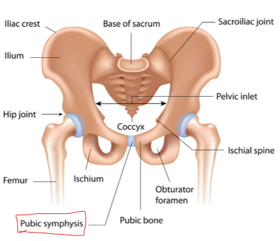
Fibrocartilage is tough, durable tissue found in tough, durable tissue where strong compressive and tensile (pulling/ stretching forces).
Structure:
Contains type 1 and type 2 collagen fibres arranged in parallel bundles, aligned with stress lines
Abundant in type 1 collagen fibres, provides the tensile strength
Chondrocytes are fewer and lie singly or in pairs with lacunae, often between dense collagen bundles
No perichondrium, as fibrocartilage merges directly with tendons or ligaments
Locations:
Intervertebral discs (annulus fibrosus)
Pubic symphysis
Temporomandibular and sternoclavicular joints
Sites where tendons attach to bone
Functions:
Provides tensile strength and resistance to compression
Acts as shock absorber and maintains joint stability
Clinical notes:
Since fibrocartilage has no perichondrium, healing after injury is poor.

-
Understand the key role water plays as a structural and functional component in bone and cartilage?
Cartilage:
Cartilage matrix contains proteoglycans which have negatively charged GAG chains, the negative charges attract water into cartilage matrix.
So, when cartilage is compressed, water is squeezed within, hydrated matrix resists compression and absorbs shock. Also allows nutrients, oxygen and waste products to diffuse through matrix (important since cartilage is avascular).
Bone:
Water contributes to hydration and mechanical properties of bone, helps prevent bone from being excessively brittle, provides a medium for ions/ nutrients and metabolic processes involving bone cells.
What causes achondroplasia, what happens to bone growth and what the person’s skeletal features are:
Achondroplasia is a genetic disorder of bone growth, and is the most common cause of disproportionate short stature.
Caused by mutation in FGFR3 gene = fibroblast growth factor receptor 3, becoming overactive
Where too much inhibition of chondrocyte proliferation in growth plate, leads to reduced cartilage growth, reduced endochondral ossification, leading to short limbs and disproportionate short stature
Features:
Disproportionately short limbs
Large head (macrocephaly)
Prominent forehead (frontal bossing)
Appreciate how limited nutrient diffusion affects chondrocyte metabolism and ECM maintenance:
Since, cartilage does not have blood vessels, substances have to diffuse through the extracellular matrix to reach.
Synovial fluid → oxygen + glucose/nutrients → Chondrocytes
Since oxygen supply is relatively limited, chondrocytes adapt to low-oxygen environment, so rely on anaerobic glycolysis to make ATP.
Chondrocytes are responsible for producing and maintaining cartilage ECM the collagen and proteoglycans/ GAGs. So, reduced nutrients/ oxygen leads to reduced synthesis and repair of ECM.
Describe how Hyaline cartilage acts as a template to long bone growth in endochondral ossification
The key idea: hyaline cartilage model is made first, this acts as a template that is gradually replaced by bone, this process known as endochondral ossification.
Hyaline cartilage model forms
Chondrocytes divide and produce cartilage matrix
Chondrocytes enlarge (hypertrophy), surrounding cartilage matrix becomes calcified. Chondrocytes die because nutrients can no longer diffuse through the calcified matrix effectively
Blood vessels invade calcified cartilage, bringing osteoprogenitor cells, which differentiate into osteoblasts
Osteoblast deposit bone matrix, and cartilage model becomes bone.
About Red bone marrow:
Full of developing blood cells
Rich blood supply
Only found in spongy bone
Function: To replenish cells in blood (haemopoiesis)
About White bone marrow:
Full of adipocytes
Poor blood supply
Function: Shock absorber and energy source, during shock such as haemorrhage can convert to red marrow
What are the constituents of bone marrow:
Haematopoietic stem cells: Multipotent cells that give rise to all blood cell lineages.
Progenitor cells: Committed precursors for erythroid, myeloid, and lymphoid cells.
Maturing blood cells: Developing erythrocytes, leukocytes, and megakaryocytes at various stages.
Stromal cells: Provide structural support and secrete growth factors.
Sinusoidal capillaries: Allow mature cells to enter the bloodstream
The different layers present in veins and arteries and their functions:
Tunica = coat
Tunica intima (inner layer): Is a single layer of endothelial cells lining the lumen, supported by a thin layer of connective tissue and sometimes an internal elastic lamina. Function: Provide a smooth surface to reduce friction and acts as a selective barrier for exchange between blood and tissues
Tunica media (middle layer): Layers of smooth muscle cells and elastic fibres. Functions: Controls vessel diameter through vasoconstriction and vasodilation, regulating blood flow and pressure.
Tunica adventitia (outer layer): Connective tissue with collagen and elastic fibres, may contain small blood vessels and nerves. Function: Provide structural support to anchor to surrounding tissue tissues


About capillary function and the 3 different types:
Capillaries are single endothelial cells with a basement membrane, their function is to exchange gases, nutrients and waste between blood and tissues.
Types:
Continuous: Tight junctions (e.g. muscle, brain)
Fenestrated: Pores for filtration (e.g. kidney, endocrine glands)
Sinusoidal: Large gaps for cell movement (e.g. liver, bone marrow)

Name the major blood vessels of the body and determine whether they contain oxygenated or deoxygenated blood or a mixture of both
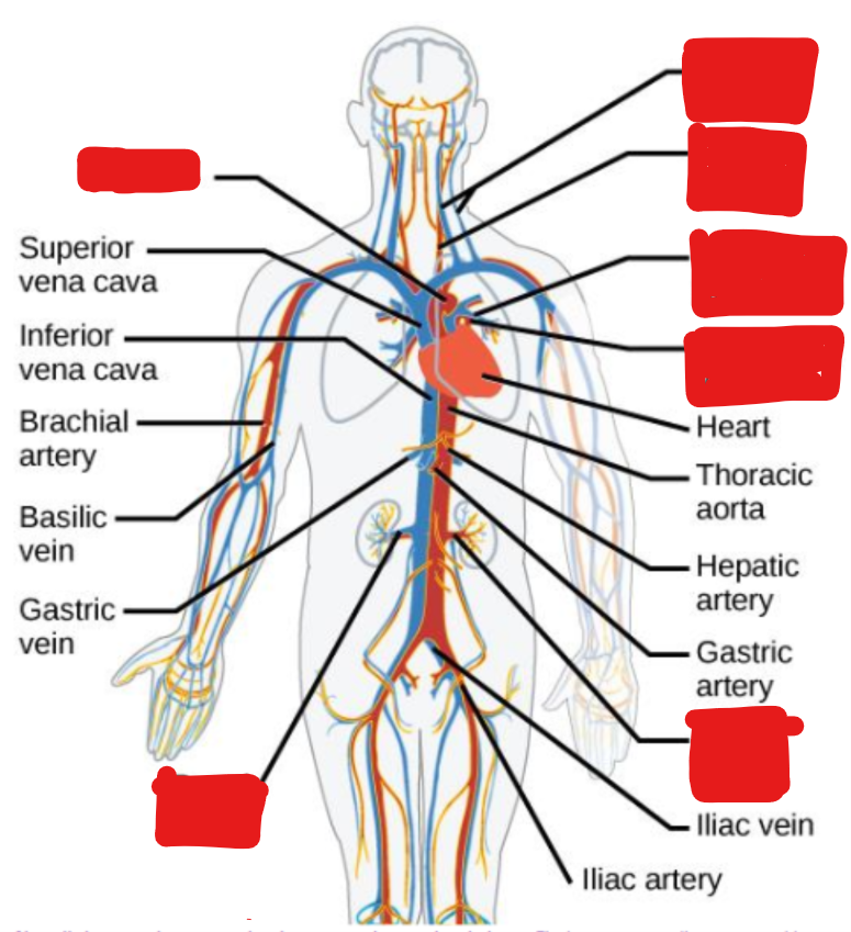
Artery = carries blood away from heart, important, since pulmonary artery contains deoxygenated blood, but is known as artery.
So, key points:
arteries = away from heart // veins = towards heart
Systemic circulation: arteries = oxygenated // veins = deoxygenated
Pulmonary circulation: arteries = deoxygenated // veins = oxygenated

