Life Sci 7a - Week 5 Question Bank
 Call Kai
 Learn
 Practice Test
 Spaced Repetition
 Match
 Flashcards
 Knowt Play
1/37

There's no tags or description
Looks like no tags are added yet.
38 Terms
What are three mechanisms of RNA processing in eukaryotes?
Three mechanisms of RNA processing (chemical modification of the primary transcript to generate the finished mRNA) in eukaryotic cells are as follows:
A) Addition of the 5' cap. This modified nucleotide (7-methylguanosine) allows the mRNA to be recognized by the translation initiation complex and helps stabilize the mRNA.
B) Addition of the poly(A) tail. This modification is important in the export of the mRNA into the cytoplasm, and the poly(A) tail, like the 5' cap, helps stabilize the mRNA.
C) RNA splicing. Splicing is the process by which introns are removed from a primary RNA transcript. A single transcript with multiple introns may be spliced in different ways to generate multiple mRNAs, each resulting in a different protein with somewhat different properties. Alternative splicing is therefore one more mechanism that increases the number of ways that the genetic information stored in DNA can contribute to the diversity of cellular proteins in eukaryotes.
What are five types of noncoding RNA and their functions?
Five types of noncoding RNA are
(1) ribosomal RNA (rRNA), which is found in all ribosomes and is essential in translation
(2) transfer RNA (tRNA), which carries individual amino acids to the ribosomes in translation
(3) small nuclear RNA (snRNA), which is involved in eukaryotic gene splicing, polyadenylation, and other processes in the nucleus
(4) microRNA (miRNA), which can destroy transcripts or inhibit translation
(5) small interfering RNA (SiRNA), which can also destroy transcripts or inhibit translation. snRNA, miRNA, and SiRNA have other regulatory functions as well, and these are still not fully understood.
When a region of DNA that contains the genetic information for a protein is isolated from a bacterial cell and inserted into a eukaryotic cell in a proper position between a promoter and a terminator, the resulting cell usually produces the correct protein. But when the experiment is done in the reverse direction (inserting eukaryotic DNA into a bacterial cell), the correct protein is often not produced. Why is this the case?
The eukaryotic DNA sequence contains introns, which the bacterial cell cannot splice out properly. Thus, the correct protein is not produced from the information in the bacterial RNA transcript.
Alternative splicing allows for:
different polypeptides to be made from a single gene.
Alternative splicing means that:
different spliced forms contain different combinations of exons.
An exon is:
an RNA sequence that is maintained in the mature mRNA.
Draw one of the 20 amino acids and label the amino group, the carboxyl group, the R group, and the a carbon.

What are the three major groups of amino acids as categorized by the properties of their R groups? How do the chemical properties of each group affect protein shape?
Amino acids can be categorized into three major groups based on the properties of their side chains. The first group consists of the hydrophobic (water-fearing") amino acids, whose side chains are nonpolar, usually found buried in the interior of the folded proteins, and typically form bonds with other hydrophobic amino acids (for example, valine) or nonpolar solvents. The second group consists of the hydrophilic ("water-loving") amino acids. Hydrophilic amino acids include ones with polar side chains, usually found on the outside surface of folded proteins, and typically form bonds with other hydrophilic amino acids or water. Hydrophilic amino acids also include basic amino acids with side chains that are positively charged at intracellular pH (for example, lysine) and acidic amino acids with side chains that are negatively charged (for example, aspartic acid).
The third group includes "special" amino acids, whose unique structures and chemical properties have significant effects on higher levels of protein structure because they allow more or less flexibility around the peptide bond (glycine and proline) or allow covalent interactions between side chains (cysteine).
How do peptide bonds, hydrogen bonds, ionic bonds, disulfide bridges, and noncovalent interactions (van der Waals forces and the hydrophobic effect) define a protein's four levels of structure?
The way in which amino acids interact in a polypeptide chain is important for the structure and function of the protein. Peptide bonds maintain the primary structure of a polypeptide chain. These bonds form between the carboxyl group of one amino acid and the amino group of the next amino acid in the chain. Hydrogen bonds are important in maintaining the secondary structure of the polypeptide chain. These bonds form between the oxygen in the carbonyl group (C=O) of one peptide bond and the hydrogen in the amide group (N-H) of another. Two common types of secondary structures formed by hydrogen bonds are a helices andß sheets.
Four types of bonds or interactions are important in creating tertiary and quaternary structures. Ionic bonds form between a negative charge and a positive charge. For example, an ionic bond forms between a basic amino acid and an acidic amino acid because they have oppositely charged side chains. Hydrogen bonds form between the oxygen of one amino acid's side chain and the hydrogen of another amino acid's side chain. Disulfide bridges are covalent bonds that form between two cysteines in the same polypeptide chain or between two cysteines in two different chains. Finally, noncovalent interactions, such as van der Waals forces and hydrophobic interactions, maintain interactions with different domains of the protein.
What ultimately determines the three-dimensional shape of a protein?
The order of amino acids in the polypeptide chain determines the way in which proteins fold because of the various interactions and bonds formed between the amino acids. These interactions, depending on the type and location, will give rise to specific secondary and tertiary structures.
A mutation leads to a change in one amino acid in a protein. The result is that the protein no longer functions properly. How is this possible?
The sequence of amino acids in a protein determines how a protein folds, so a change in even a single amino acid can affect the way the protein folds and can disrupt its function. For example, if the hydrophobic R groups of two amino acids must aggregate for proper structure and function, then a mutation that changes one of the hydrophobic amino acids to an acidic or a basic amino acid will prevent this aggregation and disrupt structure and function. Similarly, if proper folding requires interaction between the R groups of an acidic and a basic amino acid, then changing either one of them to a hydrophobic amino acid means that proper folding will not take place.
What are the relationships among the template strand of DNA, codons in
MRNA, anticodons in tRNA, and amino acids?
The codons of mRNA are groups of three nucleotides that code for a particular amino acid. Each is transcribed from the template strand of DNA according to the normal rules of base pairing (but in RNA, U replaces T).
The sequence of the codons in the mRNA gives rise to the order of the resulting amino acid polypeptide chain. The codons are translated on ribosomes by tRNAs. The sequence of each tRNA includes a group of three nucleotides called an anticodon that is complementary in sequence and, therefore, can recognize and bind to a specific codon in the mRNA. Because of the complementary and antiparallel nature of nucleic acid structures, an anticodon in a tRNA has the same 3' to 5' sequence as the template DNA, except with U's instead of T's. Each tRNA is also bound to a specific amino acid, affiliated with a particular anticodon-codon pair, on the 3' end of the molecule. When the mRNA is being "read" on the ribosome, the order of the amino acids in the polypeptide chain is dependent upon the sequential interaction of the mRNA codon with the correct tRNA anticodon-amino acid pair. See Fig. 5.15.
Which polypeptide sequences would you expect to result from a synthetic mRNA with the repeating sequence 5'-UUUGGGUUUGGGUUUGGG-3'?
The three reading frames are as follows:
VUU GGG UUU GGG..., which codes for repeating Phe-Gly-Phe-Gly...
VUG GGUUUGGGU..., which codes for repeating Leu-Gly-Leu-Gly...
UGG GUU UGG GUU..., which codes for repeating Trp-Val-Trp-Val...
What are the steps of translation? Name and describe each one.
Translation of mRNA by ribosomes can be divided into three steps.
- Initiation: Initiation factors bind to the 5' cap of the mRNA (in eukaryotic cells) or at the Shine-Dalgarno sequence (for prokaryotes) and recruit the small subunit of the ribosome and a tRNA charged with methionine. This complex then moves along the mRNA until it finds a start codon (AUG, coding for methionine). The large ribosomal subunit then joins the complex and causes the initiation factors to be released. The tRNA Met is then bound in the P site of the ribosome. The next tRNA, determined by the codon of the mRNA, binds in the A site of the ribosome. This elicits a coupled reaction in which the bond between the Met and its tRNA is broken and a new bond is formed between the carboxy! group of the Met and the amino group of the next amino acid (a peptide bond). The ribosome complex then slides to the next codon on the mRNA, shifting the now-uncharged tRNA Met to the E site, where it is released from the ribosome complex, and moving the peptide-bearing tRNA to the P site. The A site is now free for the next charged tRNA. See Fig. 5.17.
- Elongation: The ribosome continues in this fashion, shifting down the mRNA one codon at a time, adding amino acids to the growing peptide chain. Elongation factors provide the energy needed for these reactions to happen. See Fig. 5.18.
- Termination: When the ribosome complex comes across a stop codon (UAA, UAG, or UGA), a protein release factor binds in the A site of the ribosome and causes the bond between the polypeptide chain and the last tRNA to break. Once the polypeptide chain is released, the ribosomal subunits disassociate from the mRNA and each other, and translation is complete. See Fig. 5.19.
Bacterial DNA containing an operon encoding three enzymes is introduced into chromosomal DNA in yeast (a eukaryote) in such a way that it is properly flanked by a promoter and a transcriptional terminator. The bacterial DNA is transcribed and the RNA correctly processed, but only the rotein nearest the promoter is produced during translation. Why?
With proper eukaryotic processing, the RNA transcript from the bacterial DNA will be capped at the 5' end. The initiation complex will form at the 5' cap and move along the mRNA until the first AUG codon is encountered. At that point, translation begins. When one of the termination codons is encountered, the polypeptide is released. Translation of the downstream polypeptides cannot take place because the Shine-Dalgarno sequences preceding them are not recognized by the eukaryotic translational machinery.
If you were to denature a protein in a lab, you'd most likely be:
Unfolding the protein with the help of heat or a chemical treatment
The smaller sub-structures such as helices and sheets within a single polypeptide chain are referred to as:
Secondary
T/F: Most proteins have their metabolic activity disrupted when denatured.
True
The interactions between amino acids are major factors in determining the shape of a protein. These interactions can be affected by the environment surrounding a protein.
Which factor would have an effect on the shape of a protein?
whether the other molecules in the environment are predominantly hydrophilic or hydrophobic
the temperature of the environment
the pH of the environment
the concentrations of ions present in the environment
Which ribosomal unit is recruited before the translational machinery finds its start codon?
The small ribosomal subunit
Assuming A‑U and G‑C pairing between the anticodon and the codon, what anticodon in tRNATyr would pair with the codon 5′‑UAC‑3′?
5'-GUA-3'
Ribosomes in prokaryotes and eukaryotes are:
similar in structure and translate using the same genetic code.

The figure below depicts two eukaryotic genes, Gene A and Gene B. For each gene, exons are depicted as white boxes, and introns are indicated by lines. The corresponding DNA sequence is shown below the gene diagrams. The bolded and underlined base pair represents the +1 transcription start site for each gene. The three dots on either side of the DNA sequence indicate that this DNA sequence can be assumed to extend beyond this figure on both sides. Use this figure to answer the following questions.
T/F: The promoter sequence for Gene B is located to the right of the figure.
False
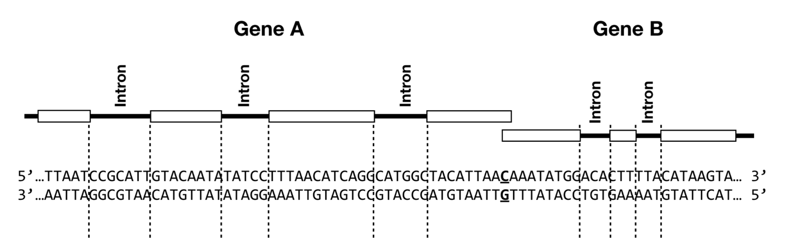
The figure below depicts two eukaryotic genes, Gene A and Gene B. For each gene, exons are depicted as white boxes, and introns are indicated by lines. The corresponding DNA sequence is shown below the gene diagrams. The bolded and underlined base pair represents the +1 transcription start site for each gene. The three dots on either side of the DNA sequence indicate that this DNA sequence can be assumed to extend beyond this figure on both sides. Use this figure to answer the following questions.
T/F: The template strand for Gene A is the top strand as shown in this diagram.
True

The figure below depicts two eukaryotic genes, Gene A and Gene B. For each gene, exons are depicted as white boxes, and introns are indicated by lines. The corresponding DNA sequence is shown below the gene diagrams. The bolded and underlined base pair represents the +1 transcription start site for each gene. The three dots on either side of the DNA sequence indicate that this DNA sequence can be assumed to extend beyond this figure on both sides. Use this figure to answer the following questions.
T/F: You would expect to find a long stretch of adenosines (a polyA tail) to the right of the figure in the DNA sequence for Gene B.
False

The figure below depicts two eukaryotic genes, Gene A and Gene B. For each gene, exons are depicted as white boxes, and introns are indicated by lines. The corresponding DNA sequence is shown below the gene diagrams. The bolded and underlined base pair represents the +1 transcription start site for each gene. The three dots on either side of the DNA sequence indicate that this DNA sequence can be assumed to extend beyond this figure on both sides. Use this figure to answer the following questions.
Which of the following probes would bind to the mRNA for Gene B?
5'-ATATTT-3'
If you made a change in the promoter sequence in the DNA that inactivates the promoter, what would happen at the RNA level?
The RNA polymerase would not be able to recognize and bind the DNA, so no RNA would be made.

A double-stranded DNA molecule, only part of which is shown, is being transcribed. If the molecule is transcribed from left to right, one of the nucleotides shown in bold red font would be the first transcribed in this small molecule.
Which sequence is the correct RNA produced from the transcription of this DNA molecule?
5′‑AUGAUCGGAUCGAUCCAU‑3′

Each of the questions below describes a different mutation that could occur in a eukaryotic gene. Determine which RNA gel below [A, B, C, or D] you would be most likely to observe as a consequence of each mutation. On each gel, “WT” is the normal wild type version of the mature mRNA and “MUT” is the mutated version of the mature mRNA. Answer choices may be used more than once or not at all.
Gel that would most likely result from a mutation in the promoter that prevents a general transcription factor from binding:
Gel D

Each of the questions below describes a different mutation that could occur in a eukaryotic gene. Determine which RNA gel below [A, B, C, or D] you would be most likely to observe as a consequence of each mutation. On each gel, “WT” is the normal wild type version of the mature mRNA and “MUT” is the mutated version of the mature mRNA. Answer choices may be used more than once or not at all.
Gel that would most likely result from a mutation that prevents the spliceosome from recognizing an intron:
Gel B

Hemoglobin is a protein found in the cytosol of red blood cells. In humans, it is coded for by two different genes: HBA and HBB. The HBA gene encodes a protein subunit called alpha-globin and the HBB gene encodes a protein subunit called beta-globin. Each of these subunits binds a heme molecule that binds oxygen. A diagram of human hemoglobin is shown below. Use this information to determine whether each of the following statement is true or false.
T/F: An individual alpha-globin polypeptide chain lacks quaternary structure.
True

Hemoglobin is a protein found in the cytosol of red blood cells. In humans, it is coded for by two different genes: HBA and HBB. The HBA gene encodes a protein subunit called alpha-globin and the HBB gene encodes a protein subunit called beta-globin. Each of these subunits binds a heme molecule that binds oxygen. A diagram of human hemoglobin is shown below. Use this information to determine whether each of the following statement is true or false.
T/F: A DNA mutation that affects the primary structure of the alpha-globin protein subunit would also affect the primary structure of the beta-globin subunit.
False

Arginine, 3'-UCU-5'

The diagram below shows an imaginary eukaryotic gene containing two exons. A portion of the intron is not shown to save space.
Exon nucleotides are numbered beginning at the transcription start site (+1), and they do not include the nucleotides in the intron. Exon 1's first nucleotide is nucleotide 1, and exon 2's first nucleotide is nucleotide 19. Use this information to answer the following questions.
The product of this gene contains how many amino acids?
6
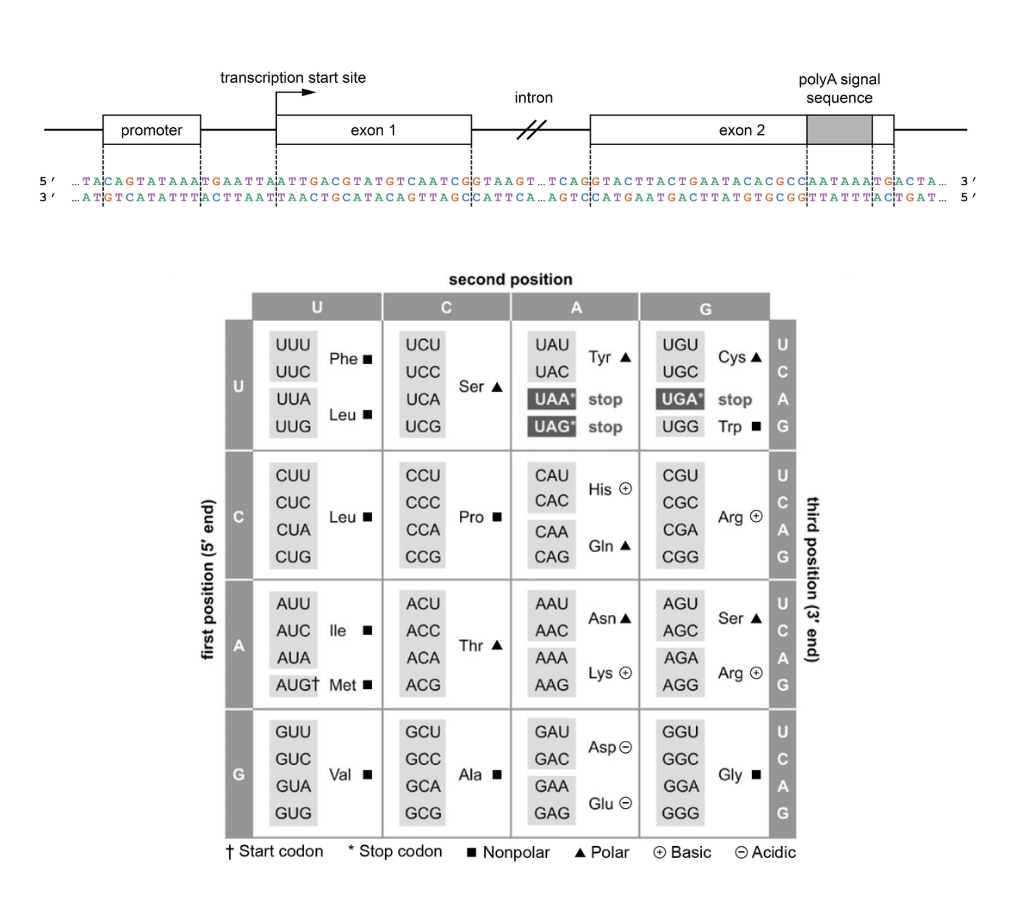
The diagram below shows an imaginary eukaryotic gene containing two exons. A portion of the intron is not shown to save space.
Exon nucleotides are numbered beginning at the transcription start site (+1), and they do not include the nucleotides in the intron. Exon 1's first nucleotide is nucleotide 1, and exon 2's first nucleotide is nucleotide 19. Use this information to answer the following questions.
What amino acid does the second codon of this gene's open reading frame encode?
Serine (Ser)

The diagram below shows a gene with four exons. The reading frame that encodes the protein is shaded and begins in exon 1 and ends in exon 3. Use this diagram to determine whether each of the following statements is true or false.
T/F: The start codon is in exon 1.
True

The diagram below shows a gene with four exons. The reading frame that encodes the protein is shaded and begins in exon 1 and ends in exon 3. Use this diagram to determine whether each of the following statements is true or false.
T/F: The stop codon is in exon 3.
True

The diagram below shows a gene with four exons. The reading frame that encodes the protein is shaded and begins in exon 1 and ends in exon 3. Use this diagram to determine whether each of the following statements is true or false.
T/F: The polyA signal sequence is in exon 4.
True