Anatomy 2300 Unit 4: The Special Sense Organs Pt2
 Call Kai
 Learn
 Practice Test
 Spaced Repetition
 Match
 Flashcards
 Knowt Play
1/30

There's no tags or description
Looks like no tags are added yet.
31 Terms
General Senses
Senses associated with touch. They lack special sense organs and their receptors are spread through the body, such as in the skin.
Thermoreceptors
General sense receptors that detect temperature.

Nociceptor
A general sense receptor that detects pain.

Baroreceptor
A general sense receptor that detects pressure.

Proprioceptors
General sense receptors that detect the position of the body in space.

Special Senses
The five special senses are vision, smell, taste, hearing, and equilibrium.
Each has a dedicated sense organ, and each is tied to a receptor type: vision uses photoreceptors, smell and taste use chemoreceptors, and hearing and equilibrium use mechanoreceptors.

Chemoreceptors
Receptors that detect chemicals.
They are the receptor type for taste (gustatory/taste cells in taste buds) and smell (olfactory receptor cells).

Mechanoreceptors
Receptors that detect mechanical movement.
They are the receptor type for hearing and equilibrium, both of which are housed in the inner ear.

Tongue (Sensory Innervation?)
Taste is a chemoreceptor sense. The taste cells sit in taste buds on the papillae (circumvallate and fungiform) on the tongue's surface. Special sensation (taste) and general sensation (touch, temperature, pain) travel in different nerves depending on the region:
Anterior 2/3: Taste is carried by the facial n. (CN VII).
General sensation is carried by the mandibular division of the trigeminal n. (CN V3).
Posterior 1/3: Both taste and general sensation are carried by the glossopharyngeal n. (CN IX).

Extrinsic muscles of the tongue
Muscles that attach the tongue to structures outside it (chin, styloid process, hyoid bone) and move it.
All three are innervated by the hypoglossal n. (CN XII).

Styloglossus
Attaches to the styloid process.
Action: Pulls the tongue posteriorly (retract) and superiorly (elevate)
Innervation: Hypoglossal nerve
(CN XII)

Hyoglossus
Attached to the hyoid bone.
Action: Pulls the tongue down (depression)
Innervation: Hypoglossal nerve
(CN XII)

Genioglossus
Attaches to the chin ("genio" means chin).
Action: Pulls the tongue out of the mouth (protrudes)
Innervation: Hypoglossal nerve
(CN XII)

Smell (Pathway for smell?)
The receptor type is the chemoreceptor. The sensory receptor organ is the olfactory epithelium, which lines the roof of the nasal cavity, where inhaled odor molecules dissolve in mucus and bind the olfactory cilia. The pathway runs as follows:
Olfactory receptor cell axons (CN I) pass through the cribriform plate of the ethmoid bone.
They reach the olfactory bulb, then the olfactory tract.
The tract has two destinations (only need to know one). The primary olfactory cortex, mostly in the medial temporal lobe, handles conscious awareness of odors.
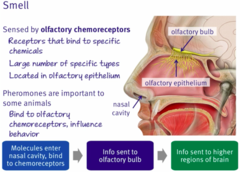
Olfactory bulb
The structure that sits on top of the cribriform plate, in the anterior cranial fossa.
It receives the CN I axons coming up through the plate and passes the signal on to the olfactory tract.

Olfactory tract
The pathway leaving the olfactory bulb and traveling back toward the brain.
It carries smell information to the primary olfactory cortex and the limbic structures listed above.

Primary Olfactory Cortex
Located mostly at the medial aspect of the temporal lobe; conscious awareness of odor.
Receives the olfactory tract.

Ear (External, Middle, Inner Ear) (Main Functions? Componets?)
The ear is divided into three regions.
The external and middle ear conduct sound. The inner ear, which sits in the temporal bone, holds the mechanoreceptors for both hearing and equilibrium.
The cochlea is the hearing organ and the semicircular canals are the equilibrium organs. Both send signals through CN VIII (vestibulocochlear) to the brain.
External ear: The auricle, external auditory canal, and tympanic membrane.
Middle ear: The air-filled space containing the auditory ossicles and the opening of the pharyngotympanic tube.
Inner ear: The semicircular canals, cochlea, and the oval and round windows.

Auricle (Ear Region? Function?)
The visible, cartilage-supported outer part of the ear. It funnels sound into the external auditory canal.

(External) Auditory canal (Also known as? Function?)
(external acoustic meatus): The canal that runs from the auricle inward to the tympanic membrane. It funnels sound waves to the membrane.

Tympanic membrane (Also known as? Function?)
The eardrum, which separates the external ear from the middle ear. Sound waves make it vibrate.

Auditory Ossicles (Ear Part? Function? Order?)
Three tiny bones in the middle ear that pass the tympanic membrane's vibrations to the inner ear and amplify sound about 20X. In order from the tympanic membrane inward:
Malleus (Hammer)
Incus (Anvil)
Stapes (Stirrup)

Malleus (Also known as? Function?)
(Hammer) Attached to the tympanic membrane; receives and transmits its vibrations to the incus.

Incus (Also known as? Function?)
("anvil"): Middle ossicle; transfers vibrations from the malleus to the stapes.

Stapes (Also known as? Function?)
("stirrup"): Innermost ossicle; pushes against the oval window, transmitting vibrations into the inner ear.

Pharyngotympanic tube (Ear Part? Function?)
A passage connecting the middle ear to the nasopharynx that equalizes air pressure on both sides of the tympanic membrane, allowing it to vibrate properly.
It also helps drain mucus and fluid from the middle ear into the pharynx.

Semicircular canals (Ear Part? Function? Contain?)
Looped canals in the inner ear.
They are the sensory receptor organ for equilibrium and contain mechanoreceptors.
They send signals through the vestibular part of CN VIII.

Cochlea (Ear Part? Function? Contain?)
The spiral-shaped structure in the inner ear.
It is the sensory receptor organ for hearing and contains mechanoreceptors.
It sends signals through the cochlear part of CN VIII.

Oval window (Function?)
The opening in the inner ear where the stapes sits. Vibrations enter the inner ear here ("vibrations in").

Round window (Ear Part? Function?)
A second opening of the inner ear, located below the oval window, covered by a flexible membrane that relieves pressure in the cochlea as vibrations leave the inner ear ("vibrations out").

Pathway of sound and balance signals
The cochlear and vestibular nerves join to form the vestibulocochlear n. (CN VIII).
It leaves the inner ear through the internal acoustic meatus and enters the brainstem at the pons/medulla.
The hearing signal then reaches the auditory cortex in the temporal lobe. The slides do not go into further detail on the equilibrium route.
Cochlea → CN VIII → Brainstem → Thalamus → Auditory cortex (temporal lobe)
