bchm exam 2
 Call Kai
 Learn
 Practice Test
 Spaced Repetition
 Match
 Flashcards
 Knowt Play
1/94

There's no tags or description
Looks like no tags are added yet.
95 Terms
6 bonds of hemoglobin (Hb) and when they occur
all covalent
4 bonds between the Fe2+ and the nitrogen atoms in the 4 pyrrole rings of the porphyrin ring
1 proximal histidine bond with His F8
1 bond to oxygen, which bonds with distal histidine His E7
how do we know myoglobin binds more tightly to o2 than Hb?
Kd = P50 = [L] at Y = 0.5 : Kd is equal to the ligand concentration [L] when exactly half of the binding sites are bound by ligand (Y = 0.5).
lower Kd means less substrate needed to occupy the same number of sites (50%)
what is the shape of Mb vs Hb curve? draw both. why is Hb shape that way?
mb = hyperbolic
hb = sigmoidal - favors release of O2 at low conceentations (left side of graph) and binding at high concentrations (rigth side of graph). this shape allows for change in binding affinity.
Kd and Ka: what does each represent (eqn), relationship, and units
Ka: association constant, for P + L → PL. Ka = [PL] / [P][L]. units: 1/M
Kd: dissoc constant, for PL → P + L. Kd = [P][L] / [PL]. units: M
Kd = 1/Ka
strong binding and weak binding in terms of Kd
strong: Kd < 10 nM
weak: Kd > 10 uM
theta or Y = ?
[L] / Kd + [L]
Hb and Mb have similar ______ because of similar _____ but not similar ______ (only _____ identical).this is because there are: (define each)
invariant residues
conservative substitution
simialr teritary structure due to similar function, but not similar primary structure (only 18% identical).
invariant residues: exactly the same between the two
conservative subsitution: substitued with a similar amino acid (ex. asp and glu are both acidic)
what three conformational chanegs happen when O2 binds and Hb swtiches from T to R state
15 degree rotation of a1b1 subunit from a2b2 subunit
central cavity becomes smaller
interfacial subunit interactions (salt bridges) are broken
where does O2 bind in Hb
at the 4 heme groups
what residues are involved in interfacial interactions
His146 (β), Asp94 (β), Lys40 (α), and Arg141 (α)
does lower pH favor r or t state? explain mechanism
lower pH → more H+ → residues get protonated (ex. His146 is positive) → neutral/positive AAs become more positive and can form salt bridge with Asp94 on the same B subunit → t state favored
what is the bohr effect
the response to an increase in CO2 or lower pH: favors the t state and lowers the binding affinity of O2, favoring release of O2
does changing His146 to Glu favor t or r state? explain
Glu = - → unable to form salt bridge with Asp94 → r state favored
what makes the t state stable? what makes the r state stable? what makes the r state more stable than t
t state: interfacial subunit interactiosn
r state: fe-o2 bonds
fe-o2 bonds with 4 ligands (o2) bound is more stable than t state
carbonic anhydrase rxn, why it happens, and its effect on Hb
CO2 + H2O → H+ + HCO3- . turns CO2 in blood (which owuld cause bubbles) into H+.
more H+ / lower pH → Hb protonated → t stare favored → lower bidng affinity
homotropic vs heterotropic effector
positive vs negative effector and how each shifts the curve and what state is favored
homo - binds to same site as ligand (active site)
hetero - binds to an allosteric/different site
positive - increases enzyme activity, leftward shift (decreases P50), T state favored
negative - decreases enzyme acitivity, rightward shift, R state favored
what kind of effector and where does it bind?
Oxygen:
H+
CO2
2,3-BPG
CO
O2 = ligand = positive homotropic allosteric eff
H+ = negative hetertropic allosteric eff - binds to basic amino acids in subunit interface and casues them to be more pos → stronger interaction w their acidic AA pair
CO2 = neg hetero. reacts with uncharged R-NH2 groups and forms NH-COO- which is charged, created more interfacial subunit interactions/salt bridges and favoring t state
BPG = neg hetero. binds in cavity, keeping it in t-state and lowering O2 affinity. when O2 binds, it forces r-state and expels BPG.
CO = neg homo
write Hb and H+ reaction and explain what happens if pH increases.
Hb4O2 + nH+ → ← HbnH+ + 4O2
if pH increases, H+ decreases, rxn shifts left, R state favored + binding affinity increased
what three amino acids does BPG bind to and what makes fetal Hb different
H143, Lys82, His2
fetal Hb has Ser143 instead of His143, so BPG binding affinity is decresed. its called a2g2 instead of a2b2
how can u seen pos cooperativtiy in a binding curve
sigmoidal
hill coefficinet n possibile numbersand what they mean
n = 1 → no cooperativty
n > 1 = pos cooperativity, ie binding of one ligand increases bindnig affinity at other sites
n < 1 → negative cooperativity
hill plot x and y axis
logpO2 and log (Y/ 1 - Y), Y or theta is the fractional saturation
concerted model vs sequential model: what do they model, how are they diff, and what is the limitation of each
models for cooperativity in o2 binding
concerted model: Hb goes back and forth (in equilibrium) between t state and r state, with r state favored as more O2 binds
limitate: t state w o2 bound doesnt acc exist
sequential: when o2 binds, the subunit gets coverted to t state
limitation: Hb with some subunits in r state and some in t state (ie an inbetween r and t state) doesnt acc exist
what is a ponit mutation
change in one amino acid
what mutation causes sickle cell disease and how
Glu6 → Val6
glu is - val is NP → causes it to want to be inside → causes sickling and binding to hydrophobic pocket of other rbcs
explain why each mutation leads to inc or dec in O2 binding affinity
Glu6 → Val6
His F8 → Tyr
Arg141 → His141
His146 → Asp146
sickle cell - glu6 to val6 - sickling due to val being NP and binding to hydrophobic pocket of other RBCs
dec affinity, as His F8 is necessary in binding to Fe2+ in heme so its Fe3+ instead
inc aff, as his is less positive and does not form salt bridges as much, so t state is less stable
inc affinity, as His146 is part of bohr effect and Asp is neg and cannot form salt bridge. without bohr effect and salt bridge, r state is favored.
classes of enzymes: what do they do?
oxidoreductase
trasnferase
hydrolase
lyase
isomerase
ligase
redox rxns
transfer function group (ex. kinases)
hydrolysis reaction
group elimination to form double bonds
isomerization
bond formation coouples w atp hydrolysis
what is the purpose of catalysis
to stabilize transition state, reducing TS energy and making rxn faster
what do the followign do and what does it make at the end
acaid base catalysis
covalent catalysis
metal ion/electrostatic catalysis
usses acid or base (H, asp, glu, lys) to transfer/remove a proton from TS to stabilize it, generates CB which can regenerate acid/base with water
uses a nucleophilic group (ex. RNH2, so ser, Lys, cys) to form a covalent bond with the substrate to stabilize TS
metal ion used to stabilize TS
active site amino acids of chymotrypsin and their role in catalysis
Asp102: stabilizes His57 through H-bonding
His57: general base catalysis
Ser195: covalent catalysis
what does chymotrypsin do and what makes it specific to that substrate?
cleaves polypeptide at the peptide bond prior to the aromatic amino acid
araomatic AA substrates becasue chymotryp has a three AA hydrphobic active site, so it wants large NP substrate, and it has to be a polypeptide because there needs to be a peptide bond
chymotrypsin mechansim
His57 acts as a base and removes H from ser195 (-OH) to make it a good nucleophile
Ser195 attacks the carbonyl carbon of the scissile (to-be-cleaved) peptide bond) → tetrahedral intermediate 1 w/ oxyanion (C—O-)
His57 acts as an acid and donates H to NH (H- -NH). Since NH2 is a good LG. carbonyl C=O reforms, breaking C-N peptide bond to form NH2.
the C-terminal half of the polypeptide diffuses away.
the N-terminal half of the substrate is covalently stuck to the enzyme via Ser 195 = acyl-enzyme intermediate.
His57 acts as a base and takes H from H2O, forming OH-. OH- attacks C=O to form tetrahedral intermediate 2 w/ oxyanion
His57 acts as an acid and donates H to Ser195, allowing oxyanion to reform C=O and break Ser195-enzyme bond. → reform enzyme + cleaved polypeptide
how does chymotryp stabilize TS?
how does the oxyanion hole stabilize the oxyanion in tetrahedal intermediates?
4 hydrogen bonds:
3 primary H bonds
1 low barrier h bond
specificty pockets of serine proteases:
chymotrpsin
trypsin
elastase
chym: 3 uncharged AAs: GSG → likes large NP side chains
try: neg charge: GDG, D is neg → like pos side chains
elastase: only two AAs, VT → likes small NP aliphatic (like alanine)
Km = ?
[E total] = ?
[ES] / [E] [S]
[E tot] = [E free] + [ES]
MM eqn: v0 = ?
what is Km? units?
what is kcat? units?
v0 = Vmax[S] / Km + [S]
Km = [S] at V = 0.5Vmax. units = M
kcat = k2 (rate constant for ES → E + P) = rate of turnover. units = 1/s
lineweaver burk eqn: 1/v0 = ?
slope = ?
y int =?
x int = ?

three assumptions of michaelis menten eqn
1 substrate → 1 product
k2 = kcat (meanign step 2 is the rate determining step)
rate of change of [ES] = 0, i.e. steady state process, rate of formation = rate of depletion
kcat = ?
efficiency = ?
kcat = Vmax / [E tot]
eff = kcat/Km
types of non MM enzymes:
1) ordered sequential
2) random sequential
3) ping pong
A + B → C + D, where enzyme E binds to A first, then B, and releases C then D. always in this order
alcohol dehydrogenase
A + B → C + D, when E binds to A or B, then the other, and releases C or D, then the other
glycolysis
A into E, C out of E, E turns into F. B into F, D out of F, F back into E. \
ex. chymotrypsin
reversible vs irreversible inhibition
reversible: noncovalent - compeititve, noncompetitive, uncompeititve, mixed
irreversible: covalent
how do each of the following affect Km and Vmax?
compeietive inhib
decribe change in rate/concentraaiton grpah and linweav-burk graph
competitive: increase Km, unchanged Vmax because with enough substrate Vmax can still be reached but affinity/speed of binding is decreased
[S] vs v: graph moves right
LB: slope increases, x int move right. y int the same

how do each of the following affect Km and Vmax?
noncompeittive inhib
decribe change in rate/concentraaiton grpah and linweav-burk graph
noncompetiitve:
how do each of the following affect Km and Vmax?
uncompetitive inhib
decribe change in rate/concentraaiton grpah and linweav-burk graph
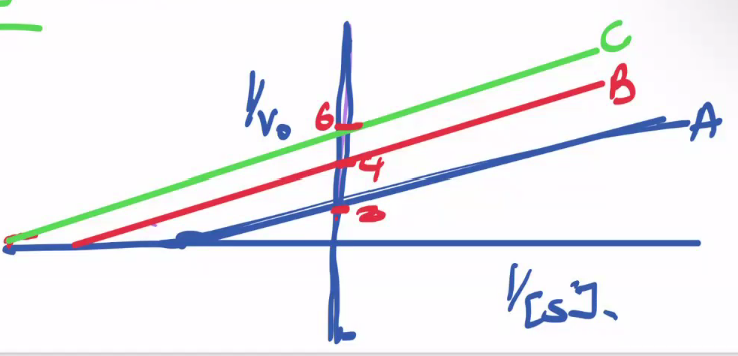
uncompeititve: inhib only binds to ES not E. Vmax decreases, Km decreases becasue making ES → EI decreases ES which means more E + S → ES, increasing affinity for substrate.
[S] vs v: moves left and down
LB: parallel slope but y-int moves upward, x int moves left
how do each of the following affect Km and Vmax?
mixed inhib
decribe change in rate/concentraaiton grpah and linweav-burk graph
mixed: inhib binds to both E and ES. Vmax decreases, Km can inc or dec
alpha < alpha’, ie inhibitor has greater affinity for ES → Km inc (typical)
alpha > alpha’, ie inhibitor has greater affinity for E → Km dec
[S] vs v: moves right and down
LB: slope increases, y int moves up, x int moves right

Km vs Ki
Km = substrate affinity for enzyme
Ki = inhibitor affinity for enzyme
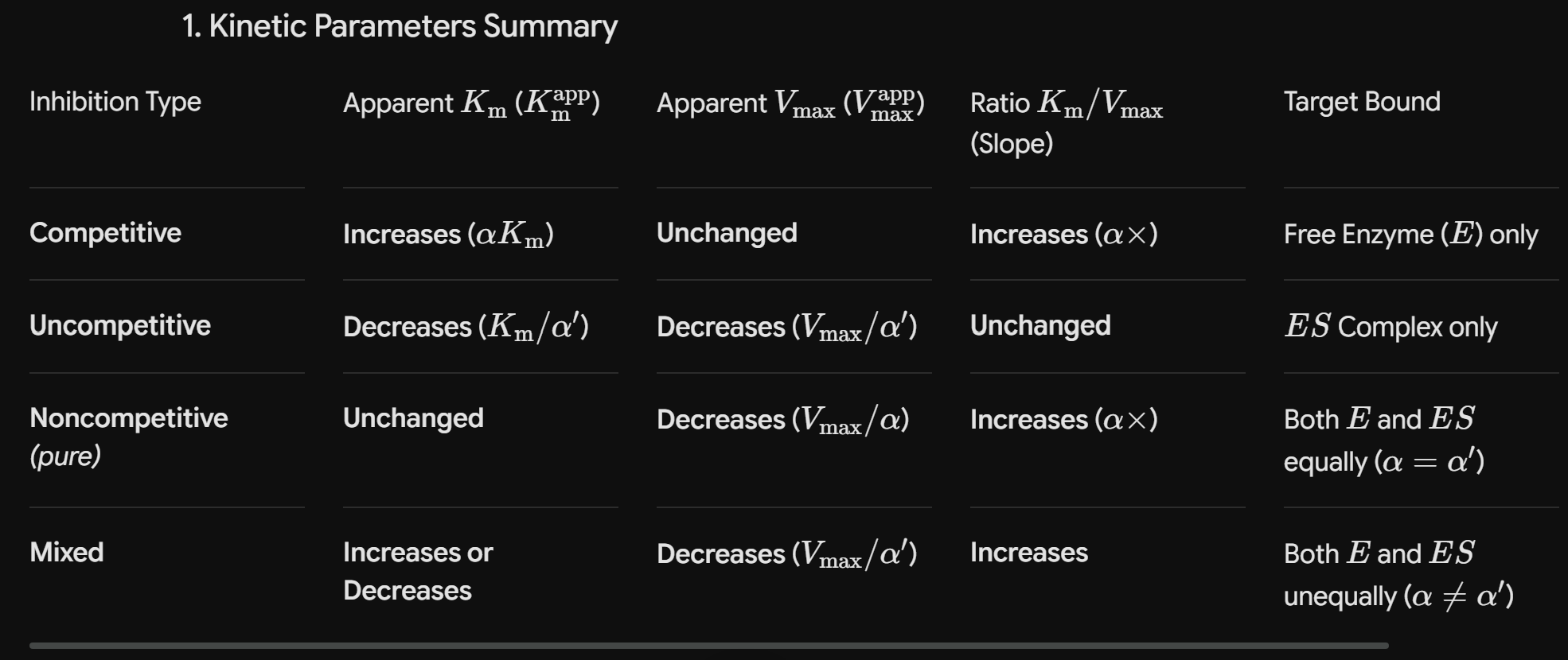
alpha factor = ? (3 eqns)
Km apparent / Km, when Km app = Km with inhibitor
= 1 + ([I] / Ki)
Vmax / Vmax app


tagret bound meaning does it bind to E or ES
three forms of enzyme regulation:
substrate level
feedback inhibition
covalent modification
substrate level: changing substrate concentration
feedback inhibition: product formation at one step regulates the acitivity of another step
covalent modification:
reversible: phosphorylation on serine, tyrosine, or theronine (AAs with OH)
of ACTG:
pyrimidines
purines
pairs and # H bonds
pyr: CT
pur: AG
pairs: AT (2 H bonds), CG (3 H bonds)
how does ATCase respond to ATP? CTP?
allosterically
ATP: decreases Km
CTP: increases Km
differences between DNA and RNA
double vs single stradned
thymine vs uracil
deoxyribose (2 OH groups) sugar vs ribose sugar (3 OH groups)
difference between deoxyribose and ribose
ribose - 2’ OH
deoxy - 2’ H → more stable due to not being susceptibel to hydrolsysis
nucleotide vs nucleoside
nucleotide: has O—phosphate (ex. adenoside mono phosphate)
nucleoside: has OH group instead (ex. adenoside)
what is a zymogen
an enzyme that requires a biochemical change to become active, usually by cleavign a certain peptide bond.
ex. Chymotrypsinogen → Chymotrypsin due to peptide bond cleavage by Trypsin
what three structures form a nucleic acid unit
sugar (deoxy/ribose)
phosphate group
nucleotide (ACTG)

DNA:
what is the angle between bp
how many bp per turn
rise of helix/distance between bp
36 degrees
10 bp/turn
0.34 A between bp
carbohydrate general formula
simplest carb formula
(CH2O)n
simplest: n = 3, C3H6O3

aldose vs ketose - draw
see green
aldehyde = O=CRH
ketone = O=CRR


name the following

most stable form of carb: D or L
D
how to know D or L carbon
look at last chrial carbon
if OH on right = D (r config). OH on left = L (s config)


draw the L glucose
flip ALL chiral groups and top group - mirror image

what is an epimer? if the regular is D, what is the epimer?
carbons that differ in confirmation at a single chiral carbon
also D

#of epimers possible for a heptamer
4 chiral centers, so 2^4 = 16
Anomer
differs in configuration about the anomeric carbon, which is the one bound to two oxygens (alpha vs beta)
steps of cyclization of carbs, ex glucose
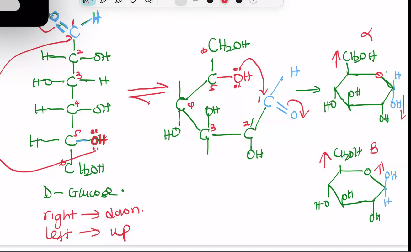
OH group on last chiral center uses electrons to attack the aldehyde at top → ring with carbon 6 (nonchiral) left out
on each carbon in the ring, if OH was on the right, it becomes down. if OH was on the left, it becomes up
the attacked carbon can have OH up or down (racemic mix)
alpha vs beta
alpha: if the OH at carbon 1 is in opposite direction to the last carbon, CH2OH
beta: if the OH at carbon 1 is in same direction to the last carbon, CH2OH
if noCH2OH group was used in cycle formation → in plane, not up/down:
if D enantiomer:
OH on carbon 1 = up = alpha
OH on carbon 21 = down = beta
if L enan: switched, up = beta
two types of pentose ketose: name and sturcutre
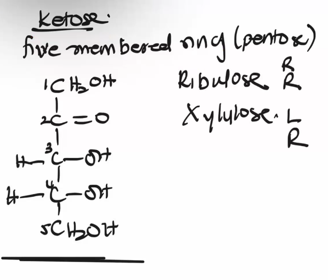
ribulose - RR (pictured)
xylulose - RL
3 and 4 are chiral
hexose ketose: name and sturcutre
fructose - LRR
3 4 5 are chiral

pentose aldoses: name and structure
ribose: RRR
Arabinose: LRR
xylose: RLR
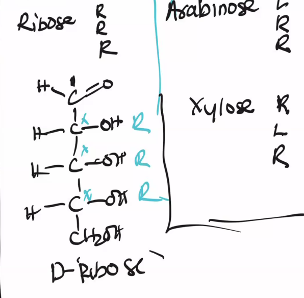
six membered aldoses
glucose: RLRR
mannose: LLRR (epimer of glu)
galactose: RLLR (epimer of glu)

pyran vs furan
6 = pyran
5 = furan

is alpha or beta most stable? what is the exception? what is changing between the two called?
beta; mannose; mutarotation
naming carbs
alpha or beta
D or L
fructo or whatever else (based on ketose or aldose and how many members)
pyranose or furanose
how to tell reducing vs non reducing suger. whihc can be oxidized?
reducign: has a hemiacetal or hemiketal, formed from :OH attacking carbonyl in aldehyde or ketone
idenntify anomeric carbon
check if it has OH, ie not involved in linkage
reducing can be oxidized back to carbonyl bc the OH is not involved in linkage

how to number carbons on a ring
start from the CH2OH group sticking out of ring = 6, then 5 is the first carbon from there on the ring, the go around away from the O until u get to 1.
how to name disaccharadies
alpha-D-glycopyranoSYL (1-4) alpha-D-glycopyranose
if anomeric carbon involved: …-glycopyranoSIDE
naming monosaccharide and D/L and aldose/ketose from a ring
starting from TWO until FIVE, use convention down = right, up = left
on the last one, OH is at the opposite position of CH2OH
ketose if CH2OH group attached to C on ring with OH. ketose can have same number of carbons but smaller ring
where cna oxidation happen in glucose and what are the resulting forms called
at carbon 1 in a reducing sugar, turning OH back into C=O → gluconic acid
at carbon 6, CH2OH becomes COO- → glucaronic acid
addition of a phosphate group to a monosacch

what are reduced forms of carbs called and what is reduced?
called -ol for alcohol, ex glycerol, ribitol, xylitol
the aldehyde or ketone C=O is reduced to OH
what monosaccharides and disaccharides can mutarotate?
monosaccharides: anything with 4 or more carbons. 3 carbons cannot form a ring so theres no alpha/beta anomers
disacch: if its reducing
At least one anomeric carbon is free (it retains a free OH group, making it a hemiacetal or hemiketal) → can open into an aldehyde/ketone (non ring) and re-close into alpha or beta configuration.
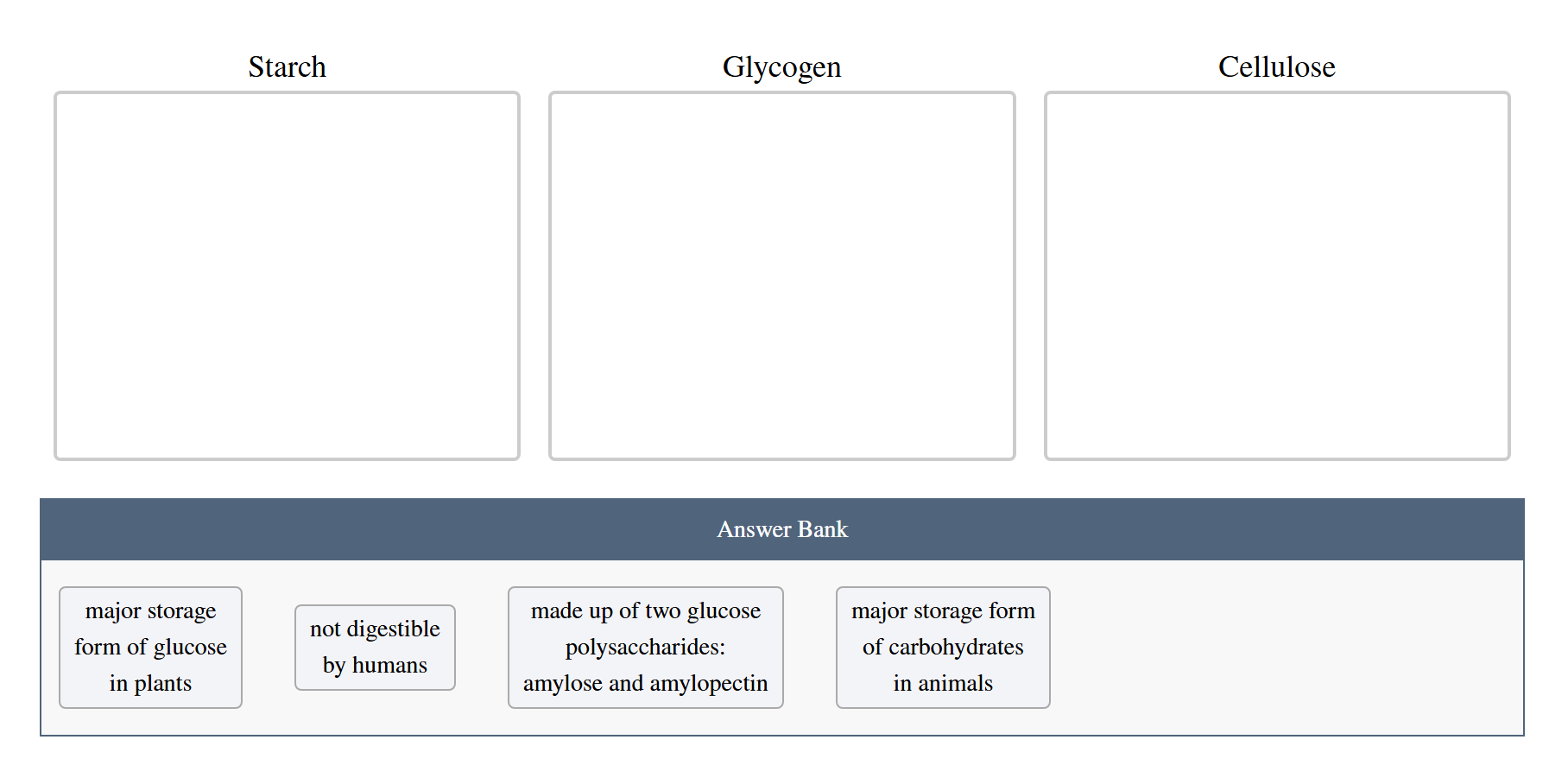


homopolysacch or heteropolysacch?
all HOMO: all except chitin are made of D-Glucose, chitin is N-amino whatever
why cant we digst cellulose? what part is not digestable?
dont have enzyme cellulase
cant hydrolyze the linkage (BETA 1-4 between glucose)


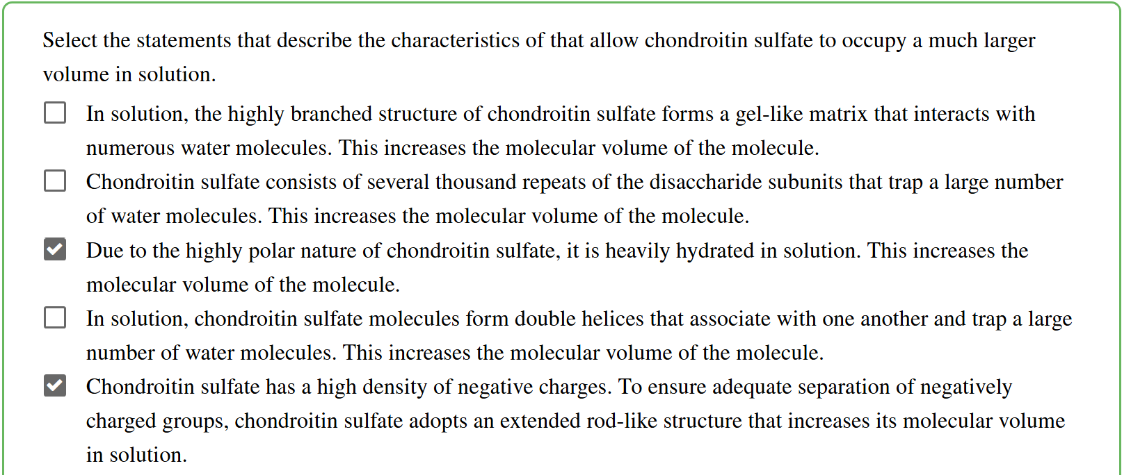
GAGs structure
short, rod like linear chains (not branched, not double helix bc the charges repel)


compare linkages in the two polymers in starch
which one is glycogen like what makes it different?
Starch contains both amylopectin, α(1-4) glucose polymer with α(1-6) branches, and amylose, α(1-4) unbranched polymer. Glycogen (animals/microbes) is like amylopectin, but higher MW with shorter and more frequent branch point
structure, linkages, abd binding in cellulose
cellulose is a linear glucose polymer with β(14) linkages.
has hydrogen bonding between linked glucose monosaccharides
glycosaminoglycan - what kind of heteropolysacchride?
proteoglycans
proteoglycan vs glycoprotein
proteoglycan - mostly carb
glycoproteins - 95% protein, small amnt carb, blood group identification (certain carb unit makes certain blood type), Serine/Threonine?
how would you change B blood type so everyone could receive it
cleave the B oligosacchride on the glycoprotein?
tamiflu?
competitive inhibitor for sialic acid, inhibiting neuraminidase and blocking release of viral particles