Skull: Headwork part 1: positioning notes & image critique
 Call Kai
 Learn
Learn Practice Test
 Spaced Repetition
 Match
Match Flashcards
 Knowt Play
1/38

There's no tags or description
Looks like no tags are added yet.
Name | Mastery | Learn | Test | Matching | Spaced | Call with Kai | Chat |
|---|
No analytics yet
Send a link to your students to track their progress
39 Terms
Skull ROUTINE views
AP axial towne
PA axial caldwell
Right & left laterals
Skull 3 views or less charge
AP/PA & left lateral
AP axial towne technique
85 kVP @10 mAs, center cell
PA axial caldwell technique
85 kVp @7.1 mAs, center cell
Lateral skull techniques
85 kVp @3.2 mAs, center cell
AP axial towne overview:
10×12 LW
MSP is perpendicular to midline of the IR, flex pt neck so OBML is perpendicular to the plane of the IR. Position IR so its upper margin is at the level of the highest point of the vertex
angle 30 degrees caudad to OML (or 37 to IOML)
center 2.5” above glabella & pass through the EAM
R or L marker
if patient has difficulties flexing neck far enough to get OML perpendicular in AP axial towne, do this
place IOML perpendicular
breathing for all of headwork
suspended respiration
what view does this purpose describe: “symmetric image of petrous pyramids, the posterior portion of the foramen magnum, dorsum sellae and posterior clinoid processes projected WITHIN THE FORAMEN MAGNUM, the OCCIPITAL BONE, and the posterior portion of the parietal bones”
ap axial towne
AP axial towne criteria:
entire cranium w/o rotation or tilt demonstrated by:
equal distance from lateral border of skull to lateral margin of foramen magnum on both sides
symmetric petroud pyramids
MSP of cranium aligned w/ long axis of collimated field
dorsum sellae & posterior clinoid processes visible within foramen magnum
AP axial towne help:
if you turn the head, the foramen magnum goes in the OPPOSITE direction
THE LONGER SIDE ON THE AP IS THE WAY THE HEAD IS TURNED!!
Ex:
(if there is longer distance from the foramen magnum to the R, the head is turned to the R)
occipital bone is best visualized on what skull view?
AP axial towne
if you see a “bridge” within the foramen magnum this means
over tucking or too much tube angle (posterior arch of C1 is visualized)
if you cannot see the foramen magnum clearly, this indicates
not enough angle or not enough chin tuck
if you have the foramen magnum but it is not clear, this indicates
not enough angle or not enough tuck
degree differences for skull views
30 (ap axial towne)
15 (pa axial caldwell)
0 (lateral)
0 (lateral)
frontal bone is best visualized on what view
PA axial caldwell
PA axial caldwell overview
10×12 LW
flex pt’s neck so OML is perpendicular to the plane of the IR (generally, the patient’s forehead & nose will be resting against the IR). MSP perpendicular to the IR
check for rotation by feeling the mastoid tips
center 15 degrees caudad exiting the nasion
R or L marker
what view has the purpose of petrous pyramids within the lower third of the orbits, the anterior & posterior ethmoidal air cells, crista galli, frontal bone, and frontal sinuses
PA axial caldwell
PA axial caldwell evaluation criteria
entire cranium w/o rotation or tilt demonstrated by:
equal distances from lateral border of the skull to the lateral border of the orbits on both sides
petrous ridges should be symmetric
entire cranial perimeter showing 3 distinct tables of squamous bone
if petrous ridge is at top of orbits in a PA axial caldwell, this indicates
chin is tucked too much or not enough angle
if you want the petrous ridges to come down, what can you do?
bring chin up or increase the angle
if you want petrous ridges to come up higher because they are too low, what can you do?
tuck chin or decrease angle
PA axial caldwell helpful hint
shorter distance side from foramen magnum is the side it is rotated towards
bilateral laterals overview
12×10 CW
place PT in anterior oblique, MSP must be parallel to IR, flex PT neck so IOML is perpendicular to front edge of IR & parallel to IR long axis. the interpupillary line should be perpendicular to IR
center 2” superior to the EAM
mark side closest to IR & anterior to pt
what view shows superimposed halves of the cranium showing the detail of the side AGAINST the IR. the sella turcica, anterior clinoid processes, dorsum sellae, and posterior clinoid processes are visualized
bilateral laterals
biltateral laterals evaluation criteria
entire cranium w/o rotation or tilt demonstrated by
superimposed orbital roofs & greater wings of sphenoid
superimposed mastoid regions & EAMs
superimposed TMJs
sella turcica seen in profile
no overall of cervical spine on mandible
rotation on bilateral laterals example (if you can see the mandible)
the side that is magnified is the side that is farthest away
Skull special view PA (no angle) purpose
frontal bone is area of interest
orbits are filed by petrous ridges

what special skull view is this?
PA no angle
skull special view: SMV full basal
hyperextend neck so IOML is nearly parallel w/ IR, rest head on its vertex, & adjust head so MSP is perpendicular to IR
direct CR through sella turcica perpendicular to the IOML between the angles of the mandible ( ¾” anterior to EAM)

what special view of the skull is this?
SMV full basal
skull special view: trauma x-table lateral
12×10 CW
supine w/ head elevated on sponge
MSP parallel to IR & IP is perpendicular to IR
center 2” superior to EAM
mark side against!!!
visualize cranium details of side against IR
skull special view: recumbent (AP AXIAL, reverse caldwell)
10×12 LW
center 15 degrees cephalad entering nasion
SAME AS PA CALDWELL EXCEPT ORBITS ARE MAGNIFIED
skull special view: recumbent trauma AP
center entering the nasion- NO ANGLE
SAME AS PA EXCEPT ORBITS ARE MAGNIFIED

what skull special view is this?
recumbent AP axial/ Reverse caldwell

what skull special view is this?
recumbent trauma AP
skull special view: recumbent trauma AP axial towne
angle CR to go through the OML, then add another 30 degrees, enter 2.5” above glabella
ex: if pt is collared
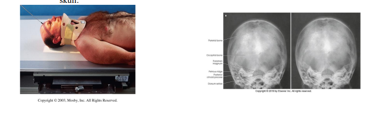
what skull special view is this?
recumbent trauma AP axial towne