WBC abnormalities
 Call Kai
 Learn
 Practice Test
 Spaced Repetition
 Match
 Flashcards
 Knowt Play
1/33

There's no tags or description
Looks like no tags are added yet.
34 Terms
% of wbc in peripheral blood
Neutrophils: 50-70%
3-5% are band forms
Lymphocytes: 25-35%
Monocytes: 4-6%
Eosinophils: 1-3%
Basophils: 0.4-1%

Absolute count of wbc in peripheral blood
Total leukocytes: 4-11 e9 /L
Neutrophils: 2.5-7.5 e9/ L
Lymphocytes: 1.5-3.5 e9/ L
Monocytes 0.2-0.8 e9/ L
Eosinophils 0.04-0.4 e9/ L
Basophils: 0.01-0.1 e9/ L

Suffix for increase in cell line
-Osis
Leukocytosis
-Philia
Eosinophilia
Suffix for decrease in cell line
-penia
neutropenia, leukopenia
Causes of increase in WBC

Causes of decrease in WBC

Stages of WBC phagocytosis
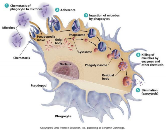
WBC qualitative defects
May happen in cytoplasm or nucleus
May be inherited or acquired
Acquired more common
Results as stress during maturation and activity in circulation or tissue
toxic changes
Toxic granulation
Human ehrlichiosis
Nuclear abnormalities
Toxic changes in WBC
Visible changes in the response of WBC to infection and inflammation occurs along the path
Usually increased number of segmented neutrophils in peripheral smears
Neutrophilia: shift to the left. Younger cells appear

Toxic granulation (in neutrophil)
More granules in segmented and bands
vivid blue-black color
Direct response to enhanced lysosome enzyme production
due to severe stress or immune activation
Need to carefully differentiate between toxic granulation of neutrophils from basophils

Toxic vacuolization (in neutrophils)
Small and large vacuoles in cytoplasm
Large, uneven vacuoles distributed= serious infections, maybe sepsis
Causes: drugs (sulfonamides, chloroquine), prolonged blood storage from phagocytosis of cytopalsmic granules
10% of neutrophils affected
Indicates prognosis

Döhle Bodies
Ribosomal RNA
Cytoplasmic inclusions near membrane
Pale blue-grey, rod-shaped
Transient inclusions in neutrophils
may also appear in monocytes and bands
Can be due to non-pathological conditions like pregnancy

Human Ehrlichiosis
Ehlrlichia bacteria is from tics
Human Monocytic Ehrlichiosis (HME)
Ehrlichia chaffeensis
Southwest and Mid-atlantic US
Flu-like symptoms
Human Granulocytic Anaplasmosisi (HGA)
Anaplasma Phagocytophilum
Midwest US
Acute high fever, chills, headaches
Hypersegmented
Abnormality in neutrophils where it has more than 5 lobes
usually in megaloblastic anemia
accompanied by ovalocytes

Hereditary WBC disorders
May-Hegglin Anomaly
Alder’s Anomaly
Pelger-Huët Anomaly
Chediak-Higashi Syndrome

May-Hegglin Anomaly
thrombocytopenia (low number of thrombocytes)
giant platelets
Dohle bodies in neutrophils
Larger than normal dohle bodies in infections
nmemonic: Hegglin→ thrombocytes hagglin away

Alder’s Anomaly (Alder-Reilly Anomaly)
Decreased mucopolysaccharide production
Causes lipid deposits in cytoplasm
Alder-Reilly granules: Coarse dark granules in all WBC cell lines
Differentiated from toxic granulation by staining with toluidine blue
Toxic granulation: in clusters
only in neutrophils and monocytes
Surrounded by vacuoles
does not affect cell fxn
Inclusions seen in BM, not in peripheral
nmemonic: O’Reilly oil change→ oil is fat

Pelger-Huët Anomaly
hyposegmentation in 70-95% of neutrophils
Heterozygous nucleus: peanut, dumbbell, pince-nez- shaped
Homozygous nucleus: spherical, no lobes
neutrophils have normal fxn

Chédiak-Higashi Syndrome
Defective fusion protein that affects lysosomal secretion
Giant lysosomes in cytoplasm
Green-grey peroxidase-positive bodies
Fusion of primary, secondary, non-specific, specific granules
Autosomal disorder of neutrophilic granules
Cytoplasmic granules are giant purple-grey
Lymphocytes and monocytes may also have a single large granule
WBCs not fully functional
shows reduced chemotaxis→ poor phagocytosis of pathogens
S/S:
Neutropenia
Albinism, photophobia due to melanosome abnormality
Staph Aureus recurrent infections
Usually causes childhood death

Chronic Granulomatous Disease (CGD)
inherited
65% x linked
35% autosomal recessive
Defects in respiratory burst oxidase system
recurrent infections leading to granulomas
Affected cells are morphologically normal but cannot generate antimicrobial oxygen metabolites like H2O2
Neutrophils can phagocytize but cannot kill them
Catalase-positive organisms destroy the H2O2 of their own metabolism→ continues to grow intracellularly where they are protected from antibiotics
Catalase-negative organisms kill themselves by generating H2O2 that the phagocyte uses as antimicrobial defense mechanism
Peripheral neutrophil count is normal but increases with infection
IGs increased due to chronic infection
Treatment: prophylactic Abs
NBT test detects abnormal oxygen metabolism of neutrophils
Myeloperoxidase Deficiency
Benign autosomal recessive disorder
Absence of myeloperoxidase in neutrophils and monocytes
MPO is used in bactericidal process but there’s no increase in infections in this deficiency
Neutrophils use alternative antimicrobial systems (just more slowly)
May experience disseminated fungal infections: usually with C albicans
Leukocyte Adhesion Deficiency
Two forms: LAD I, LAD II
LAD I:
Rare autosomal recessive
Decreased/ absent leukocyte cell-surface adhesion proteins
β-integrins, AKA CD11/ CD18 complex
Neutrophils have functional defects
impaired adhesion to endothelial cells, chemotaxis, phagocytosis, respiratory burst activation, degranulation
LAD II:
Failure of endothelial cells to synthesize ligand for leukocyte L-selectin
Leukocyte fxn is normal but cannot adhere to vessel wall endothelium and cannot exit the circulation to enter tissues
Features:
Recurrent soft tissue bacterial and fungal infections
persistent leukocytosis and granulocytosis from increased stimulation of BM
Reactive lymphocytosis in common disease states
1-4 yrs: lymphocytosis is normal
Normal WBC differential: shows a reversal in numbers of lymphocytes to neutrophils
Lymphocytes have normal fxn
Lymphocytosis: infectious mononucleosis
Caused by Epstein-Barr virus
Most common disease showing lymphocytosis
Enters orally→ infects B lymphs
normal lymphs transform to reactive lymphs
Enlarged lymph nodes
hepatosplenomegaly
Lymphocytosis: infectious mononucleosis smear
10-60% are reactive lymphocytes
Reactive lymphs are enlarged
has royal blue cytoplasm that scallops red cells
Must be recorded separately from non-reactive normal lymphocytes
Viral disorders often affect CBCs similarly

HIV/AIDS on hematology parameters
AIDS: Acquired immunodeficiency syndrome
Mainly targets T-helper lymphocytes (CD4)
Normal CD4:CD8 ratio is 2:1
in HIV infection, CD4 drastically decreased to 1:2→ failure in immune response
CD4 lymph count reduced to <200/ uL
Lymphocytes show extreme basophilic cytoplasm, possible folds, vacuolization
Lipid storage disorders
Missing or inactive strategic metabolic enzyme in the WBCs from single gene deletion
Undigested metabolic products accumulate in cells→ affects cell integrity
RES (reticuloendothelial system) cells most affected: histiocytes, monocytes, macrophages, BM cells, liver, spleen, lymph nodes
BM contains easily-identifiable cells specific to each disease
Not usually seen in laboratory
Most common Lipid Storage Diseases
Gaucher’s Disease
Niemann-Pick’s Disease
Tay-Sachs Disease
All autosomal recessive from single gene mutation
Abnormal facial features in Gaucher’s and Niemann-Pick
CNS involvement in infant forms
BM transplant and enzymes produced through biotechnology available
Gaucher’s Disease (pronounced Go-shay)
Missing Enzyme: β-Glucocerebrosidase
crinkled paper appearance of cytoplasm
Glucocerebrosides build up in liver, spleen, lungs, BM
macrophage cannot digest stroma of ingested cells→ lipid glucocerebrosides accumulate
Abnormal facial features
Splenohepatomegaly→ may result in anemia
nmemonic: Goshay→ glucocerebrosidase

Niemann-Pick disease
Missing enzyme: Sphingomyelinase (breaks down lipids)
Excessive sphingomyelin storages
Niemann-Pick type A and Type B disease
Macrophages are large and have foamy appearance in lymphoid tissue and BM
Off center nucleus, cytoplasmic inclusions
Splenohepatomegaly from increased sequestration
Blood lymphocytes have vacuoles that are lipid-filled lysosomes
Abnormal facial features
Fatal by 3 years
nmemonic: Niemyelinase
Tay-Sachs disease
Missing enzyme: Hexosaminidase A
Accumulation of GM2 ganglioside in brain and spinal cord→ breaking them down
Early childhood fatality
or neurological issues in adulthood
Bacteria in Blood stream
Bacteremia→ sepsis
Intra and extracellular bacteria must be recognized in peripheral blood smear ASAP

Platelet Satellitism
blood reacts with EDTA (lav tops)
Platelets form a ring around neutrophils
Causes low platelet count
Can be corrected only with blood collected in sodium citrate instead (light blue tops)

Pyknosis
Seen in degenerated neutrophils
Segmented nucleus becomes amorphous, smooth, bloblike structure
Chromatin shrinks, gets darker, breaks apart
No clear segments
Not counted in WBC peripheral smear
