Lec. 3 Erythrocytes and variations
 Call Kai
 Learn
Learn Practice Test
 Spaced Repetition
 Match
Match Flashcards
 Knowt Play
1/41

Earn XP
Description and Tags
Exam 1
Name | Mastery | Learn | Test | Matching | Spaced | Call with Kai | Chat |
|---|
No analytics yet
Send a link to your students to track their progress
42 Terms
How are erythrocytes viewed under a scope? Explain the steps getting to the scope.
1 drop of EDTA blood → stain w/ Wright stain → microscope
Who discovered the first stain used to see blood cells? This stain is the backbone stain for what 2 stains?
Romanowsky
Backbone stain for Wright stain and Giemsa stain
What does the Romanowsky stain have in it?
Methylene blue and eosin (Y or B)
Romanowsky stains are alcoholic bases and have basic and acidic components. Because of this what are these stains also called? What colors do they stain?
Polychromatic stains
cell nucleus → purple
cytoplasm → pink & blue
The Wright stains is made of the Romanowsky stain base which has acidic and basic dyes (eosin/methylene blue), what color do they dye parts of the cell?
Acidic dye: eosin → stain basic part of cell red/pink (hemoglobin)
Basic dye: methylene blue → stains acidic part of cell
List everything that makes an RBC normal. (size, shape, color, distribution, inclusions, Mean cell weight (hemoglobin), Mean cell [hemoglobin])
size: 6-7 um
No nucleus, disc-shaped/bi-concave, pink w/ central pallor (1/3 of cell)
Cell weight: 28-34 pg
Cell [hemoglobin]: 32-36 g/dL
Distribution under scope: even & separated/slightly touching
Inclusion: none
How does the cell weight and [hemoglobin] affect the cell visually?
Affects the color → more red or less red
What term is used to describe an RBC of being of normal size? What is the Histogram of a normal RBC?
Normocytic - 80-100fL
Define Anisocytosis
Random variation in size (cells can be a mix of large & small)
What type of measurement is used to check for size variation of an RBC? What does it have to be to be normal?
Red cell distribution width (RDW), normal is 14.5%
What is microcytic and macrocytic? Give their histogram numbers.
micro: small RBC’s ; <80fL
macro: large RBC’s ; >100fL
What is the word for random variation of shape for RBC’s?
Poikilocytosis

What kind of RBC are these? Describe how it looks.
Schistocyte
Fragments of RBCs, small & irregular (can look triangular)

What kind of RBC is shown here? Describe the way it looks. What is this a subtype of?
Blister cell - subtype of schistocyte
Vacuole creates blister shape in cell.

What kind of RBC is this? Describe it and what is this a subtype of?
Helmet cell, subtype of schistocyte
Fragments of whole RBC that resembles a helmet.

What kind of RBC is this? Describe and explain it.
Acanthocyte
Spur shaped (spiky projections) RBC, have no central pallor.

What kind of RBC is this (2 names)? Describe it. and explain echinocyte and draw it.
Echinocyte (Burr cell) - has short & evenly spaced projections, no central pallor.

What kind of RBC are seen here? Describe it.
Spherocyte - Very round and slightly darker than other RBC, no central pallor.
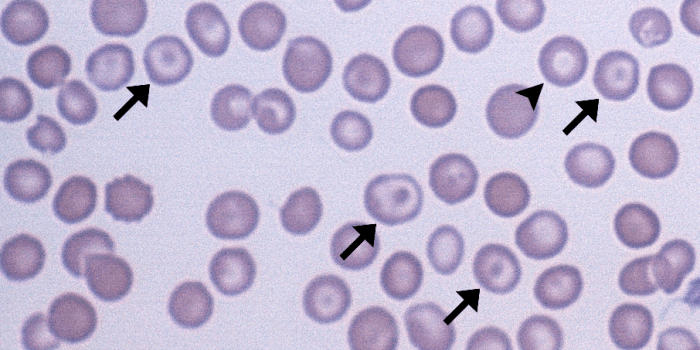
What kind of RBCs are these (2 names)? Describe it.
Codocyte (Target cell): looks like a target w/ bulls eye center

What kind of RBC is seen here (2 names)? Describe it.
Drepanocyte (Sickle Cell) - looks like a crescent shape.

What kind of RBC is seen here (2 names)? Describe it.
Elliptocyte (Cigar-shaped) - looks a bit like a thick rod.

What kind of RBC are seen here (2 names)? Describe it.
Dacryocyte (teardrop): Teardrop shaped

What kind of RBC are seen here? Describe it.
Stomatocyte (mouth): RBC has slit-like area in central pallor
What is the word for a cell that is normal in color?
Normochromic
Define hypochromic. Know what it looks like.
increased central pallor and lowered hemoglobin production

Define polychromic/polychromasia. Know what it looks like.
RBC has blue tint when Wright stained due to residual RNA in cytoplasm (due to reticulocyte stage)
Supravital stain has a chain of blue dots

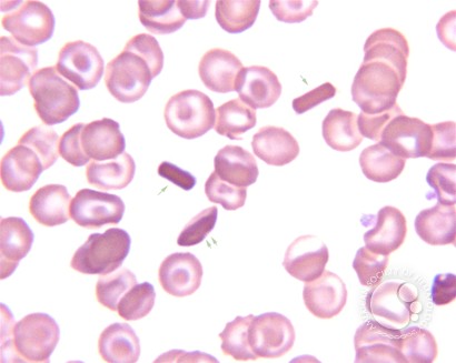
RBC inclusions are present when diseased. What kind of RBC is this? Describe it.
Hemoglobin C crystal: Dark red, hexagonal crystal w/ blunt ends. caused by C hemoglobin.


RBC inclusions are present when diseased. What kind of RBC is seen here (hint: crystal)? Describe it.
Hemoglobin SC crystal: Dark red, 1-2 fingerlike projections produced by SC hemoglobin.

RBC inclusions are present when diseased. What kind of RBC is this? Describe it.
Howell-Jolly Bodies: Round & 1-3 deep blue dots (DNA) in cell

RBC inclusions are present when diseased. What kind of RBC is this? Describe it. What could this be a sign of?
Basophilic stippling: multiple tiny dots (RNA) w/ even distribution.
Could be sign of PB poising & alcoholism

RBC inclusions are present when diseased. What kind of RBC is this? Describe it.
Pappenheimer bodies: small cluster of blue dots (iron) around edge of cell.

RBC inclusions are present when diseased. What kind of RBC is this? Describe it.
Cabot ring: ring-shaped, figure-eight/loop shaped (mitotic spindle).
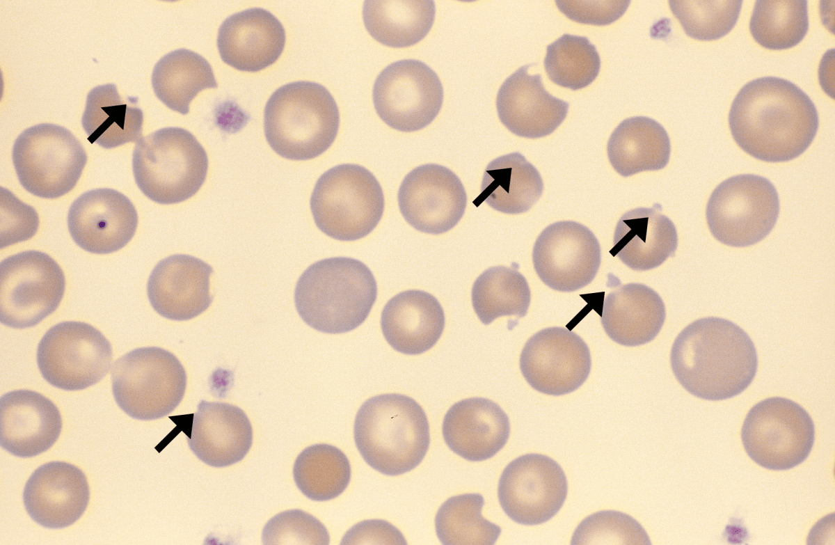
RBC inclusions are present when diseased. What kind of RBC is this? Describe it.
Heinz bodies: Blue & round bodies (denatured/broken-down hemoglobin) ONLY seen w/ supravital
RBCs can have variation in their distribution. Define/describe autoagglutination and know what it looks like. How does this effect a CBC? How can this be resolved?
RBCs are clumped/grouped together.
CBC will show an increased MCV (abg. cell size) and a decreased RBC count due to clumped cells being counted as one cell
resolved by warming blood to 37°C

RBCs can have variation in their distribution. Define/describe Rouleaux and know what it looks like. How/why does it occur and how can this be resolved?
RBCs look like a stack of coins
Occurs due to blood staying in tube for too long or pathologic state → seen through thick blood smear
Fix: suspend RBCs in saline

Microorganisms such as parasites, bacteria & toxins produced by infectious agents can do what to RBCs?
destroy them
Malarial parasites can infect RBCs. What are the 4 species of malaria and what RBC stage/age does it infect? Know what it looks like.
Plasmodium vivax: young RBCs or reticulocytes, predominant species
Plasmodium malariae: older RBCs, mild infection
Plasmodium falciparum: young & old RBCs, very pathogenic
Plasmodium ovale: young RBCs or reticulocytes

What blood smear is used to see malaria in peripheral blood? Where is malaria prevalent and what insect transmits it? What is the treatment for it?
Thick blood smear (3 drops of blood)
tropical Africa
Anopheles mosquito
Treatment: chlorquine or quinine sulfate
What is babesia/what does it cause on/in RBCs? What transmits it and where is it usually found in the world? What blood smear is used to see Babesia in peripheral blood? What is the treatment for it?
Small ring-like protozoa in RBCs
Transmitted by tick from wild deer mice
Found in Nantucket/costal regions
Thick blood smear
Treatment: clindamycin + quinine

What is Ehrlicia? What transmits it and what cells does it effect and how?
Small, intracellular bact.
transmitted via ticks
Effects neutrophils & monocytes & grows as a cluster in them

What is clostridium perfringens? What does it do?
Gram + spore forming bacillus (bact.)
Can cause septicemia

What is Bartonellosis and what is the infection called that it causes? What is it transmitted by and where is it found?
A bacteria that infects RBCs w/ bartonella bacilliformis
Transmitted via sandfly, common in Colombia, Peru & Ecuador
