Coronal polishing
 Call Kai
 Learn
 Practice Test
 Spaced Repetition
 Match
 Flashcards
 Knowt Play
1/41

There's no tags or description
Looks like no tags are added yet.
42 Terms
What should you use if the patient has Ortho appliances?
Use of a small rubber cup, and a pointed or tapered brush, especially around the brackets
What should you use if the patient has Hypersensitive teeth?
Use a light stroke and an abrasive designed for two sensitivity
What should you use if the patient has a Green chromogenic bacterial stain?
Before beginning to coronal polish use a solution of equal parts 3% hydrogen peroxide and water applied to the stained areas with a cotton tip applicator for a few seconds and then rinse
What should you use if the patient has Minor, oral irritations? (EX: cold sores)
Avoid the areas when polishing and retracting the tissues or establishing a fulcrum. Apply a protective coating of lubricant
What should you use if the patient has lack of stains?
No need for a coronal polish
What should you use if the patient has minimal staining?
Selective polishing would be done
What are Soft deposits?
Include dental pellicle, material alba, food debris and plaque.
What is Materia Alba?
Soft cottage cheese-like mass of food debris and bacterial growth that collects in the grooves and spaces of the teeth, gums, and appliances. Provides a source for plaque development.
What is Dental Pellicle?
Thin clear film of insoluble proteins, fats, and other materials from saliva that forms within minutes of removal. it may protect enamel or provide a braiding, ground for plaque and calculus.
What is Plaque?
sticky mass that contains bacteria and grows in colonies on the teeth if not removed will form calculus.
Materia Alba/ food debris is removed by
Rinsing, brushing and flossing
Dental Pellicle is removed by
Polishing with an abrasive
Plaque starts building on the Dental Pellicle
Within 1-2 hours
Plaque is removed by
Brushing, flossing and coronal polish
What is Calculus/ tartar?
A calcified deposit of plaque that has materialized over a period of time on the teeth, restorations or dental appliances
What is Subgingival Calculus?
Black brown, or dark green in appearance. Forms below the gums. Must be removed by dentist or hygienist.
What is Supragingival Calculus?
Chalky, white yellow, gray, or stained by food. Forms above gums. Must be removed by a dentist or hygienist.
What are Intrinsic stains?
On the inside of the tooth structure and are mostly permanent origin can be both endogenous and exogenous
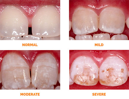
What is Dental fluorosis? (endogenous)
Occurs as a result of high concentrations of fluoride received systematically during tooth development. Other names include brown stain and mottled enamel.
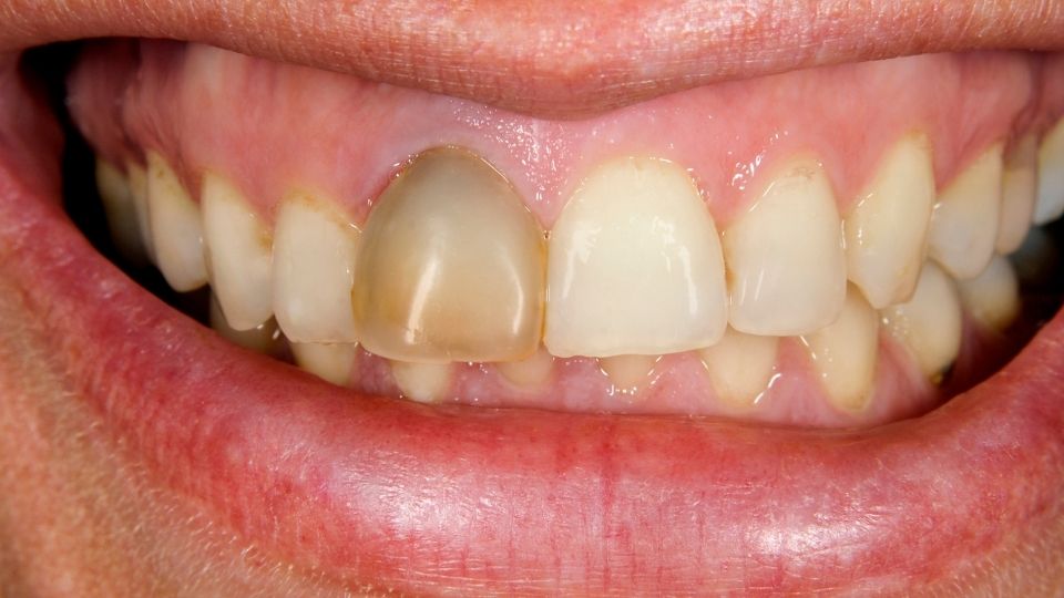
Non-vital Tooth or Pulp Damaged (endogenous)
Occurs when the pulp is damaged or removed. Is caused by blood and pulp tissues seeping into the dentinal tubules
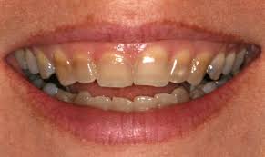
Tetracycline (endogenous)
Occurs as a result of high concentrations of tetracycline antibiotics during the time the tooth was developing
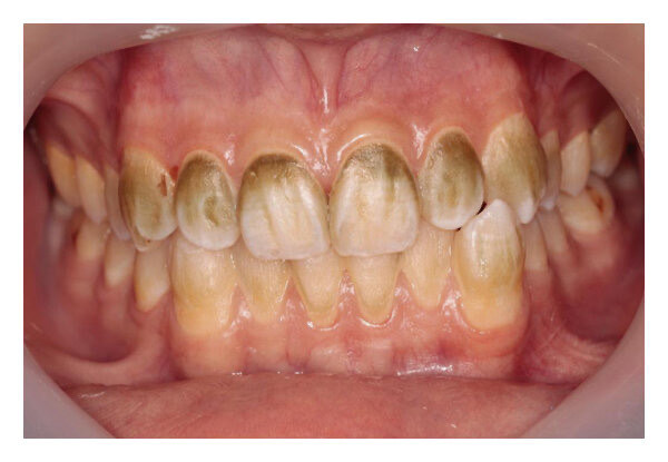
Metallic (intrinsic or extrinsic)
Metals and metallic salts may be inhaled in industrial settings or taken orally in certain drugs or make compromise part of the materials used to restore teeth. The metals adhere to the pellicle and other soft deposits.
Extrinsic Stains
On the outside of the tooth structure and can be removed by scaling and polishing. The origin of extremes are exogenous only.
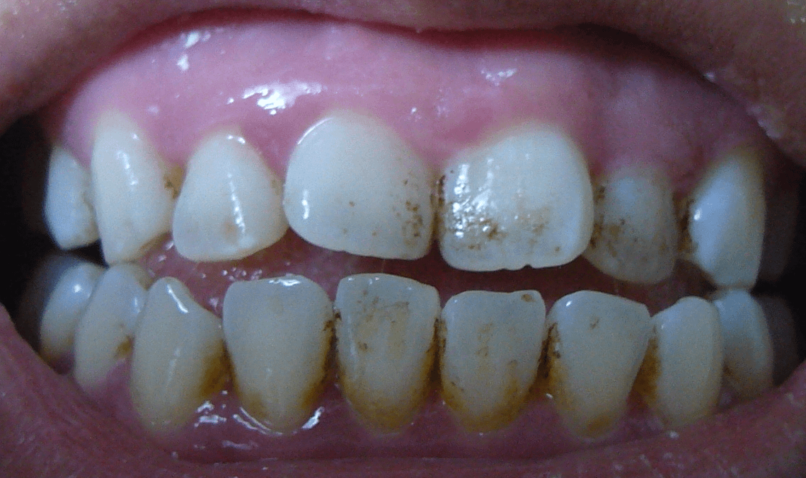
Yellow and brown stains (exogenous)
Usually associated with poor oral hygiene and are dull yellow to light brownish found on the buckle surface of the maxillary molars and lingual surface of the mandibular incisors
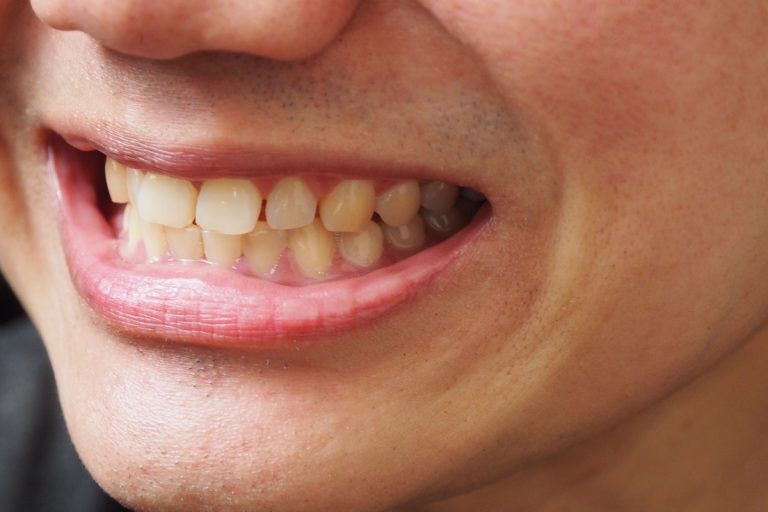
Tobacco stain (exogenous)
Pigments from chewing tobacco, penetrating the pits and fissures on the enamel and Dentin surfaces, light brown to black in color
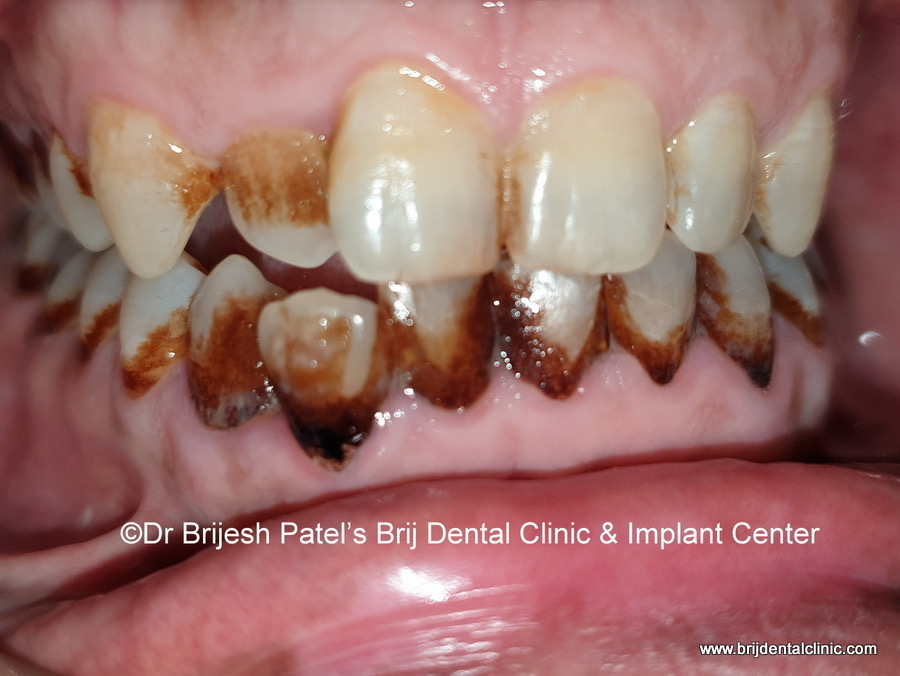
Green stain (exogenous)
Varies in color from light to dark, green or yellowish green is found most frequently in children found on the facial surface of the maxillary anterior teeth at the cervical third and contains chromogenic, bacteria and fungi
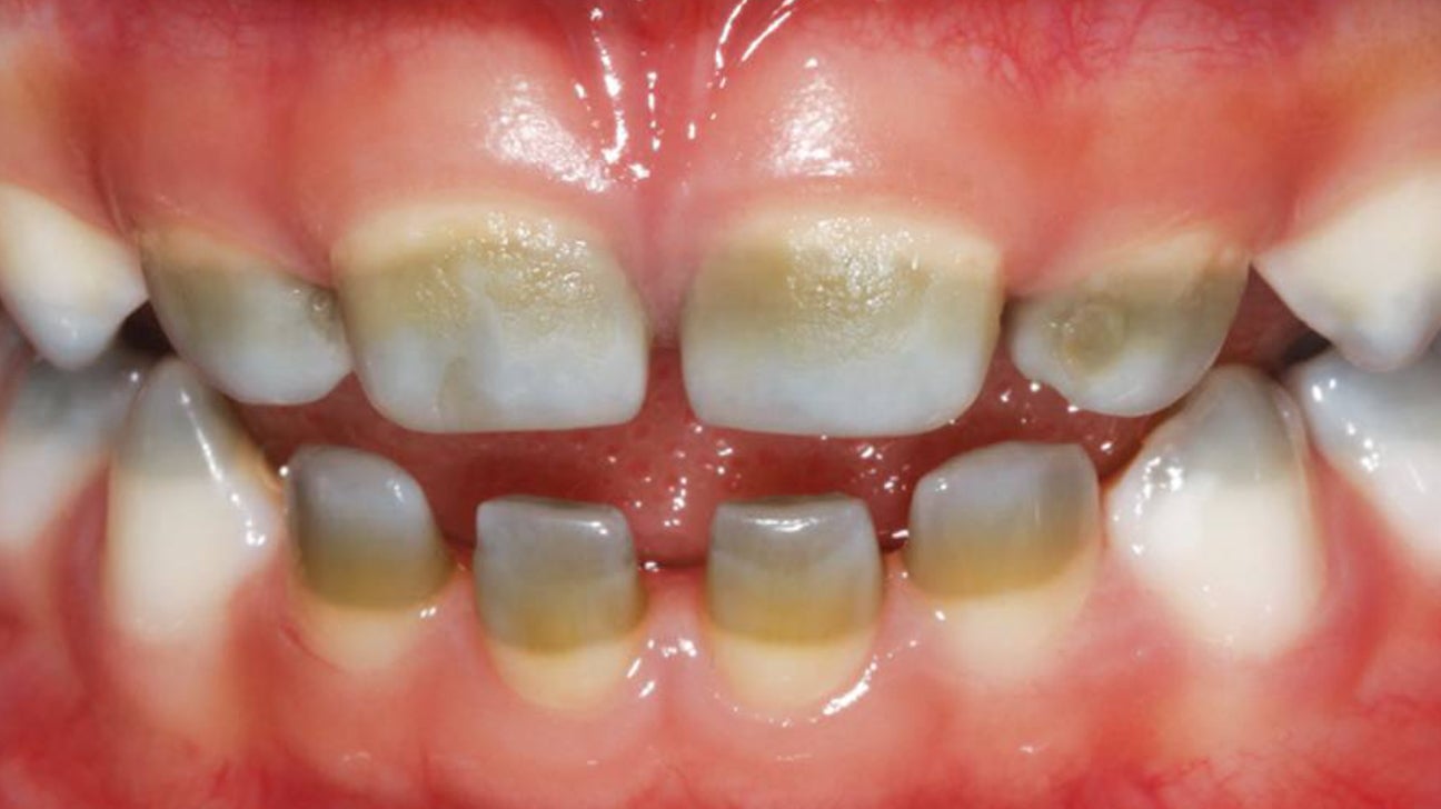
Black line stain (exogenous)
Forms a thin black to dark brown line above the gingiva and follows the contour of the gingival margin found mostly and women, and often where there is excellent, oral hygiene forms on the facial and lingual surfaces of the teeth tends to reform after removal
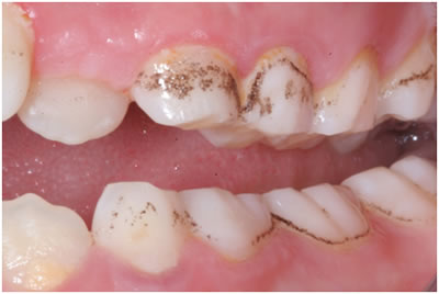
Orange stain (exogenous)
Believe to be caused by chromogenic bacteria often related to drug therapy, such as antibiotics can be found on the facial lingual surfaces of the anterior teeth near the gingival margin
Chlorhexidine stain exogenous
Caused by prolong use of chlorhexidine, which is found in chewing gum and mouth rinse. The stain is yellowish to green to brown and can appear on the cervical and proximal surfaces of the teeth can be removed with toothbrushing and corona polishing.
Zirconium silicate
Used for stain removal and polishing may be used on gold restorations exposed Denton tooth colored restorations and enamel
Silica/fine silica
Used to remove stains as a high-powered, cleaning agent and a fine polishing agent
Tin Oxides
A very fine polishing agent used on enamel and metallic restorations. Mixed with water, alcohol, or glycerin
Flower of pumice
Used to remove stains from the enamel relatively course and should be followed by a fine polishing agent NOT used on exposed Denton tooth colored restorations or gold restorations
Calcium carbonate
Mild abrasive used in some prophy paste
Fluoride prophy paste
Should not be used if sealants are being done after the coronal polish
Silex
Fairly abrasive for use on heavy stains
Diamonds
A polishing abrasive used on porcelain surfaces
Critical Items
A high risk for infection if they are contaminated with any Microorganism. This would include all instruments. This category should be sterilized with steam if possible.
Semi critical items
Contact mucous membranes or non-intact skin. This category should be free from all microorganisms however, small numbers of bacterial Spores are permissible.
Non-critical items
Items are those that come in contact with intact skin but not mucous membranes
Oral Prophylaxis
Removes plaque, stains, debris, and calculus. Done by a dentist or hygienist
Coronal Polish
Removes plaque and stains. Done by polishing the clinical crown, which may involve both enamel and exposed dentin that is visible in the mouth