Somatic Lower Extremity
 Call Kai
 Learn
 Practice Test
 Spaced Repetition
 Match
 Flashcards
 Knowt Play
1/44

There's no tags or description
Looks like no tags are added yet.
45 Terms
General rule for posterior thigh muscle innervation
Sciatic nerve (Tibial division).
General rule for posterior thigh muscle actions
Hip extension and knee flexion.

Exception to posterior thigh muscle general rules
Biceps femoris short head (origin: linea aspera, innervation: common fibular division).
Semitendinosus insertion
Pes anserinus (proximal, anterior, medial tibial shaft).
Biceps femoris long head vs. short head origin
Long head: Ischial tuberosity. Short head: Distal 1/2 of linea aspera.
Quadriceps common insertion
Tibial tuberosity via patellar tendon, patella, and patellar ligament.
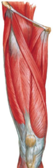
General innervation of anterior thigh muscles
Femoral nerve.
Rectus femoris origins
Anterior inferior iliac spine (AIIS) and superior acetabulum.
Sartorius actions
Hip flexion, abduction, lateral rotation, and knee flexion (figure-4 position).
Iliopsoas insertion
Lesser trochanter of femur.
Pectineus double innervation
Femoral nerve (2/3) and Obturator nerve (1/3).
Function of Articularis Genu
Pulls suprapatellar bursa and synovial membrane upward during knee extension.
General rules for medial thigh compartment
Origin on pubis, insertion on femur, action hip adduction, innervation obturator nerve.
Adductor magnus dual innervation
Adductor part: Obturator nerve. Hamstring part: Sciatic nerve (Tibial division).
Muscles attaching to the pes anserinus
Sartorius, Gracilis, and Semitendinosus.
General innervation of posterior leg compartment
Tibial nerve.
Popliteus function at the knee
Unlocks the extended knee via medial rotation of tibia (or lateral rotation of femur).
Muscles forming the Triceps Surae
Gastrocnemius (medial and lateral heads) and Soleus.
Mnemonic 'Tom, Dick, AN' Harry' for tarsal tunnel (ant to post)
Tibialis posterior, Flexor Digitorum longus, Artery (post. tibial), Nerve (tibial), Flexor Hallucis longus.
General rules for anterior leg compartment
Action dorsiflexion/digit extension, innervation deep fibular nerve.
Tibialis anterior insertions
Inferior base of 1st metatarsal and medial/inferior aspect of medial cuneiform.
General rules for lateral leg compartment
Action ankle eversion and plantar flexion, innervation superficial fibular nerve.
Muscles forming the anatomical stirrup of the foot
Tibialis anterior and Fibularis longus.
Foot intrinsic muscles innervated by Medial Plantar Nerve
Abductor hallucis, Flexor digitorum brevis, Flexor hallucis brevis, Lumbrical 1.
Gluteus maximus insertion
Upper fibers: IT band (Gerdy's tubercle). Lower fibers: Gluteal tuberosity.
Clinical sign of Gluteus Medius/Minimus weakness
Trendelenburg sign (unsupported pelvis drops during single-leg stance).
Gluteus medius & minimus primary innervation
Superior gluteal nerve.
Tensor Fascia Lata insertion
Gerdy's tubercle of tibia via IT band.
Deep lateral rotators of the hip (6 muscles)
Piriformis, Superior gemellus, Obturator internus, Inferior gemellus, Quadratus femoris, Obturator externus.
Most common relationship of Sciatic Nerve to Piriformis (Type A)
Entire sciatic nerve passes inferior/underneath the piriformis muscle (80-90%).
Lumbar plexus spinal segments and location
Formed by L1-L4 spinal nerves within the psoas major muscle.

Sacral plexus spinal segments and location
Formed by L4-S4 spinal nerves on the anterior surface of the piriformis muscle.

Saphenous nerve origin and cutaneous distribution
Terminal branch of femoral nerve supplying skin of the medial knee, leg, and foot.
Meralgia paresthetica cause and nerve involved
Compression of the lateral femoral cutaneous nerve causing pain and paresthesia in lateral thigh.
Key dermatomes for L3 and L4
L3 innervates anterior thigh and medial knee; L4 innervates medial leg and foot.
Key dermatomes for L5 and S1
L5 innervates anterior leg and dorsum of foot; S1 innervates lateral leg and foot.
Deep fibular nerve cutaneous innervation
Skin on the dorsum of the foot between the 1st and 2nd toes.
Femoral triangle contents (lateral to medial)
Femoral Nerve, Femoral Artery, Femoral Vein, Lymphatics (NAVL).
Femoral triangle borders
Superior: Inguinal ligament. Lateral: Medial border of sartorius. Medial: Medial border of adductor longus.

Femoral artery continuation
Passes through adductor hiatus to continue as the popliteal artery.
Branches of the deep femoral artery
Medial femoral circumflex, lateral femoral circumflex, and perforating branches.

Arteries of the cruciate anastomosis
Medial femoral circumflex, lateral femoral circumflex, 1st perforating branch of deep femoral, and inferior gluteal.

Anterior tibial artery termination
Passes under extensor retinaculum to terminate as the dorsalis pedis artery.

Great saphenous vein drainage
Drains medial foot, leg, and thigh into the femoral vein.

Small saphenous vein drainage
Drains lateral foot and leg into the popliteal vein.
