Lung PP Slides Practice Questions
 Call Kai
 Learn
Learn Practice Test
 Spaced Repetition
 Match
Match Flashcards
 Knowt Play
1/147

There's no tags or description
Looks like no tags are added yet.
Name | Mastery | Learn | Test | Matching | Spaced | Call with Kai | Chat |
|---|
No analytics yet
Send a link to your students to track their progress
148 Terms
What organs are present in the respiratory system?
- Trachea
- Right and Left Lung
- Bronchus
- Bronchioles
- Bronchi
- Aveoli
- Mouth
- Nasal cavity
Describe the route of air flow through the respiratory system
1. Nose/Mouth
2. Pharynx
3. Trachea
4. Bronchi
5. Bronchioles
6. Alveoli
7. Capillaries
Explain the process of gas exchange in the aveoli
1. Inhalation
2. Blood delivery
3. Oxygen movement
4. Oxygen binding
5. Carbon dioxide movement
6. Exhalation
What is the role of the respiratory system?
- Moves air into the body
- Removes watse products
-
Which 2 entries of the body receive air?
- Nasal cavity
- Mouth
What happens when air is inhaled?
Air is warmed up
What part of the respiratory does the air pass?
- Trachea
The trachea connects to the ____________ and _______________ cavity to the rest of the _________________ ___________________.
- mouth
- nasal
- respiratory system
What does the trachea lead too?
- To two bronchi and one bronchus lead into each lung
How does air pass through the bronchus?
- Through the bronchioles that seperate from the bronchus
What is located at each brochioles?
- Alveoli
How does alveoli facilitate gas exchange?
- They are air sacs where oxygen enters your bloodstream and carbon dioxide leaves it through a process called diffusion
How many alveoli are located in the lungs?
- Millions
Feature of the alveoli include?
- Thin walls to reduce the distance of diffusion
- Walls are one cell thick
- Large surface area so that moist gases can dissolve and diffuse across
- Surrounded by capillaries for rich blood supply
What muscle facilitates inhalation and exhalation?
- Diaphragm
In inhalation, the diaphragm will...
- Contract and move downwards
In exhalation, the diaphragm will...
- Relaxes and moves upwards during exhalation
What patient prep is needed for vent/perfusion lung scans?
- No dietary restrictions
- 2 view Chest X-Ray (PA/Lat) within 24 hours
Which radiopharmacuetical requires a negative pressure room?
- 133^Xe
133^Xe imaging requirements:
- Aquired Posistion: One - view
- Gamma energy: 80 keV
- Washout: Yes
- Negative Pressure: Yes
99m^TcDTPA imaging requirements:
- Acquire Position: Many --- Many Views
- Gamma Energy: 140 keV
- Washout: No
- Negative Pressure: No
99m^Tc-Techegas imaging requirements:
- Aquired Posistion: Many ---- Many Views
- Gamma energy: 140 keV
- Washout: No
- Negative pressure: No
What should you look out for when doing lung images?
- Difficulty breathing
- Taking breaks
- Nausea
-Coughing
What patient posistion and FOV are used for ^133Xe ventilation scan?
- Supine posistion and must include entire posterior lung field
What collimator and matrix size are standard for ^133Xe Ventilation study?
- LEHR ( Low-Energy High Resolution )
- Matrix Size: 128 x 128
What is the recommended dose for a ^133Xe ventilation study
- 5 to 20 mCi ( 200 -- 750 MBq )
In a V/Q study using ^133Xe, which phase is performed first: ventilation or perfusion?
- Ventilation is performe prior to perfusion (to avoid downscatter from the higher-energy ^99mTC perfusion tracer)
What are the acquistion parameters for the initial breath (single breath) phase of a ^133Xe scan?
- Acquire 1 image for approx 20 to 30 secs
What are the acquisition parameters for the equilibration phase of a ^133Xe scan?
- Acquire 1 to 2 images at 60 secs per images
What is the standard imaging protocol and normal duration for the washout phase of a ^133Xe scan?
- Acquire sequential 1-minute images until tracer clears, typically taking about 4 to 5 mins
How does COPD typically affect the washout phase of ^133Xe scan?
- It causes tracer retention/ gas trapping, meaning activity remains visible in the lungs for a significantly longer period (>5 mins)
What contraption is used to exel Xenon?
- Calidose Dispenser
What does the Calidose dispenser administer?
- A gas dose of ^133Xe from a vial
The first step of the Calidose despenser is:
- Placing a needle at the oppisite end and inserting into the spirometer
The second step of the Calidose despenser is:
- plunger is pushed, which pierces the rubber stopper on the Xenon vial
The third step of the Calidose despenser is:
- When the bulb is squeezed, it forces the gas into the xenon delivering system

1 is:
- Needle that is inserted in the mask
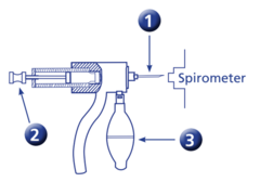
2 is:
- Insertions that pushes vial

3 is:
- Bulb that pushes air in and out of the needle
What is the patient position and detector placement during the initial breath phase of a Xenon-133 lung ventilation study?
- Upright position, imaging the posterior lung field
What does the patient do immediately after Xenon-133 gas is injected during the initial breath phase?
- Holds their breath for 20-30 seconds
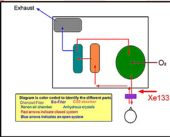
What type of breathing system is utilized during the Initial Breath phase of a Xenon ventilation study?
- A closed system

In the Xenon delivery diagram, what do red arrows vs. blue arrows represent?
- Red = Closed system
- Blue = Open system (leading to exhaust)

What are the 5 main color-coded components inside the Xenon ventilation system diagram?
- Charcoal Filter
- Bio-Filter
- CO2 Absorber
- Xenon Air Chamber
- Anhydrous Crystals
What type of breathing system is used during the equilibration phase of a ¹³³Xe ventilation study?
- A closed system
What is the purpose of sodium lime in a closed equilibration system?
- It absorbs CO₂ and prevents acidosis
What component removes moisture from the exhaled air during equilibration?
- Anhydrous crystals
What imaging parameters are typically acquired during the equilibration phase of a Xenon study?
- 1-2 images at 60 seconds each
Exhaled air passes through a _______________ before moisture is removed by _________________
- biofilter
- anhydrous crystals
During equilibration, ________________ absorbs CO₂ to prevent ________________.
- sodium lime
- acidosis
Xenon enters a large chamber where additional ______________ is added before returning to the patient
- O2
During the equilibration phase, you should acquire ____________ images at ____________ each.
- 1-2
- 60s
List the first path and processing steps of exhaled air during the closed-system equilibration phase of a Xenon study.
1. Passes a biofilter
List the second path and processing steps of exhaled air during the closed-system equilibration phase of a Xenon study.
2. Anhydrous crystals remove moisture
List the third path and processing steps of exhaled air during the closed-system equilibration phase of a Xenon study.
3. Sodium lime absorbs CO₂ (prevents acidosis)
List the fourth path and processing steps of exhaled air during the closed-system equilibration phase of a Xenon study.
4. Enters large chamber where O₂ is added
List the fifth path and processing steps of exhaled air during the closed-system equilibration phase of a Xenon study.
5. Filtered air and ¹³³Xe return to the patient
What is the gas flow path during the Wash Out (Open System) phase?
- Anhydrous → sodium lime → charcoal filter → exhaust
Is new Xenon administered to the patient during the wash-out inhalation?
- No. The patient only breathes in room air while Xenon washes out of the lungs.
What happens to Xenon gas that escapes the charcoal filter?
- It must be vented out of the department
What is the imaging frequency and duration for the wash-out phase?
- One-minute images are acquired until Xenon is cleared from the lungs

These are called?
- Xenon Ventilation Systems
Why must Xenon-133 (¹³³Xe) be administered in a negatively pressured room?
- To ensure that if any radioactive gas escapes during administration, it is contained and does not leak into the rest of the facility
What ventilation requirement must be calculated for a ¹³³Xe administration room in case of a gas leak?
- A 100% air turnover should be computed to handle potential gas escapes
How often must a ¹³³Xe administration room be tested for negative pressure?
- It must be tested quarterly (every 3 months) to ensure the negative pressure is still present
What color change occurs in the anhydrous crystals as they absorb moisture?
- They start blue and turn pink as they absorb moisture
When should the anhydrous crystal tube be discarded and refilled?
- Once all materials turn pink, it must be discarded and the tube refilled
What is the rule regarding the patient connection during the washout phase?
- The patient must stay connected until all the xenon leaves their lungs
What happens if a patient is discharged prematurely while ¹³³Xe activity is still noted?
- Xenon gas will be exhaled directly into the room and the surrounding environment, causing potential radioactive contamination.

What are the main components of a Ventilation-Aerosol delivery setup?
- Injection port
- Nebulizer
- Oxygen (O₂) port (connected to the O₂ source)
- 1-way valves
- Filter
- Connection for mouthpiece (or extra tubing for a ventilator patient)
- Aerosol Shield (to house the delivery system)
What radiopharmaceutical and dose are used for a Ventilation-Aerosol lung scan according to this protocol?
- 25 - 35 mCi of 99𝑚Tc DTPA, which is placed into the injection port and contained within the nebulizer
What matrix and collimator settings are used for the Ventilation-Aerosol scan?
- Matrix: 128 × 128 or 256 × 256
- Collimator: LEHR (Low Energy High Resolution)
Which views must be acquired during a Ventilation-Aerosol lung scan?
* 8 standard projections:
- ANT (Anterior) & POST (Posterior)
- LPO (Left Posterior Oblique) & RPO (Right Posterior Oblique)
- LAO (Left Anterior Oblique) & RAO (Right Anterior Oblique)
- RT LAT (Right Lateral) & LT LAT (Left Lateral)
What is the stopping condition for each image, and what is the next step in the overall study?
- Stopping Condition: Acquire 150K counts per image.
- Next Step: Once ventilation is completed, continue immediately to the PERFUSION phase.
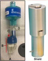
What part of the Areosol system is labeled A?
- O₂ port

What part of the Areosol system is labeled B?
- Nebulizer

What part of the Areosol system is labeled C?
- Injection port

What part of the Areosol system is labeled D?
- Single use tubing that goes to the patient
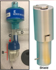
What part of the Areosol system is labeled E?
- Filter trap to the exhaled droplets

What part of the Areosol system is labeled F?
- Filter exhalation
What should the volume be for DTPA in the nebulizer?
- 3-5 mL
The ______________ the O2 per min, the ______________ - suggest ____________L/min
- higher
- better
- 10
How long should the administartion of DTPA be?
- No more than 5 minutes

This is ^99mTC DTPA
True

This is ^133XE
True

Both of these normal ^99m TC DTPA Aerosol Scans show:
- Mouth
- Oral cavity
- Aerosol deposition in the bronchi stomach uptake that occurs from swallowing areosol
What is the primary clinical reason to ideally administer
99m Tc-MAA first in a V/Q lung scan?
- If the 99mTc-MAA perfusion scan is normal, there is no clinical need to perform the 133Xe ventilation scan, saving time and radiation exposure.
What technical complication occurs if 99mTc-MAA is given before 133Xe?
- Compton Scatter (downscatter) from the higher-energy 99mTc will spill over into the lower-energy 133 Xe gamma camera window, degrading image quality.
What are the respective photopeak energies of 133Xe and 99m Tc?
- 133Xe: 81 keV (centered around 80 keV)
- 99mTc: 140 keV
What does the acronym MAA stand for in the context of a lung perfusion scan?
- Macro-Aggregated Albumin
What is the standard particle diameter range for Macro-Aggregated Albumin (MAA) used in lung perfusion procedures?
- 10 to 90 micrometers
What is the collimation setting for 99m Tc MAA lung perfusion?
- LEHR
- 20% energy window
Standard dose and route for 99m Tc MAA
- 4 to 5 mCi administered intravenously (IV)
Patient position required during radiopharmaceutical injection
- Supine position
Reason for injecting the patient while supine
- Ensures homogeneous particle distribution across the lungs
Effect of gravity if injected while standing
- Particles concentrate heavily in the lower lobes
Required step immediately before injecting the dose
- Agitate or shake the syringe to distribute particles
Critical technique to avoid during the injection
- Do not pull back the syringe

Why must the number of 99mTc-MAA particles be reduced in patients with right-to-left shunts during a lung perfusion scan?
- To prevent the particles from entering the systemic circulation, which can cause microembolisms in critical organs like the brain, kidneys, and heart
For which three patient conditions should you reduce the particle number in a 99mTc-MAA lung perfusion procedure?
- Severe pulmonary hypertension (due to a reduced pulmonary vascular bed)
- Right-to-left shunts
- Pneumonectomy (prior surgical removal of a lung)