Cardiovascular system
 Call Kai
 Learn
Learn Practice Test
 Spaced Repetition
 Match
Match Flashcards
 Knowt Play
1/95

There's no tags or description
Looks like no tags are added yet.
Name | Mastery | Learn | Test | Matching | Spaced | Call with Kai | Chat |
|---|
No analytics yet
Send a link to your students to track their progress
96 Terms
What is the function of arteries?
Transport blood from the heart to organs and tissues.
What are the two types of arteries?
Elastic arteries and muscular arteries.
What is the function of arterioles?
Deliver blood into the capillary network.
What is the function of capillaries?
Allow gas exchange and the exchange of substances between blood and tissues.
What is the function of capillaries?
Allow gas exchange and the exchange of substances between blood and tissues.
What is the function of venules?
Drain blood from capillaries.
What is the function of veins?
Receive blood from venules and return it towards the heart.
Put the blood vessels in order from the heart to the tissues and back.
Arteries → arterioles → capillaries → venules → veins.
Which blood vessels connect arterioles to venules?
Capillaries.
What are the three layers of blood vessel tissue?
Tunica externa, tunica media and tunica intima.
1. What is the tunica externa (adventitia) made of, and what is its function?
Elastic and collagen fibres. Provides protection and structural support to surrounding anatomy.
2. What is the tunica media made of, and what is its function?
Muscle, elastic and connective tissue. Allows vasodilation and vasoconstriction.
3. What is the tunica intima (interna) made of?
A lining of endothelial cells.
What layers do arterioles have?
Tunica media (muscle), endothelial layer and basement membrane.
What layers do venules have?
Tunica externa and tunica intima.
3. What is the wall structure of capillaries?
A single-cell-thick endothelial layer and a basement membrane.
Which blood vessel has a wall only one cell thick?
Capillaries — allowing efficient exchange of gases and substances.
What are the main structural differences between arteries and veins?
• Tunica externa: thicker in veins
• Tunica media: thicker in arteries, with more muscle and elastic tissue
• Lumen: generally larger in veins
• Valves: present in veins
Describe the pathway of systemic circulation, including where gas exchange occurs.
Left ventricle → aorta → arteries → capillaries (gas exchange) → venules → veins → right atrium. Oxygenated blood delivers oxygen to tissues; deoxygenated blood returns to the heart.
Describe the pathway of pulmonary circulation, including where gas exchange occurs.
Right ventricle → left and right pulmonary arteries → lung capillaries at alveoli (external respiration/gas exchange) → pulmonary veins → left atrium. Blood changes from deoxygenated to oxygenated.
List the great vessels associated with the heart in the thorax.
• Ascending aorta
• Aortic arch
• Descending thoracic aorta
• Brachiocephalic trunk
• Left common carotid artery
• Left subclavian Artery
List the main arteries of the upper limb in order.
→Left subclavian
→ left axillary
→ left brachial
→ left radial and ulnar arteries
Name the main veins of the left upper limb.
Left subclavian,
Left axillary,
Left cephalic,
Left brachial,
Left basilic veins.
List all the abdominal vasculature .
ARTERIES: Celiac trunk; right renal artery; superior mesenteric artery; inferior mesenteric artery; right common iliac artery.
VEINS: Right and left hepatic veins; hepatic portal vein; right and left renal veins; IVC; common iliac veins; cystic vein; gastric vein; splenic vein; superior and inferior mesenteric veins.
List the five main arteries of the lower limb in order.
→ External iliac
→ femoral
→ popliteal
→ anterior tibial
→ posterior tibial arteries.
List the five main veins of the left lower limb.
→Left external iliac
→femoral
→great saphenous
→popliteal
→tibial veins.
Where is the heart located?
In the mediastinum, between the lungs, within the thoracic cavity.
How is the heart positioned, and where is its apex?
Obliquely, slightly left; the apex is about 9 cm left of the midline.
What is the base of the heart?
The flattened posterior-superior surface.
What is the heart’s structure and approximate weight?
A hollow muscular organ; 250 g in females and 300 g in males.
What are the four inner chamber of the heart?
1.Right atrium
2.Right ventricle
3.Left atrium
4.Left ventricle
What lies inferior to the heart?
The diaphragm, specifically its central tendon.
What structures lie superior to the heart?
Ascending aorta, SVC, pulmonary artery and pulmonary veins.
What lies anterior to the heart?
Sternum, ribs and intercostal muscles.
What structures lie posterior and lateral to the heart?
Posterior: oesophagus, trachea, main bronchi, descending aorta, IVC and thoracic vertebrae.
Lateral: both lungs.
How is the heart divided internally?
Into right and left halves by the septum, made of myocardium lined with endocardium.
What are the chambers of the heart?
Each half has two chambers: an atrium and a ventricle.
What separates each atrium from its ventricle?
An atrioventricular (AV) valve, made of folds of endocardium and fibrous tissue.
What are the right and left AV valves called, and how many cusps do they have?
Right: tricuspid (3 cusps).
Left: bicuspid/mitral (2 cusps).
Which vessels bring blood into the right atrium?
Superior vena cava (SVC), inferior vena cava (IVC) and coronary sinus.
How thick are the right atrium’s walls?
Approximately 2–3 mm.
How do the anterior and posterior walls of the right atrium differ internally?
Posterior wall: smooth.
Anterior wall: muscular ridges.
What separates the atria, and how does blood leave the right atrium?
The interatrial septum separates them. Blood passes into the right ventricle through the tricuspid valve.
(Right ventricle) How thick are its walls, and what surface does it form?
Walls are approximately 4–5 mm thick. It forms most of the anterior surface of the heart.
(Right ventricle) What are trabeculae carneae?
Raised bundles of muscle fibres lining the inner walls; some contribute to the heart’s electrical activity.
(Right ventricle) How are the tricuspid valve cusps attached to papillary muscles?
Chordae tendineae connect the tricuspid valve cusps to papillary muscles.
(Right ventricle) What separates the ventricles, and where does blood exit?
The interventricular septum separates them. Blood exits through the pulmonary valve into pulmonary circulation.
(Left atrium) What is its size, and what part of the heart does it form?
About the same size as the right atrium. Forms most of the base of the heart.
(Left atrium) Which blood vessels deliver blood into it?
Four pulmonary veins.
(Left atrium) How does blood pass into the left ventricle?
Through the bicuspid (mitral) valve, also called the left atrioventricular valve.
(Left ventricle) How thick are its walls, and what part of the heart does it form?
The thickest chamber, with walls approximately 10–15 mm thick. It forms the apex of the heart.
(Left ventricle) What internal structures attach to the bicuspid valve cusps?
Trabeculae carneae line the inner walls. Chordae tendineae connect the bicuspid valve cusps to papillary muscles.
(Left ventricle) How does blood leave the chamber?
Blood passes through the aortic valve into the ascending aorta.
(Myocardial thickness) Why are the atrial walls thin?
They only need to push blood into the adjacent ventricles.
(Myocardial thickness) Which ventricle has the thicker wall, and why?
The left ventricle; it pumps blood around the entire body at higher pressure against greater resistance.
(Myocardial thickness) Why is the right ventricle wall thinner?
It pumps blood a shorter distance to the lungs, requiring less pressure than the left ventricle.
(Pericardium) What are the two sacs surrounding the heart?
The fibrous pericardium and the serous pericardium.
(Fibrous pericardium) What is it made of, and what are its functions?
Connective tissue.
Protects the heart from over-distension and anchors it in the mediastinum. It is continuous with the great vessels’ tunica adventitia and the central tendon of the diaphragm.
(Serous pericardium) What are its two layers?
Parietal layer: fused to the fibrous pericardium. Visceral layer
(epicardium): lies directly on the heart and forms part of its wall.
(Pericardium) What is the function of pericardial fluid, and how much is present?
Approximately 20 mL of serous fluid lubricates the space between the serous pericardium’s layers, reducing friction.
(Pericardium) What are its four main functions?
Prevents over-distension, anchors the heart, protects against infection, and lubricates.
(Pericardium) How does it prevent over-distension?
Limits excessive filling and stretching of the heart chambers.
(Pericardium) How does it protect and support the heart?
Anchors the heart in place and helps protect it from infection.
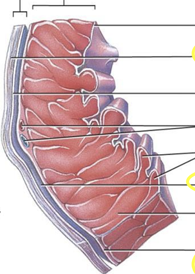
Label the the heart wall/ Pericardium
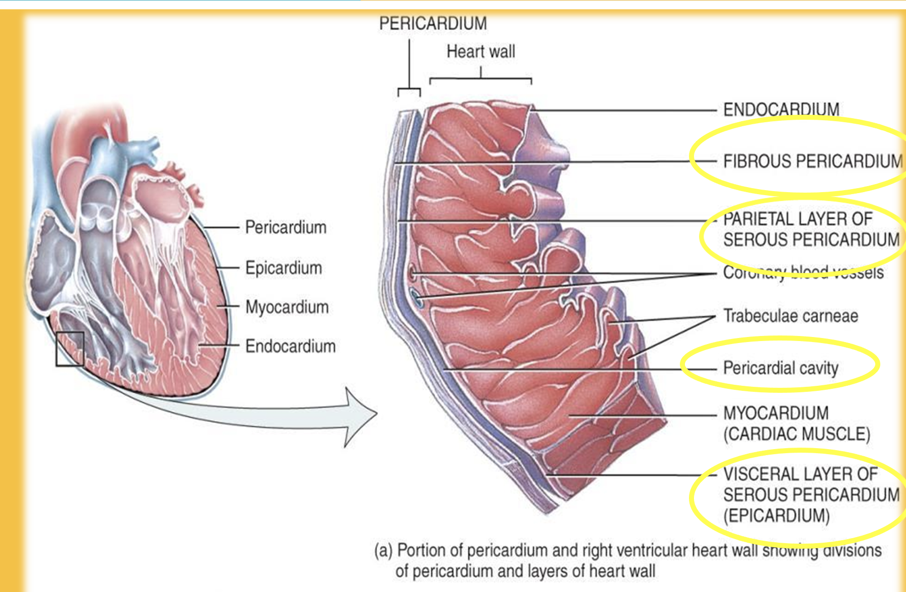
(Heart wall) What are its three layers?
Epicardium
myocardium
endocardium.
(Epicardium) Where is it located?
The outermost layer of the heart wall.
(Epicardium) What is its other name, and what does fluid do?
Visceral pericardium. Serous fluid lubricates the surfaces to reduce friction as the heart moves.
(Myocardium) What type of muscle forms it?
Specialised, striated cardiac muscle found only in the heart.
(Myocardium) How do branched cells support coordinated contraction?
Closely connected cells allow electrical activation to spread easily, coordinating contraction.
(Myocardium) Where is it thickest?
At the apex, particularly around the left ventricle.
(Endocardium) What is its structure and function?
A thin layer of flattened cells lining the chambers; provides a smooth surface for blood flow and continues into vessel endothelium.
(Heart valves) What are the two main types?
Atrioventricular (AV) valves and semilunar valves.
(AV valves) Which valves separate the atria and ventricles?
Right: tricuspid valve.
Left: bicuspid (mitral) valve.
(Semilunar valves) Which valves connect ventricles to great vessels?
Right ventricle: pulmonary valve.
Left ventricle: aortic valve.
(Heart valves) Where is each valve located?
Tricuspid: right atrium–ventricle.
Mitral: left atrium–ventricle.
Pulmonary: right ventricle–pulmonary trunk.
Aortic: left ventricle–aorta.

Label the image

(Heart valves) What causes valves to open and close?
They open when pressure rises on one side and close when the pressure gradient reverses.
(Heart valves) How do they ensure one-way blood flow?
They open and close in response to pressure differences, preventing backflow.
( Atrioventricular valve/AV valves) What causes them to open?
Atrial pressure rises above ventricular pressure, forcing the valves open.
(AV valves) What happens during filling?
Blood flows from the atria into the relaxed ventricles; cusps extend into the ventricles.
(AV valves) What causes them to close?
Ventricular pressure rises above atrial pressure, closing the cusps.
(AV valves) What do papillary muscles and chordae tendineae do?
Papillary muscles contract, tightening chordae tendineae to prevent cusps turning back into the atria.
(AV valves) What do papillary muscles and chordae tendineae do?
Papillary muscles contract, tightening chordae tendineae to prevent cusps turning back into the atria.
(Chordae tendineae) How do they prevent backflow through AV valves?
They prevent the valve cusps from flipping into the atria during ventricular contraction, stopping blood flowing backwards.
(Semilunar valves) What are they made of?
Three crescent-moon-shaped cusps attached to the arterial wall.
(Semilunar valves) What happens during ventricular contraction?
Ventricular pressure rises, opening the valves so blood flows into the great vessels.
(Semilunar valves) How do they prevent backflow?
When ventricular pressure falls, blood briefly flows back and fills the cusps, closing the valve.
(Coronary circulation) Why is it needed, and how does blood enter the coronary arteries?
Diffusion alone cannot supply all heart wall layers quickly enough. Coronary arteries branch from the ascending aorta; during relaxation, increased aortic pressure drives blood into them.
(Coronary arteries) Where do they originate, how much blood do they receive, and where does most flow?
Two arteries branch from the ascending aorta, just below the aortic valve. They supply about 5% of cardiac output; most coronary blood flow goes to the left ventricle.
(Coronary arteries) What do the left and right coronary arteries supply?
Left coronary artery divides into:
• LAD: supplies both ventricles
• Circumflex: supplies left atrium and ventricle
Right coronary artery: supplies right atrium; its marginal branch supplies right ventricle.
(Coronary veins) Where does deoxygenated blood from the myocardium drain?
Most drains into the coronary sinus, a thin-walled vein on the posterior surface of the right atrium, which empties into the right atrium.
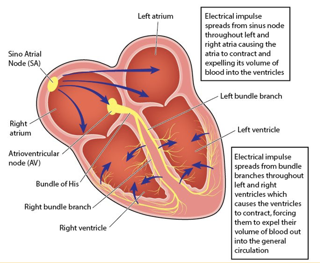
Route of conduction (Stage 1–2) Where does the heartbeat impulse start, and what is its role?
The SA node in the right atrium near the SVC. It acts as the natural pacemaker and triggers both atria to contract.
Route of conduction. (Stage 3) What is the AV node's role?
Receives the impulse from the atria and transmits it towards the ventricles, with a brief delay.
Route of conduction (Stage 4–5) What happens after the AV node?
The impulse travels through the AV bundle (Bundle of His), which divides into right and left bundle branches in the ventricular septum.
Route of conduction. (Stage 6) What do Purkinje fibres do?
Spread the electrical impulse through the ventricular myocardium, coordinating ventricular contraction.
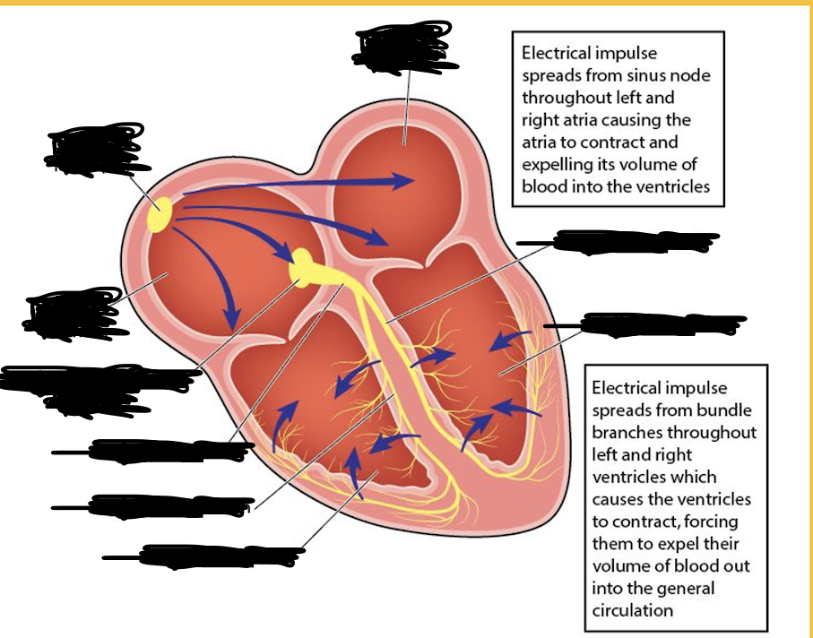
Label the image of the Route of conduction