Upper GI Grossing
 Call Kai
 Learn
 Practice Test
 Spaced Repetition
 Match
 Flashcards
 Knowt Play
1/64

There's no tags or description
Looks like no tags are added yet.
65 Terms
What are the anatomic segments of the esophagus?
Cervical — 15-20 cm
Upper thoracic — 20-25 cm
Middle thoracic — 25-30 cm
Lower thoracic/EGJ — 30-40 cm
Abdominal esophagus — 40-45 cm
What segment is the first 15-20 cm of the esophagus?
Cervical esophagus
What segment is 20-25 cm of the esophagus?
Upper thoracic esophagus
What segment is 25-30 cm of the esophagus?
Middle thoracic esophagus
SCC common location
What segment is 30-40 cm of the esophagus?
Lower thoracic esophagus (EGJ)
Adenocarcinoma common location
Z-line ~40 cm
What segment is 40-45 cm of the esophagus?
Abdominal esophagus
What important feature of the esophagus affects margin assessment?
Most of the esophagus lacks a true serosal layer
The adventitial/circumferential margin is particularly important
What does the gastroesophageal (GE) junction represent grossly?
The transition from esophageal-type mucosa to gastric-type mucosa
Why is an attached gastric cuff important on an esophagectomy specimen?
It provides anatomic landmarks from both the esophagus and stomach
Helps establish the orientation of the specimen
What specimen may contain both esophagus and proximal stomach?
An esophagogastrectomy/esophagectomy with attached gastric cuff
What are three ways to distinguish the proximal esophageal end from the distal gastric end?
Overall specimen length/anatomic appearance
Mucosal color change at the GE junction
Make note of the relationship with the Z-line
Surgical sutures/designations
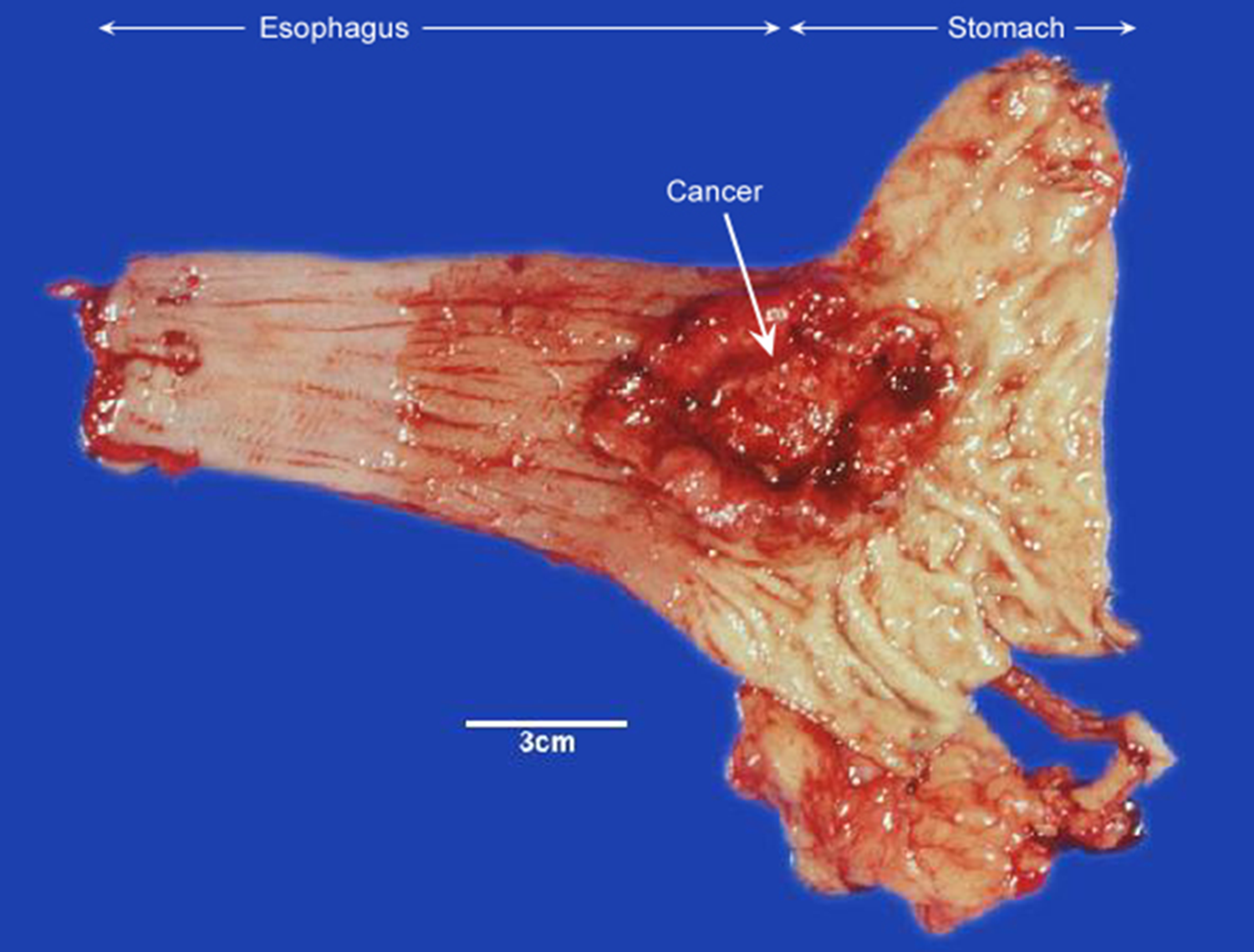
What margin should be inked on an esophagectomy specimen?
The circumferential/radial margin
Especially the area adjacent to the tumor
What should you document about the resection margins?
Whether the margins are stapled or open
Document them accordingly
What measurements should be taken on an esophagectomy specimen BEFORE sectioning?
Overall specimen length
Length of any attached gastric cuff
What should be documented regarding the tumor's location of esophageal lesion?
Its location relative to:
The GE junction
Each resection margin
In how many dimensions should an esophageal lesion be measured?
Three dimensions
What gross growth patterns should be considered when describing an esophageal lesion?
E-U-I-F
Exophytic/polypoid
Ulcerated
Infiltrative
Flat
What should you estimate regarding tumor invasion of esophageal lesion?
The depth of invasion
How should the distance from an esophageal lesion to the margins be measured?
Measure the distance to each margin
Preferably on the fresh specimen when possible
How should sections be taken through an esophageal tumor?
Take perpendicular sections through the lesion at its deepest point of invasion
Deepest invasion = perpendicular section
Which esophageal margins should be sampled in esophagogastrectomy?
Proximal
Distal
Radial/circumferential
What uninvolved tissue should be sampled from an esophagogastrectomy?
Representative uninvolved esophagus and stomach
How should regional lymph nodes be handled for esophagogastrectomy?
Sample them separately
Why are esophagogastrectomies commonly performed?
They are usually performed for:
Severe dysplasia
Adenocarcinoma arising in a background of Barrett's esophagus

Why is Barrett's esophagus important when grossing an esophagogastrectomy?
Adenocarcinoma can arise in a background of Barrett's esophagus
Makes evaluation of the esophageal mucosa and tumor relationship to the GE junction important
What are the five major anatomic regions of the stomach?
Cardia
Fundus
Body
Antrum
Pylorus
What are two major gross landmarks of the stomach?
Greater curvature
Lesser curvature
Why are the omental and mesenteric attachments important?
They can contain regional lymph nodes
What are common gastric specimens received in surgical pathology?
Endoscopic biopsy
Wedge/local excision
Partial gastrectomy
Total gastrectomy
What are common reasons for gastric resection?
Primary tumors
Less commonly benign ulcers
As part of larger resections, such as
Whipple procedures
Esophagogastrectomies
Morbid obesity, particularly sleeve gastrectomy
What types of gastrectomy can be performed?
Total
Partial
What two types of mucosa should you specifically look for on a gastrectomy specimen?
Esophageal mucosa and/or duodenal mucosa
Depends on what portions are attached
What landmarks can be used to orient a stomach specimen?
Greater curvature
Lesser curvature
Pylorus/duodenal cuff
Attached esophagus
Which gastric margins should be inked?
Proximal margin
Distal margin
What additional surface should be inked when evaluating a gastric tumor?
The serosa overlying or nearest the tumor
How should gastric lymph nodes be handled in gastric tumor?
Separate and label them
What attached tissue should be measured in gastric tumor?
The attached omentum
Any separately submitted tissue
What should be documented when describing a gastric lesion?
Size in three dimensions
Location by:
Region
Curvature
Gross growth pattern
Wall thickness
Presence/Absence of rugae
Distance to margins
Distance to nearest serosal surface
Include omentum
What are the major gross growth patterns of gastric tumors listed in the lecture?
Fungating
Ulcerated
Infiltrative
What is linitis plastica?
A diffusely infiltrative gastric tumor that may present grossly as wall thickening rather than as a discrete mass
(Linitis plastica → diffuse infiltration → thickened gastric wall)

What wall measurements should be taken when evaluating a gastric lesion?
Measure wall thickness at the lesion and in an uninvolved area
What two distances should be particularly documented for a gastric lesion?
Distance to each margin
Distance to the nearest serosal surface
How should a gastric tumor be sectioned?
Submit full-thickness sections through the lesion
Include the area of deepest invasion
Which gastric margins should be submitted for a gastric lesion specimen?
Proximal margin
Distal margin

What uninvolved tissue should be submitted from a gastrectomy?
Representative uninvolved stomach
How should lymph nodes from a gastrectomy be submitted?
Submit lymph nodes by station
Record the total number of nodes
What gross finding should never be overlooked when grossing a stomach?
ANY thickened area of the gastric wall
What cell type do GISTs arise from?
The interstitial cells of Cajal in the muscularis propria
What locations within the wall can a GIST occupy?
Subserosal
Intramural
Submucosal
How do GISTs usually grow grossly?
Generally well-circumscribed with a pushing border
(rather than an infiltrative border)
What is the typical cut surface of a GIST?
Fleshy
Tan-white
Whorled
What gross secondary changes should be assessed in a GIST?
Necrosis
Hemorrhage
Cystic change
What is an important adverse prognostic gross finding in a GIST?
Capsular disruption/tumor rupture
Why must GIST rupture be carefully documented?
Capsular disruption/tumor rupture is a significant adverse prognostic finding
What is the most common location of a GIST?
The stomach
Accounts for approximately 47–60% of GISTs
What symptoms can result from a GIST?
Symptoms are related primarily to mass effect
Includes mucosal ulceration that can result in blood loss and anemia
GISTs may also be discovered incidentally
What factors correlate with GIST prognosis?
Tumor size
Mitotic index
Tumor location
How does gastric GIST behavior compare with small intestinal GIST?
Gastric GISTs tend to be LESS aggressive than small intestinal GISTs
What margin-related features are particularly important when grossing a GIST?
Distance to the margin
Completeness of the serosal surface
What are satellite nodules in relation to a GIST?
Nodules separate from the main tumor mass
Should be documented with their size and distance from the main mass
What should you do if lymphoma is in the differential diagnosis of a GI mass?
Reserve a fresh tissue sample in RPMI
What should be included in GIST sampling?
Margins
Full-thickness sections of the mass
Any regional lymph nodes
What sampling guideline is provided for a GIST mass?
Approximately 1 piece per centimeter of tumor
What is the major difference in the external surface of the esophagus vs. stomach regarding serosa?
Esophagus — Most of it lacks a true serosal layer → circumferential/adventitial margin is important
Stomach — Has a serosal surface that should be evaluated, particularly near a tumor
What are these showing?


Examples of GISTs