Week 8-9: Nucleic Acids

0.0(0)
Studied by 0 people
call kaiCall Kai
learnLearn
examPractice Test
spaced repetitionSpaced Repetition
heart puzzleMatch
flashcardsFlashcards
GameKnowt Play
Card Sorting

1/20

encourage image

There's no tags or description

Looks like no tags are added yet.

21 Terms

1
New cards

What are nucleic acids, and what is their primary structure?

  • DNA (deoxyribonucleic acid) and RNA (ribonucleic acid) are biological macromolecules that encode genetic information.

  • Nucleic acids are linear polymers composed of repeating nucleotide units.

  • Each nucleotide contains three components: a pentose sugar, nitrogenous base, and phosphate group.

  • Nucleotides are connected through phosphodiester bonds.

  • The alternating sugars and phosphates form the sugar-phosphate backbone of nucleic acids.
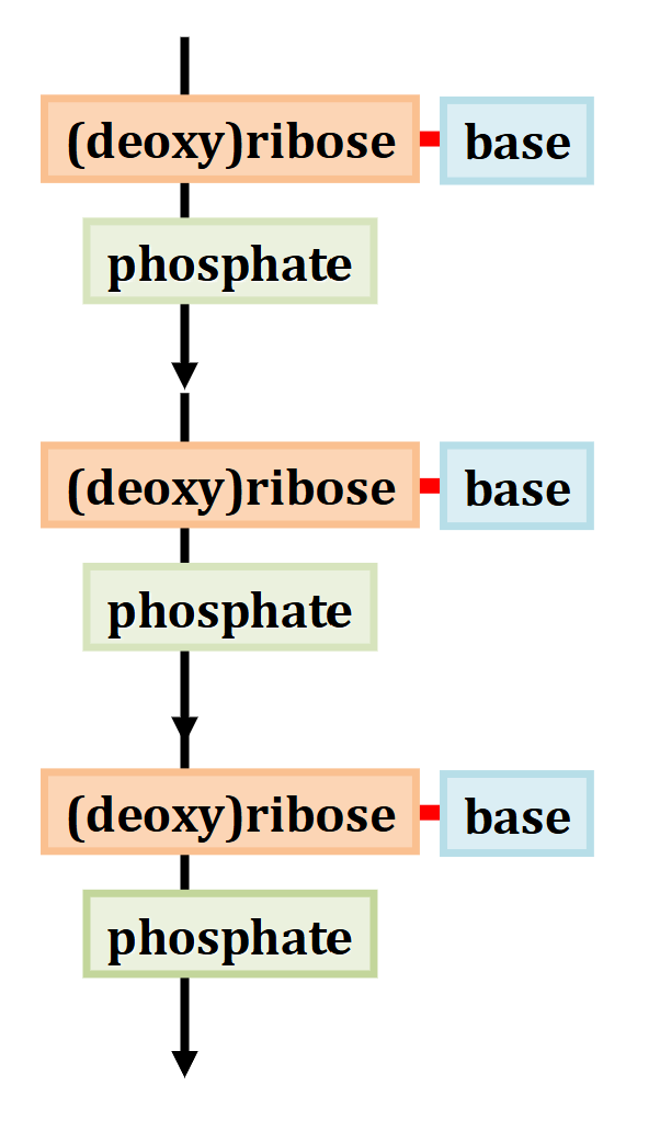

<ul><li><p><strong>DNA (deoxyribonucleic acid)</strong> and <strong>RNA (ribonucleic acid)</strong> are biological macromolecules that encode genetic information.</p></li><li><p>Nucleic acids are <strong>linear polymers</strong> composed of repeating <strong>nucleotide units</strong>.</p></li><li><p>Each nucleotide contains three components: a <strong>pentose sugar, nitrogenous base, and phosphate group</strong>.</p></li><li><p>Nucleotides are connected through <strong>phosphodiester bonds</strong>.</p></li><li><p>The alternating sugars and phosphates form the <strong>sugar-phosphate backbone</strong> of nucleic acids.</p></li></ul><p></p>
2
New cards

How do the sugars in DNA and RNA differ, and how are their carbon atoms numbered?

  • RNA contains D-ribose, while DNA contains D-2-deoxyribose.

  • Both sugars are pentoses containing five carbon atoms.

  • Ribose: Contains an –OH group at C-2′.

  • Deoxyribose: Has –H instead of –OH at C-2′.

  • Both sugars occur in their β-furanose forms, containing five-membered rings.

  • Sugar carbons are numbered using prime symbols (1′, 2′, 3′, etc.) to distinguish them from the numbered atoms of nitrogenous bases.


<ul><li><p><strong>RNA</strong> contains <strong>D-ribose</strong>, while <strong>DNA</strong> contains <strong>D-2-deoxyribose</strong>.</p></li><li><p>Both sugars are <strong>pentoses</strong> containing five carbon atoms.</p></li><li><p><strong>Ribose:</strong> Contains an <strong>–OH group at C-2′</strong>.</p></li><li><p><strong>Deoxyribose:</strong> Has <strong>–H instead of –OH at C-2′</strong>.</p></li><li><p>Both sugars occur in their <strong>β-furanose forms</strong>, containing five-membered rings.</p></li><li><p>Sugar carbons are numbered using <strong>prime symbols (1′, 2′, 3′, etc.)</strong> to distinguish them from the numbered atoms of nitrogenous bases.</p></li></ul><p></p>
3
New cards

What are purines and pyrimidines, and which nitrogenous bases are found in DNA and RNA?

  • Nitrogenous bases are derivatives of two parent compounds: purines and pyrimidines.

  • Purines: The purine ring is a fused (joined together), bicyclic (two rings) heterocycle.

    • Adenine (A) and Guanine (G) occur in both DNA and RNA.

  • Pyrimidines: Contain one six-membered ring.

    • Cytosine (C) occurs in both DNA and RNA.

    • Thymine (T) occurs in DNA.

    • Uracil (U) occurs in RNA.

  • DNA bases: A, G, C, T.

  • RNA bases: A, G, C, U.


<ul><li><p>Nitrogenous bases are derivatives of two parent compounds: <strong>purines and pyrimidines</strong>.</p></li><li><p><strong>Purines:</strong> <span>The purine ring is a fused (joined together), bicyclic (two rings) heterocycle</span>.</p><ul><li><p><strong>Adenine (A)</strong> and <strong>Guanine (G)</strong> occur in both DNA and RNA.</p></li></ul></li><li><p><strong>Pyrimidines:</strong> Contain one six-membered ring.</p><ul><li><p><strong>Cytosine (C)</strong> occurs in both DNA and RNA.</p></li><li><p><strong>Thymine (T)</strong> occurs in DNA.</p></li><li><p><strong>Uracil (U)</strong> occurs in RNA.</p></li></ul></li><li><p><strong>DNA bases:</strong> A, G, C, T.</p></li><li><p><strong>RNA bases:</strong> A, G, C, U.</p></li></ul><p></p>
4
New cards

What are tautomers, and what is the difference between keto–enol and amino–imino tautomerism?

  • Tautomers are isomers that differ by the movement of a hydrogen atom and a double bond.

  • Keto–enol tautomerism: Involves interconversion between a carbonyl (C=O) group and an enol (C–OH with an adjacent double bond).

  • Amino–imino tautomerism: Involves interconversion between an amino (–NH₂) group and an imino (=NH) group.

  • These rearrangements change the positions of hydrogen atoms and double bonds without changing the overall molecular formula.


<ul><li><p><strong>Tautomers</strong> are isomers that differ by the movement of a <strong>hydrogen atom and a double bond</strong>.</p></li><li><p><strong>Keto–enol tautomerism:</strong> Involves interconversion between a <strong>carbonyl (C=O)</strong> group and an <strong>enol (C–OH with an adjacent double bond)</strong>.</p></li><li><p><strong>Amino–imino tautomerism:</strong> Involves interconversion between an <strong>amino (–NH₂)</strong> group and an <strong>imino (=NH)</strong> group.</p></li><li><p>These rearrangements change the positions of hydrogen atoms and double bonds without changing the overall molecular formula.</p></li></ul><p></p>
5
New cards

What tautomeric forms of cytosine are shown in the lecture, and which form predominates?


  • Cytosine can undergo both keto–enol and amino–imino tautomerism.

  • The three forms shown are:

    • Enol + Amino

    • Keto + Amino

    • Keto + Imino

  • The keto + amino form predominates.

  • Tautomeric interconversion involves the movement of a hydrogen atom and a double bond.

  • You should understand how to identify the changing functional groups and recognize the predominant cytosine form.
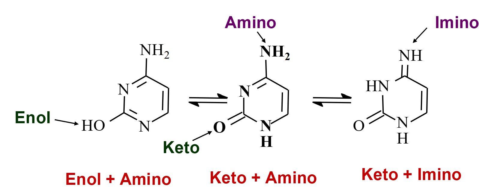

<p></p><ul><li><p>Cytosine can undergo both <strong>keto–enol</strong> and <strong>amino–imino tautomerism</strong>.</p></li><li><p>The three forms shown are:</p><ul><li><p><strong>Enol + Amino</strong></p></li><li><p><strong>Keto + Amino</strong></p></li><li><p><strong>Keto + Imino</strong></p></li></ul></li><li><p>The <strong>keto + amino form predominates</strong>.</p></li><li><p>Tautomeric interconversion involves the movement of a <strong>hydrogen atom and a double bond</strong>.</p></li><li><p>You should understand how to identify the changing functional groups and recognize the predominant cytosine form.</p></li></ul><p></p>
6
New cards

What are nucleosides, how are they formed, and where do purine and pyrimidine bases attach to the sugar?

  • Nucleosides consist of a nitrogenous base and a pentose sugar (ribose or deoxyribose).

  • The base attaches to the sugar's anomeric carbon (C-1′) through a C–N glycosidic bond.

  • This bond is also called a glycosylic bond

  • Purines (A and G): Attach through N-9.

  • Pyrimidines (C, T and U): Attach through N-1.

  • Nucleosides are special types of glycosides and contain no phosphate group.


<ul><li><p><strong>Nucleosides</strong> consist of a <strong>nitrogenous base and a pentose sugar</strong> (ribose or deoxyribose).</p></li><li><p>The base attaches to the sugar's <strong>anomeric carbon (C-1′)</strong> through a <strong>C–N glycosidic bond</strong>.</p></li><li><p>This bond is also called a <strong>glycosylic bond</strong></p></li><li><p><strong>Purines (A and G):</strong> Attach through <strong>N-9</strong>.</p></li><li><p><strong>Pyrimidines (C, T and U):</strong> Attach through <strong>N-1</strong>.</p></li><li><p>Nucleosides are special types of <strong>glycosides</strong> and contain <strong>no phosphate group</strong>.</p></li></ul><p></p>
7
New cards

What are the names of the nucleosides derived from the five nitrogenous bases?

knowt flashcard image
8
New cards

What is the difference between nucleosides and nucleotides, and what is their role in nucleic acids?

  • Nucleoside: Contains a nitrogenous base + pentose sugar.

  • Nucleotide: Contains a nitrogenous base + pentose sugar + phosphate group.

  • Nucleotides are phosphorylated nucleosides.

  • Example: Cytidine is a nucleoside, while cytidine 5′-phosphate is a nucleotide.

  • Nucleotides are the repeating subunits that form DNA and RNA.


<ul><li><p><strong>Nucleoside:</strong> Contains a <strong>nitrogenous base + pentose sugar</strong>.</p></li><li><p><strong>Nucleotide:</strong> Contains a <strong>nitrogenous base + pentose sugar + phosphate group</strong>.</p></li><li><p>Nucleotides are <strong>phosphorylated nucleosides</strong>.</p></li><li><p>Example: <strong>Cytidine</strong> is a nucleoside, while <strong>cytidine 5′-phosphate</strong> is a nucleotide.</p></li><li><p>Nucleotides are the repeating subunits that form <strong>DNA and RNA</strong>.</p></li></ul><p></p>
9
New cards

How are nucleotides connected through phosphodiester bonds, and why do nucleic acids have directionality and negative charge?

  • A phosphate group bridges between the 5′ OH of one nucleotide unit and the 3′ OH of another forming a phosphodiester linkage.

  • These linkages form the repeating sugar-phosphate backbone of DNA and RNA.

  • Each linear nucleic acid strand has a specific 5′ end (lacks a nucleotide at the 5′ position) and a 3′ end (lacking a nucleotide at the 3′ position).

  • Nucleotide sequences are conventionally written 5′ → 3′.

  • At pH 7, phosphodiester phosphate groups are completely ionized and carry a −1 charge.

  • This makes nucleic acids negatively charged.


<ul><li><p><span>A phosphate group bridges between the 5′ OH of one nucleotide unit and the 3′ OH of another forming a <strong>phosphodiester linkage</strong></span><strong>.</strong></p></li><li><p>These linkages form the repeating <strong>sugar-phosphate backbone</strong> of DNA and RNA.</p></li><li><p><span>Each linear nucleic acid strand has a specific 5′ end (lacks a nucleotide at the 5′ position) and a 3′ end (lacking a nucleotide at the 3′ position)</span>.</p></li><li><p>Nucleotide sequences are conventionally written <strong>5′ → 3′</strong>.</p></li><li><p>At <strong>pH 7</strong>, phosphodiester phosphate groups are completely ionized and carry a <strong>−1 charge</strong>.</p></li><li><p>This makes nucleic acids negatively charged.</p></li></ul><p></p>
10
New cards

How do you calculate the net charge of a linear DNA or RNA strand at neutral pH?

  • Count the number of nucleotides (n) in the strand.

  • A linear strand contains n − 1 phosphodiester linkages.

  • Each phosphodiester linkage contributes approximately −1 charge at neutral pH.

  • For a strand with unphosphorylated –OH ends, the net charge is: -1(n-1)

  • Example: 5′-UGCUAC-3′ contains 6 nucleotides and 5 linkages, giving a net charge of −5.

  • Terminal phosphate groups, if present, must be considered separately.
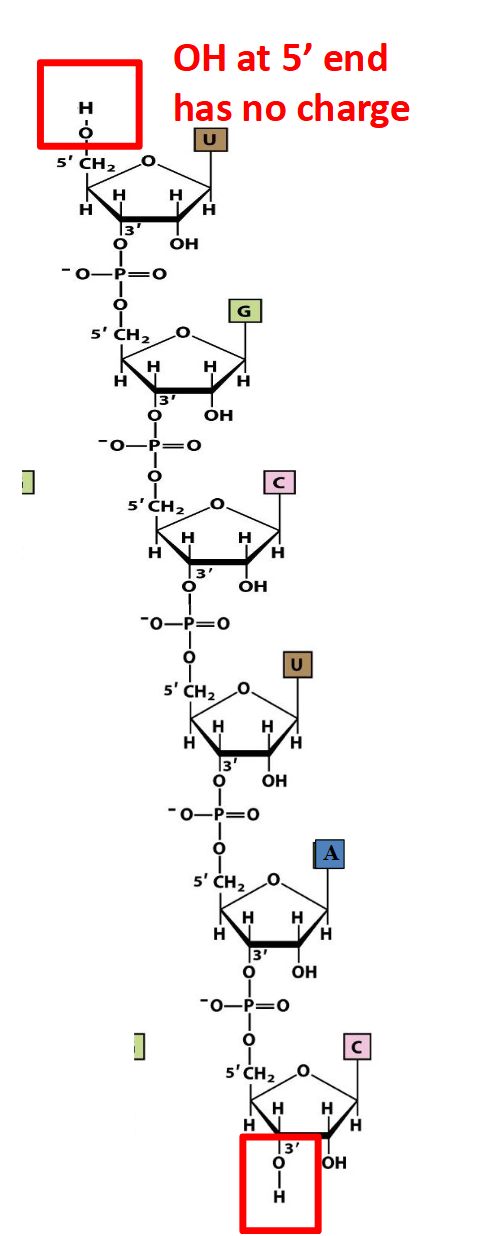

<ul><li><p>Count the number of nucleotides (<strong>n</strong>) in the strand.</p></li><li><p>A linear strand contains <strong>n − 1 phosphodiester linkages</strong>.</p></li><li><p>Each phosphodiester linkage contributes approximately <strong>−1 charge</strong> at neutral pH.</p></li><li><p>For a strand with unphosphorylated <strong>–OH ends</strong>, the net charge is: -1(n-1)</p></li></ul><ul><li><p>Example: <strong>5′-UGCUAC-3′</strong> contains 6 nucleotides and 5 linkages, giving a net charge of <strong>−5</strong>.</p></li><li><p>Terminal phosphate groups, if present, must be considered separately.</p></li></ul><p></p>
11
New cards

Why is RNA rapidly hydrolyzed under alkaline conditions, while DNA is much more stable?

  • RNA contains a 2′-OH group, whereas DNA has –H at C-2′.

  • Under alkaline conditions, RNA's 2′-OH group can act as a nucleophile.

  • It undergoes intramolecular attack on an adjacent phosphate group, breaking the phosphodiester linkage.

  • The reaction can produce a 2′,3′-cyclic monophosphate intermediate, followed by further hydrolysis.

  • DNA lacks the 2′-OH group, preventing this reaction and making it much more resistant to alkaline hydrolysis.
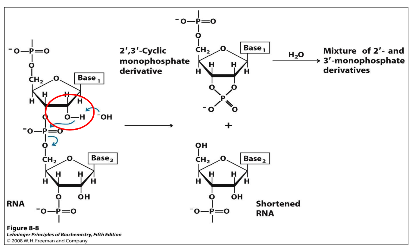

<ul><li><p><strong>RNA contains a 2′-OH group</strong>, whereas DNA has <strong>–H at C-2′</strong>.</p></li><li><p>Under <strong>alkaline conditions</strong>, RNA's 2′-OH group can act as a <strong>nucleophile</strong>.</p></li><li><p>It undergoes <strong>intramolecular attack</strong> on an adjacent phosphate group, breaking the <strong>phosphodiester linkage</strong>.</p></li><li><p>The reaction can produce a <strong>2′,3′-cyclic monophosphate intermediate</strong>, followed by further hydrolysis.</p></li><li><p><strong>DNA lacks the 2′-OH group</strong>, preventing this reaction and making it much more resistant to alkaline hydrolysis.</p></li></ul><p></p>
12
New cards

What are Chargaff's rules, and what do they tell us about DNA base composition?

  • Chargaff's rules were established by Erwin Chargaff and colleagues in the late 1940s.

  • In double-stranded DNA:

    • A = T (adenine equals thymine).

    • G = C (guanine equals cytosine).

  • The relative percentages of GC and AT can vary among organisms.

  • These rules apply to double-stranded DNA overall, not necessarily to each individual strand.

  • Example: If a double-stranded DNA molecule contains 30% A, it must contain 30% T, 20% G, and 20% C.


13
New cards

How did Rosalind Franklin, Maurice Wilkins, Watson, and Crick contribute to the discovery of DNA's secondary structure?

  • Rosalind Franklin and Maurice Wilkins used X-ray diffraction to investigate DNA structure.

  • Franklin obtained the important diffraction image known as Photo 51.

  • The diffraction data demonstrated that DNA has a helical structure with repeating distances of 3.4 Å and 34 Å.

  • Watson and Crick used accumulated structural evidence to propose their DNA double-helix model in 1953.

  • The model helped explain complementary base pairing and how DNA could serve as a template for replication.
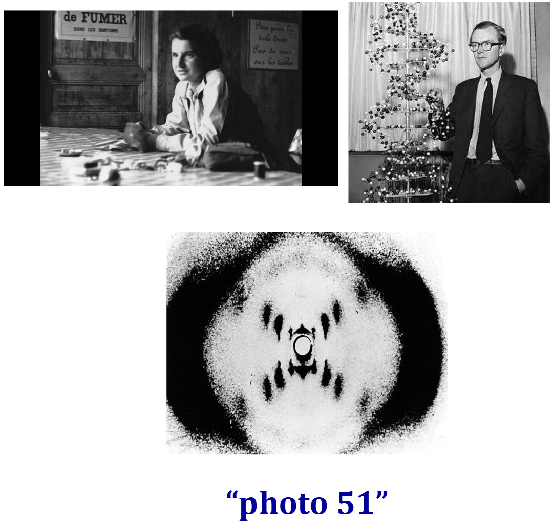

<ul><li><p><strong>Rosalind Franklin and Maurice Wilkins</strong> used <strong>X-ray diffraction</strong> to investigate DNA structure.</p></li><li><p>Franklin obtained the important diffraction image known as <strong>Photo 51</strong>.</p></li><li><p>The diffraction data demonstrated that DNA has a <strong>helical structure</strong> with repeating distances of <strong>3.4 Å and 34 Å</strong>.</p></li><li><p><strong>Watson and Crick</strong> used accumulated structural evidence to propose their <strong>DNA double-helix model in 1953</strong>.</p></li><li><p>The model helped explain complementary base pairing and how DNA could serve as a template for replication.</p></li></ul><p></p>
14
New cards

What is the overall structure of the DNA double helix, and how are its components arranged?

  • DNA consists of two helical strands that wind around a common axis, forming a right-handed double helix.

  • The hydrophilic sugar-phosphate backbones are located on the outside, facing the surrounding water.

  • The relatively hydrophobic nitrogenous bases are stacked inside the helix, approximately perpendicular to the helix axis.

  • Bases form complementary pairs between strands:

    • Adenine (A) pairs with thymine (T).

    • Guanine (G) pairs with cytosine (C).

  • This arrangement contributes to the stability of DNA and explains Chargaff's rules.


<ul><li><p>DNA consists of <strong>two helical strands</strong> that wind around a common axis, forming a <strong>right-handed double helix</strong>.</p></li><li><p>The <strong>hydrophilic sugar-phosphate backbones</strong> are located on the outside, facing the surrounding water.</p></li><li><p>The relatively <strong>hydrophobic nitrogenous bases</strong> are stacked inside the helix, approximately perpendicular to the helix axis.</p></li><li><p>Bases form complementary pairs between strands:</p><ul><li><p><strong>Adenine (A) pairs with thymine (T)</strong>.</p></li><li><p><strong>Guanine (G) pairs with cytosine (C)</strong>.</p></li></ul></li><li><p>This arrangement contributes to the stability of DNA and explains <strong>Chargaff's rules</strong>.</p></li></ul><p></p>
15
New cards

What does it mean for DNA strands to be antiparallel, and what are the major geometric measurements of the double helix?

  • Antiparallel: The two DNA strands run in opposite directions, with one oriented 5′ → 3′ and the other 3′ → 5′.

  • The DNA double helix is right-handed.

  • Distance between adjacent base pairs: 3.4 Å.

  • Base pairs per complete turn: Approximately 10.

  • Length of one complete turn: Approximately 34 Å.

  • Diameter of the double helix: Approximately 20 Å (2 nm).

  • These measurements describe the double-helix geometry presented in the lecture.


<ul><li><p><strong>Antiparallel:</strong> The two DNA strands run in opposite directions, with one oriented <strong>5′ → 3′</strong> and the other <strong>3′ → 5′</strong>.</p></li><li><p>The DNA double helix is <strong>right-handed</strong>.</p></li><li><p><strong>Distance between adjacent base pairs:</strong> 3.4 Å.</p></li><li><p><strong>Base pairs per complete turn:</strong> Approximately 10.</p></li><li><p><strong>Length of one complete turn:</strong> Approximately 34 Å.</p></li><li><p><strong>Diameter of the double helix:</strong> Approximately 20 Å (2 nm).</p></li><li><p>These measurements describe the double-helix geometry presented in the lecture.</p></li></ul><p></p>
16
New cards

What is DNA self-complementarity, and why is it essential for replication and repair?

  • Self-complementarity means that the sequence of one DNA strand determines the complementary sequence of the other through specific base pairing (A–T and G–C).

  • Each pre-existing DNA strand can act as a template for the synthesis of a new complementary strand.

  • This allows genetic information to be copied during DNA replication and passed on during cell division.

  • Complementarity also supports DNA repair, as the undamaged strand can help guide restoration of information in the damaged strand.

  • This was a key implication of the Watson–Crick model.
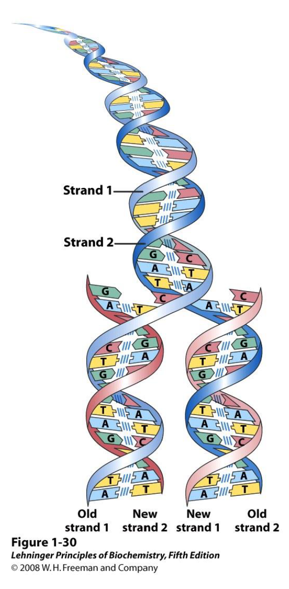

<ul><li><p><strong>Self-complementarity</strong> means that the sequence of one DNA strand determines the complementary sequence of the other through <strong>specific base pairing (A–T and G–C)</strong>.</p></li><li><p>Each pre-existing DNA strand can act as a <strong>template</strong> for the synthesis of a new complementary strand.</p></li><li><p>This allows genetic information to be copied during <strong>DNA replication</strong> and passed on during cell division.</p></li><li><p>Complementarity also supports <strong>DNA repair</strong>, as the undamaged strand can help guide restoration of information in the damaged strand.</p></li><li><p>This was a key implication of the <strong>Watson–Crick model</strong>.</p></li></ul><p></p>
17
New cards

How many hydrogen bonds form between complementary DNA bases, and how does GC content affect DNA stability?

  • Adenine–Thymine (A–T): Forms 2 hydrogen bonds.

  • Guanine–Cytosine (G–C): Forms 3 hydrogen bonds.

  • Hydrogen bonds contribute to holding complementary DNA strands together.

  • DNA with a higher GC-to-AT ratio is generally more difficult to separate into individual strands.

  • This is partly because G–C pairs form more hydrogen bonds than A–T pairs.

  • Other interactions also contribute to DNA stability.


<ul><li><p><strong>Adenine–Thymine (A–T):</strong> Forms <strong>2 hydrogen bonds</strong>.</p></li><li><p><strong>Guanine–Cytosine (G–C):</strong> Forms <strong>3 hydrogen bonds</strong>.</p></li><li><p>Hydrogen bonds contribute to holding complementary DNA strands together.</p></li><li><p>DNA with a <strong>higher GC-to-AT ratio</strong> is generally more difficult to separate into individual strands.</p></li><li><p>This is partly because G–C pairs form more hydrogen bonds than A–T pairs.</p></li><li><p>Other interactions also contribute to DNA stability.</p></li></ul><p></p>
18
New cards

What are the major and minor grooves of DNA, how do they form, and why are they biologically important?

  • DNA contains two grooves: the major groove (wider) and minor groove (narrower).

  • They form because the glycosylic bonds connecting bases to sugars are not colinear, creating unequal angles between the sugar-phosphate backbones.

  • As DNA winds into a double helix, the grooves follow the helical surface.

  • Both grooves expose potential hydrogen-bond donor and acceptor atoms of the bases.

  • These atoms enable specific interactions with DNA-binding proteins.

  • The major groove is more accessible to proteins that recognize specific DNA sequences.


<ul><li><p>DNA contains two grooves: the <strong>major groove (wider)</strong> and <strong>minor groove (narrower)</strong>.</p></li><li><p>They form because the <strong>glycosylic bonds</strong> connecting bases to sugars are not colinear, creating<strong><em> </em>unequal angles</strong> between the sugar-phosphate backbones.</p></li><li><p>As DNA winds into a double helix, the grooves follow the helical surface.</p></li><li><p>Both grooves expose potential <strong>hydrogen-bond donor and acceptor atoms</strong> of the bases.</p></li><li><p>These atoms enable specific interactions with <strong>DNA-binding proteins</strong>.</p></li><li><p>The <strong>major groove is more accessible</strong> to proteins that recognize specific DNA sequences.</p></li></ul><p></p>
19
New cards

What forces stabilize the DNA double helix, and why is its secondary structure largely independent of sequence?

  • DNA's secondary structure is largely independent of its sequence because A–T and G–C base pairs have similar shapes and properties.

  • Three major forces stabilize the double helix:

    • Hydrophobic effect: Relatively hydrophobic bases are shielded from water inside the helix.

    • Hydrogen bonding: Complementary base pairs form hydrogen bonds (A–T = 2; G–C = 3).

    • Van der Waals stacking: Favourable interactions between adjacent stacked bases.

  • Together, these interactions contribute to the stability of double-helical DNA.


20
New cards

How do Chargaff's rules differ when examining a single DNA strand versus a complete double-stranded DNA molecule?

  • Chargaff's rules: In complete double-stranded DNA, A = T and G = C.

  • These equalities do not necessarily apply to an individual strand.

  • Within a single strand, the four base percentages must total 100%.

  • Example: If one strand contains 30% A and 24% G, then C + T = 46%, but their individual values cannot be determined.

  • In the complementary strand, T = 30% and C = 24% because A pairs with T and G pairs with C.


<ul><li><p><strong>Chargaff's rules:</strong> In complete double-stranded DNA, <strong>A = T</strong> and <strong>G = C</strong>.</p></li><li><p>These equalities do <strong>not necessarily apply to an individual strand</strong>.</p></li><li><p>Within a single strand, the four base percentages must total <strong>100%</strong>.</p></li><li><p>Example: If one strand contains <strong>30% A and 24% G</strong>, then <strong>C + T = 46%</strong>, but their individual values cannot be determined.</p></li><li><p>In the <strong>complementary strand</strong>, <strong>T = 30% and C = 24%</strong> because A pairs with T and G pairs with C.</p></li></ul><p></p>
21
New cards

What is the central dogma of molecular biology, and what processes are involved?

  • The central dogma describes the general flow of genetic information from DNA to RNA to protein.

  • Replication (DNA → DNA): DNA is copied to produce another DNA molecule.

  • Transcription (DNA → RNA): Genetic information in DNA is used to synthesize RNA.

  • Translation (RNA → Protein): Information in RNA is used to synthesize a polypeptide.

  • These processes explain how genetic information is copied and expressed in cells.



