anatomy - muscular system (pt.2) 💪
 Call Kai
 Learn
 Practice Test
 Spaced Repetition
 Match
 Flashcards
 Knowt Play
1/100

There's no tags or description
Looks like no tags are added yet.
101 Terms
What are the muscle fiber types?
Slow-Twitch Oxidative (Type l)
Fast-Twitch Oxidative (Type lla)
Fast-Twitch Glycolytic (Type llx)
Slow-Twitch Oxidative
→ Contract slowly and rely on aerobic respiration (due to lots of mitochondria to produce ATP).
→ Prolonged contraction, low power, best for endurance, abundant in postural muscles (e.g back)
*”red (due to myoglobin), fatigue-resistance (due to endurance)

Fast-Twitch Oxidative
aka glycolytic fibers
→ Store a lot of glycogen, can generate strong, rapid contractions for short bursts of activity (ex/ jumping or sprinting)
→ Can fatigue quickly, appearing with a white color (due to fewer mitochondria = less myoglobin)
→ Mix of aerobic and anaerobic metabolism and common in leg muscles

Fast-twitch Glycolytic
→ Fewer mitochondria, lots of glycogen
→ Strong, rapid contractions, fatigue quickly
→ Best for short, powerful bursts
found in upper limb muscles
*white, anaerobic

What’s best for the following activity and why?
Maintain posture
Fiber type: Type l
Why?: Resistant to fatigue; aerobic
What’s best for the following activity and why?
Long-distance running
Fiber type: Type l
Why?: sustained ATP production using oxygen
What’s best for the following activity and why?
400m sprint
Fiber type: Type lla
Why?: Combines power with moderate endurance
What’s best for the following activity and why?
Jumping
Fiber type: Type llx
Why?: Produces rapid, powerful contractions
What’s best for the following activity and why?
Transferring a client once
Fiber: Type llx
Why?: Maximum force generation
How are skeletal muscles divided?
Axial muscles
Appendicular muscles
Axial muscles
Arise from the axial skeleton
About 60% of skeletal muscles
Position head, spinal cord and move rib cage

Appendicular muscles
Stabilize or move the appendicular skeleton

origin
→ the end of the muscle that stays stationary
located on the more proximal (closer to the body’s midline) or stable bone
acts as an ‘anchor’
insertion
→ the end of the muscle that moves when the muscle contracts
located on the more distal (further from the body’s midline) or mobile bone
moves toward the orgigin when the muscle contracts
action
→ what happens when the muscle contracts
ex. flexion, extension, adduction, abduction
What are the Primary Actions of Muscles
Primer mover (agonist)
Antagonist
Synergist
Muscles cannot push. They can only _____ by _______
pull, contracting
Primer mover (agonist)
→ the muscle that is chiefly responsible for producing a movement (aka. main character)
ex. when you bend your elbow, the biceps brachii is the prime mover
Antagonist
→ muscle that opposes a movement by another muscle
→ a functional opposite to the agonist
Synergist
→ muscle that helps the prime mover works efficiently
Fixators are synergists that stabilize the origin of the prime mover
Flexion of the elbow: Biceps brachii would be what?
prime mover
Flexion of the elbow: Triceps brachii would be what?
antagonist to the biceps brachii
Flexion of the elbow: Brachialis would be what?
synergist
How are muscles named?
Shape
Location
Function
Fibre Direction
Number of Heads or Divisions
Points of Attachment
Size of Muscle
Shape (state an ex.)
→ deltoid muscle is delta shaped

Location (state an ex.)
→ Brachialis (arm), gluteus (buttock), frontalis (muscle over the frontal one), lateralis (to the side), medialis (toward the midline)

Function
→ adductor muscles in the thigh adduct the leg (move it toward the midline)
Fibre Direction
→ fibres of the rectus abdominus are straight up and down and parallel (rectus means straight)

Number of Heads or Divisions
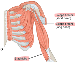
→ Biceps and triceps refer to multiple points of origin
Biceps brachii has two heads
Points of Attachment
→ points of origin and insertion may be identified in the name
sternocleidomastoid originates on the sternum and clavicle inserts on the mastoid process

Size of Muscle
→ relative to other muscles in a region
gluteus minimus, gluteus medius and gluteus maximus

Muscle-Related Terminology
Abductor
Adductor
Bi
Brevis
Extensor
Flexor
Lateralis

abductor
→ moves the bone away from the midline
adductor
→ moves the bone toward the midline
bi
two
brevis
short
extensor
→ muscle that increases the angle at the joint
flexor
→ muscle that decreases the angle at the joint
lateralis
to the outside
Muscle-Related terminology (contid.)
Longus
Maximus
Medialis
Medius
Minimus
Obqliue
Rectus
Tri
longus
long
maximus
largest
medialis
to the inside
medius
medium
oblique
at an angle
rectus
straight
tri
three
Specific Muscle Groups (header)
→ facial expressions
describe muscles of facial expression
subcutaneous tissue of the face and scalp
most insert into the skin or fascia (not bone)
→ movement of skin (NOT JOINTS!)
describe the functions of muscles of facial expression
→ Functions: control facial expressions, speech articulation, eating, blinking

Innervated by __________________
Cranial Nerve VII (Facial Nerve) (allowing us to smile, frown, raise our eyebrows or purse our lips)
Muscles of Facial Expression
frontalis
orbicularis oculi
orbicularis oris
platysma
occipitalis
zygomaticus major
depressor anguli oris

Joined by a tendon called
aponeurosis
frontalis
forehead
raises the eyebrows and wrinkles the forehead
The two muscles, frontalis and occipitalis make up….
the epicranius
occipitalis
back of the head
joined to frontalis by a flat tendon called an aponeurosis
orbicularis oculi
around the eye
circular muscle to close eyes tightly or blink
orbicularis oris
around the mouth
pucker or close the lips
zygomaticus major and minor
pulls the corners of the mouth upward
muscle most responsible for smiling
depressor anglui oris
opposes zygomaticus major
pulls the corners of the mouth downward
frowning
platysma
runs along the neck and lower jaw
tenses the skin of the neck
pulls corners of the mouth down slightly
Specific Muscle Groups (header)
→ neck muscles (responsible for actions such as chewing, swallowing and supporting the airway)
Muscles of Masticati
move the mandible for chewing, grinding and speaking
muscles action at the temporomandibular (TMJ)
Masseter and temporalis are primary elevators of the mandible (close mouth)
Pterygoids act together for side-to-side motion
What are the superficial muscles of muscles mastication?
masseter
temporalis
masseter
powerful elevator of the mandible
main chewing muscle that helps close the jaw
temporalis
fans out across the side of the skull
elevates the mandible and helps retract it
What are the deep muscles of muscles mastication?
lateral pterygoid
medial pterygoid
lateral pterygoid
protrudes the mandible
helps with side-to-side grinding movements
medial pterygoid
works with the masseter to elevate the mandible
contribute to grinding
What are the muscles of the tongue?
Intrinsic muscles
Extrinsic muscles
Innervation: cranial nerve Xll (hypoglossal)
What’s another word for tongue?
glossus
Functions of muscles that move tongue?
→ swallowing, moving food, speaking
Extrinsic muscles
orginate outside, insert into tongur
control position of tongue
What are the three parts of extrinsic muscles?
Genioglossus: sticks the tongue out and helps depress it
Hypoglossus: pulls the tongue down and back
Styloglossus: pulls the tongue up and back
pharynx
→ muscular tube that connects the nasal and oral cavities to the larynx and esophagus
protects airway, assits swallowing and maintains ear pressure
What are muscle functions?
close nasopharynx
open eustachian tubes
constrict pharynx during swallowing
Innervation for muscles of the pharynx?
→ Cranial nerves IX (glossopharynngeal) and X (vagus)
involved with the gag reflex (CN IX)
What are constrictor muscles in the pharynx?
Superior constrictor
Middle constrictor
Inferior constrictor
superior constrictor
constricts upper pharynx
closes nasopharynx during swallowing
middle constrictor
constricts middle pharynx
inferior constrictor
constricts lower pharynx
helps guide food into esophagus
_________ wave-like conttractions to push bolus down
peristaltic
________ muscles to elevate pharynx
longitudinal
What are the broad groups of anterior neck muscles?
superficial
suprahyoid
infrahyoid
scalene
deep
superficial
→ sternocleidomastoid, platysma)
suprahyoid
→ above hyoid, elevate hyoid
Which muscles are in the suprahyoid group?
mylohyoid
stylohyoid
digastric
geniohyoid
functions of muscles in suprahyoid?
swallowing and mouth movements
→ lift the larynx superiorly/anteriorly to widen pharynx
→ open the mouth
→ elevate hyoid and floor of the mouth (chewing)
→ allows backward pressure from the tonggue (push food toward pharynx)
→ form floor of oral cavity
infrahyoid
→ below hyooid, depress hyoid
What are the types of infrahyoid muscles?
superficial
deep
Describe what infrahyoid muscles are
aka strap muscles
thin, falt muscles connecting hyoid to sternum, clavicle and thyroid cartilage
what are functions of infrahyoid muscles?
swallowing
stabilizing the hyoid and larynx
depress the hyoid
superficial
sternohyoid (depress the hyoid)
omohyoid (depress and retracts the hyoid)
deep
sternothyroid (depresses the thyroid cartilage)
thyrohyoid (depresses the hyoid or elevates the thyroid)
scalene
→ lateral neck, aid respiration & neck flexion
deep
→ prevertebrak, postural support
What are functions of the anterior neck muscles?
support head, hyoid bone, larynx and assist in swallowing, breathing and movement
Most muscles attached to _______ bone
hyoid
Describe the sternocleidomastoid muscle
prominent muscle on the anterolateral neck
two inferior origins; sternum & clavicle
one suprior insertion: mastoid process
functions of the sternocleidomastoid muscle?
flexes neck
rotates head to opposite side