Electronic Imaging (RAD 1102)
 Call Kai
 Learn
 Practice Test
 Spaced Repetition
 Match
 Flashcards
 Knowt Play
1/78

There's no tags or description
Looks like no tags are added yet.
79 Terms
direct digital radiography
use detectors to convert x-ray energy to electrical energy, which is sent directly to a computer where image is digitally processed and displayed
Direct digital radiography uses one of the following:
1. charged coupled device (CCD)
2. complementary metal oxide
3. flat plate digital detectors
How does direct digital radiography convert x-ray energy to electrical energy?
directly or indirectly

charged coupled device (CCD)
indirect
complementary metal oxide (CMSO)
used in dental equipment
flat plate digital detectors
direct and indirect
What are the two types of digital radiography?
integral and portable

What is in place of (or replaced) the bucky tray?
detectors in which they can be mounted under top of the radiographic table

Detectors may be _________ in which it communicates with the computer using a cassette (IR)
wireless

What do both direct and indirect flat panels use to allow the active readout of the charge stored in the capacitor?
a thin film transistor array (TFT)

Indirect (scintillator) flat panel conversions contains:
1. cesium iodide scintillator or rare earth scintillator
2. amorphous silicon photodiodes on a glass substance
3. capacitor (stored charge)
4. TFT (open and released charge when capacitor is fully charged)
5. ADC

Flat panel indirect digital steps are as follows:
1. scintillation phosphor (fluorescent layer) or cesium iodide or rare earth phosphor converts x-rays to light
2. amorphous silicon photoconductor (photodiode) converts light from phosphor into electrons
3. TFT
4. directly to computer for processing
Indirect phosphors (which converts x-rays into light) are made of:
cesium iodide
In the indirect flat panel, the photodiode is made up of:
amorphous silicon
When the phosphor gives off light when struck by x-rays, the light is converted into a charge which is then converted into a digital image by the photodiode (amorphous silicon), this describes what type of conversion?
indirect flat panel conversions

Direct (non-scintillator) flat panel conversion contains:
1. amorphous selenium (photoconductor) on a glass substrate
2. capacitor (stored charge)
3. TFT (open and released charge when capacitor is fully charged)
4. ADC

Flat panel direct digital steps are as follows:
1. amorphous selenium photoconductor or photodetector converts x-rays directly into electronic signal (electrons)
2. TFT
3. directly to computer for processing
In the direct flat panel, the photoconductor is made up of:
amorphous selenium
amorphous
noncrystalline state of an otherwise crystalline material
Which flat panel conversion has the highest resolution?
flat panel direct (FPD)
Which flat panel conversion has a lower patient dose compared to CR?
flat panel direct digital and flat panel indirect digital
IMAGE PROCESSING
During image processing, what happens to digital data before being displayed?
digital data is evaluated and manipulated by the histogram
The histogram is constructed as the number of pixels in the entire image having _______ gray levels
same

The _________ is evaluated to determine adequacy of IP (imaging plate) exposure to x-rays
histogram

histogram
graphic (or graph) representation of the grey scale values constructed of the number of pixels in the entire image having the same grey levels of the imaged part

What is the histogram affected by?
- positioning
- centering
- processing algorithm
- scatter
- fading (CR)

Each different radiology system specifies the expected range of x-ray ___________ to produce a quality image
exposure
If the x-ray exposure error is greater or less than what percentage, it will produce a poorer quality image?
greater or less than 50%
Exposure values that are extracted from the image receptor are subject to what prior to image creation?
quantization

What indicates quantization of the signal extracted from the receptor in this graph?
the number of the blue bars

In this graph, the more discrete points means the image quality is better or worse?
better image quality
What image processor must quantize the continuous stream of electrons into unique values?
the ADC
What image processor provides a tally of each unique digital value the signal extracts from the receptor?
the histogram (graph of greys)
quantization
the number of bars that make up each histogram and represents different sampling frequency
When histograms have a higher sampling frequency, the bars in a graph are ____________
packed close together

Which graph has the highest sampling frequency?
A

Which graph has the lowest sampling frequency?
C

Which graph has the most shades of grey?
A

Which graph has the least shades of grey?
C

Which graph has low contrast?
A

Which graph has high contrast?
C
An image with many bars in the histogram will have a(n) ________ in contrast resolution
increase

What part of the histogram will correspond to the specific anatomy and technique used for an exam?
shape of the histogram

If the exposure field is not accurately recognized, the histogram will be __________
narrower
dynamic range
range of shades of grey displayed by pixels
detective quantum efficiency (DQE)
measure of receptor's ability to create an output signal that accurately represents the input signal
"values of interest" (VOI)
predetermined values that are used to create the image

automatic rescaling
the process of matching the captured image values of interest (VOI) to a standardized set of values

After looking at the image display on the monitor to check if the image is ok, where does it go?
downloaded to the computer and manipulated and transferred to MIMPS (formerly PACS) workstation where image manipulation can take place

What is the minimum matrix size that a LCD or LED monitor needs to be?
1536 x 2048
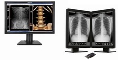
MANIPULATION OF IMAGE POST PROCESSING
after the image has popped up
subtraction
removes superimposed structures so anatomical area of interest is more visible

In the image on the far right, what post processing ability is applied?
subtraction
edge enhancement
improves visibility of small, high contrast structures (i.e. for tube placement)

What post processing ability does this image show?
edge enhancement
black/white reversal
reverses the gray scale from original radiograph

What post processing ability does this image show?
black/white reversal
image stitching
formation of a long body part image from a series of overlapping sub-images (i.e. for scoliosis)

What post processing ability does this image show?
image stitching
noise suppression (smoothing)
smoothing occurs by averaging each pixel's frequency with the surrounding pixel values to remove high frequency noise
Noise suppression is also called:
low pass filtering

Does noise suppression improve spatial resolution and visibility of an entire image?
no, it will only reduce noise in an image
What must be determined before trying to correct noise suppression?
the cause of noise

What post processing ability do these images show?
noise suppression
image equalization
useful when the anatomical part varies greatly in tissue thickness across the field of view (i.e. C-spine and foot)

What post processing ability do these images of the C-spine and foot show?
image equalization
Other Post-Processing Abilities
- annotate
- image zoom
- inversion
- rotated to achieve desired alignment
- pan
- scroll
Other Post-Processing Abilities: pan
move image around in multiple directions
Other Post-Processing Abilities: scroll
move image vertically up and down
MIMPS stands for:
Medical Image Management and Processing System
MIMPS is formally called:
PACS
PACS stands for:
Picture Archiving and Communication System
Images may be transferred into memory and/or transmitted to other monitors within or outside a facility which is along what?
a fiber optic cable

teleradiography
digital images transmitted over phone lines for remote viewing

node
each connection in a network or system
LAN (local area network)
connects all computers in a system and often uses a special wiring system
When information leaves the system to go to another location far away (teleradiography), it is over what? How is it transmitted?
wide area network (WAN) which is transmitted over fiber optic cables

Radiology is evolving into an all-digital system. What is the biggest drawback to this?
storage