Patho Lecture 4 (Inflammation)
 Call Kai
 Learn
Learn Practice Test
 Spaced Repetition
 Match
Match Flashcards
 Knowt Play
1/67

There's no tags or description
Looks like no tags are added yet.
Name | Mastery | Learn | Test | Matching | Spaced | Call with Kai | Chat |
|---|
No analytics yet
Send a link to your students to track their progress
68 Terms
Inflammation
A sequence of specific physiological events that occur in response to a non-specific agent
Functions of Inflammation?
- Neutralize or destroy the offending agent
- Restricts tissue damage to the smallest possible area
- Alerts body to threat of tissue injury
- Prepares the injured area for healing
Causes of Inflammation
- Exogenous (external cause) or Endogenous (internal cause)
- Trauma
- Surgery
- infection
- Caustic chemicals
- Extremes of heat or cold
- immune responses
- ischemic damage
What are the 2 types of Inflammation
- Acute (short-term)
- Chronic (persistent)
2 Types of Inflammation: Acute Inflammation
- Short-term response
- Hemodynamic (blood flow) changes
- Exudate formation (mass of cells and fluid that has seeped out of blood vessels or an organ)
- Presence of granular leukocytes
2 Types of Inflammation: Chronic Inflammation
- Persistent response
- Presence of nongranular leukocytes
- Results in more extensive scarring
- Initiated by non-degradable foreign matter
What are the phases of Inflammation?
1) Stimulation of cells of the innate or acquired immune system (mast cells, Th cells, monocyte/macrophages, neutrophils)
2) Release of inflammatory mediators (histamine, leukotrienes, prostaglandins) and chemotactic agents (chemokines, PGD2, C3a/C5a, N-formylated peptides)
3) Activated immune cells follow the chemotactic agents and invade the damaged tissue.
4) Removal of cell debris and secretion of growth factors which stimulate cell migration and proliferation, with tissue repair and formation of new blood cells
Pathways to Inflammation
- Direct stimulation of monocyte/macrophages, neutrophils, mast cells, or complement by components of microorganisms (PAMPS) or cell debris (DAMPS).
- Synthesis of IgE (activates mast cells and basophils)
- Direct activation (degranulation) of mast cells
- Synthesis of IgM and/or IgG (complement activation)
- Recognition of Antigen/MHC complexes by Th cells (release of cytokines)
Direct Stimulation of Leukocytes by Microbial Components: Toll-like receptors (TLR)
Bind microbial components (such as double-stranded RNA, LPS) and cause monocytes and other cells to release inflammatory cytokines (10 recognized in humans)
Direct Stimulation of Leukocytes by Microbial Components: NOD-like receptors (NLR)
Bind bacterial peptidoglycan fragments & activate NF-kB signaling, causing cytokine release
Direct Stimulation of Leukocytes by Microbial Components: Lipopolysaccharide (LPS) and Teichoic Acids
Activate the blood clotting cascade and/or complement cascade
What are the Characteristics of Acute Inflammation?
- Rapid in onset (seconds or minutes) and is of relatively short duration, lasting for minutes, several hours, or a few days
- Exudation of fluid and plasma proteins (edema) and the emigration of leukocytes, predominantly neutrophils
What are the Physiological responses?
- Release of soluble mediators
- Vasodilation
- Increased blood flow
- Extravasation of fluid (permeability)
- Cellular influx (chemotaxis)
- Elevated cellular metabolism
What are the Symptoms of Acute Inflammation?
- Heat
- Redness
- Swelling
- Pain
Acute Inflammation Components: The physiological responses of Increased blood flow & Elevated cellular metabolism lead to WHAT symptom?
Heat (calor)
Acute Inflammation Components: The physiological responses of Vasodilation & Increased blood flow lead to WHAT symptom?
Redness (rubor)
Acute Inflammation Components: The physiological responses of Vasodilation, Extravasation of fluid (permeability), Cellular influx (chemotaxis), and Elevated cellular metabolism lead to WHAT symptom?
Swelling (tumor)
Acute Inflammation Components: The physiological responses of Release of Inflammatory mediators, Cellular influx (chemotaxis), and Elevated cellular metabolism lead to WHAT symptom?
Pain (dolor)
Typical Vascular Response: Triple Response (scratch hand)
1) 3-50 sec - Thin red line(vasodilation of capillaries)
2) 30-60 sec - Flush(vasodilation of arterioles)
3) 1-5 min - Wheal (increased vascular permeability, edema)
In the Vascular response, what makes the inflamed state different from the Normal state?
- Increased blood flow (leads to warmth and redness)
- Leakage of plasma (leads to swelling)
- Neutrophil emigration
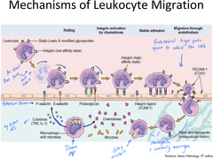
Mechanisms of Leukocyte Migration (how white blood cells leave the bloodstream and move into tissues during inflammation)
- Leukocytes roll along the blood vessel wall
- Macrophages release chemokines that signal to Leukocytes to activate their integrins (grip proteins) from a weak state to a strong state
- Stable adhesion, in which a leukocyte sticks firmly to the vessel wall using its integrins (stops the leukocyte from rolling)
- Leukocyte squeezes between the endothelial cells of the blood vessel, guided by PECAM-1 (doorways)
- Once out, the leukocyte follows the trail of microbes toward the site of infection or injury
Cellular response: Margination
The process by which leukocytes (white blood cells) adhere to the inner walls of blood vessels
Cellular response: Pavementing
The adhesion of leukocytes (white blood cells) to the endothelial lining of capillaries during inflammation
Cellular response: Emigration of Neutrophils
The process by which circulating neutrophils, a type of white blood cell, leave the bloodstream and move into surrounding tissues at a site of inflammation or infection
Cellular response: Extravasation
The leakage of fluid from its intended vessel, such as a blood vessel or IV catheter, into the surrounding tissues
Cellular response: Chemotaxis
The movement or orientation of a cell or organism in response to a chemical stimulus, guiding it towards beneficial substances like nutrients or away from harmful ones
Cellular response: Phagocytosis
The process by which a cell engulfs and destroys foreign particles, such as bacteria, viruses, and dead cells

Neutrophil Extracellular Traps (NETs)
- Extracellular fibrillar networks, produced by neutrophils in response to infectious pathogens & inflammatory mediators
- Contain a framework of nuclear chromatin with embedded granule proteins, including antimicrobial peptides & enzymes (neutrophil elastase, cathepsin G, myeloperoxidase, lactoferrin, gelatinase)
Inflammatory Exudates
- *Contain plasma, cells, and fluid
- Serous
- Fibrinous
- Purulent
- Hemorrhagic
- Catarrhal
You cut your finger: What happens in Acute inflammation?
- First responders are Neutrophils and Monocytes
- Released signals: IL-1, TNFa, Oxygen; causes redness, swelling and pain
You cut your finger: What happens in Resolution (healing)?
- Clearance of injurious stimuli
- Replacement of injured cells
- Macrophages release calming signals: IL-10, TGF-β
- Clearance of mediators and acute inflammatory cells
- Normal function
You cut your finger: What happens in post-resolution?
- Peacekeeping mode
- Resident macrophages act as guardians
- T regulatory cells make sure no one overreacts
You cut your finger: What happens in pus formation (abscess) when inflammation persists?
- Pus buildup
- Dead neutrophils and bacteria
- Body walls it off, and eventually healing happens, but it's slower
You cut your finger: When acute inflammation doesn't stop via viral infections, chronic infections, persistent injury, or autoimmune disease, and chronic inflammation takes over, what happens?
- New blood vessels grow (angiogenesis)
- Immune cells hang around
- Scar tissue starts to form (Fibrosis)
You cut your finger: What happens in Fibrosis?
- Tissue damage is too severe, and the body can't fully restore normal tissue
- Scar tissue forms
- Loss of function
What are the Major features of Chronic Inflammation?
- Persistent antigen
- Accumulation of antigen-processing cells
- Tissue destruction
- Usually distinct from acute inflammation
Acute Inflammation traits
- Initiators: Microbial surfaces and fragments. Injured tissue and tissue fragments
- Mediators: Mast cell products (histamine), Bradykinin, ROI, Complement, Lysosomal components, Lipid mediators, cytokines
- Cell populations: Neutrophils, Macrophages
- Time Course: Acute onset, days
- Outcome: Resolution, Abscess formation, or Chronic inflammation
Chronic Inflammation traits
- Initiators: Non-digestible organisms. Non-degradable foreign matter. Autoimmune reactions
- Mediators: T-cell and macrophage products => cytokines, growth factors, proteases, ROI complement, Lipid mediators
- Cell populations: T-cells, Plasma cells, Macrophages, Fibroblasts
- Time Course: Insidious onset, weeks
- Outcome: Tissue destruction; fibrosis
Types of Chronic Inflammation
- Nonspecific chronic inflammation
- Granulomatous
Types of Chronic Inflammation: Nonspecific chronic inflammation
Diffuse accumulation of macrophages and lymphocytes at the injury site
What are the 3 sources that Nonspecific chronic inflammation derives its macrophages from?
1) Recruitment
2) Local proliferation
3) Prolonged survival
Types of Chronic Inflammation: Granulomatous
- Granuloma formation: Massing of macrophages surrounded by lymphocytes
- Foreign bodies
- Deposition of antigenic material, Type IV hypersensitivity
Complement activation: What are the 3 main complement activation pathways?
- Classical
- Lectin
- Alternative pathways
Complement activation: What triggers the Classical pathway
Antigen-antibody complexes on pathogen surfaces
Complement activation: What triggers the Lectin pathway?
Mannose-binding lectin (MBL) or ficolin binds to carbohydrates on pathogen surfaces
Complement activation: What triggers the Alternative pathway?
Direct recognition of pathogen surfaces, stabilized by properdin
Complement activation: What is the key enzyme complex all three pathways generate?
C3 convertase
Complement activation: What happens when C3 is split into C3a and C3b?
- C3a: mediates inflammation and recruits phagocytes
- C3b: opsonizes pathogens, removes immune complexes, and participates in C5 convertase
Complement activation: What is the terminal outcome of complement activation?
Formation of the Membrane Attack complex (C5b, C6, C7, C8, C9)
--> death of pathogens and infected cells
The major expansion point of the complement activation cascade is the WHAT?
The ability of C3 convertase to activate multiple molecules of C3.
White blood cell products: What products do Neutrophils produce?
LTs, proteases, ROI, TXA2, PGE2,hydrolases, myeloperoxidase
White blood cell products: What products do Monocytes/macrophages produce?
ROI, RNI, LTs, IL-1, IL-4, IL-10, IL-6, TNF-α, CSFs, a-interferon, y-interferon, complement components, coagulation factor X, PGE2, chemokines
White blood cell products: What products do Lymphocytes produce?
- T helper cells (Th1, Th2, Treg, Th17): Interleukins (type depends upon Th phenotype), y-interferon, CSFs, lymphotoxin
- T cytotoxic cells: lymphotoxin, perforins, granzyme
- B cells: Immunoglobulin
White blood cell products: What products do Eosinophils control and release?
Control mediators released by mast cells, major basic protein, eosinophil peroxidase, eosinophil peroxidase, eosinophil cationic protein, and eosinophil-derived neurotoxin
Inflammatory Mediators released by Mast Cells: Preformed mediators
- Mediators that are stored in mast cell granules and released on degranulation
- Histamine
- Serotonin
- Serine Proteases
Inflammatory Mediators released by Mast Cells: Newly formed Lipid mediators
- thromboxane, prostaglandin D2, leukotriene C4, platelet-activating factor
Anti-Inflammatory Mediators: Lipids
- Arachidonic acid derived (Lipoxins, Prostaglandin D2)
- w-3 Polyunsaturated fatty acid derived (Resolvins, Protectins, Maresins)
Anti-Inflammatory Mediators: Cytokines
- IL-1ra, IL-4, IL-6, IL-10, IL-13, TGFB
What is First Intention Tissue Healing and Repair?
- Minimal tissue loss
- No or minimal granulation tissue formation
What is Second Intention Tissue Healing and Repair?
- Significant tissue loss
- Granulation tissue
- Slow healing
- Scar tissue
What is the process of Scar formation in Fibrous Tissue Substitution?
- Fibroblast and Vascular Endothelial Cell Migration
- Proliferation of fibroblasts and small blood vessels - granulation tissue
- Maturation of collagen fibers
What are Inflammatory Cytokines produced by?
Chiefly produced by macrophages & Th cells
Major pro-inflammatory cytokines include TNFa, IL-1& IL-6. What do they cause?
- Fever (w/ elevation in pulse and respiration rate)
- Elevated white cell count
- Acute-Phase Response (rise in many proteins)
- Anorexia (loss of appetite)
- Fatigue and sleepiness
- Malaise
- Pain in muscles and joints
- Catabolism of lipid and protein
What are Systemic signs of Inflammation?
- Leukocytosis
- Increase in RBC sedimentation rate, decreased iron levels in plasma (anemia)
- Fever
What does Leukocytosis cause?
- High WBC
- High %Neutrophils
- Low Lymphocyte count
- Low Monocyte count
What does Fever cause?
Causes - infection, tumors, infarction, tissue necrosis, hemolytic responses, hypersensitivity reactions, brain injury, dehydration, metabolic disturbances
What are fever-producing substances
Pyrogens
What do Pyrogens cause?
Exogenous - Bacterial products (endotoxin)
Endogenous - Leukocyte products (TNF-, IL-1, PGE2)