Lt 10 Fractures: Detection and Description
 Call Kai
 Learn
 Practice Test
 Spaced Repetition
 Match
 Flashcards
 Knowt Play
1/85

There's no tags or description
Looks like no tags are added yet.
86 Terms
What are the A.B.C.D.S of radiograph interpretation?
Alignment
Bone
Cartilage (Can't see on radiographs)
Devices
Soft Tissues
Basics of fracture detection
1. Stabilize the patient.
2. Sedation and pain control.
3. Radiographs (orthogonal)

What is the fracture being shown in this image?
Fracture of the proximal tibial epiphysis and metaphysis

How many views of the distal limb should be taken to get an accurate diagnosis of a fracture?
4 including DMPLO and DLPMO
What are the MOST COMMON reasons that fractures are missed?
Only 1 view is taken.
Minimal bone displacement.
What are other reasons to miss a fracture diagnosis?
Poor technique,
Not tangential to beam,
obscured overlying structures,
early cortical stress fracture,
Physeal fractures
What do you do if you can't find a fracture?
Oblique views,
Contralateral limb for comparison,
Repeat radiographs in 1-2 weeks,
Fracture gaps widen in 5-10 days.
Early callus formation in 10-20 days.
What radiolucent lines are considered normal?
Nutrient foramens,
Open physes,
Syndesmoses,
Superimposed metacarpal/metatarsal bones
What structures are considered Syndesmoses?
Sacroiliac joint, Mandibular and Pelvic symphysis

What is being highlighted in this image of a dog stifle?
Tibial tuberosity apophysis; NOT a fracture

What is being highlighted in this image of a dog elbow?
Apophysis of the olecranon tuber

What is occurring in this image of a dog stifle?
Proximal displacement of the tibial tuberosity of the stifle; Mineralization fragments and swelling
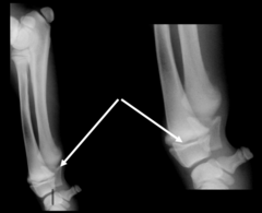
What is being highlighted in this image of a young dog's antebrachium?
Physes; normal for young dogs

What is being highlighted by the box in this image?
Nutrient foramen; normal finding commonly mistaken for fracture

What is being highlighted in this image of the femur?
Nutrient foramen

What is the abnormality in this image?
Fracture of the distal physes

What is the primary abnormality in this image?
Fracture of the distal tibial and fibular physes

What findings can help you focus your search?
Soft tissue swelling, step defects of the tibial cortex

How can you drastically improve the diagnostic value of this study?
Include an orthogonal projection
What fracture descriptions can affect the PROGNOSIS of a case?
Articular involvement,
Open vs. Closed,
Acute vs. Chronic,
Traumatic or Pathologic,
Soft tissue involvement
An OPEN fracture is _______ than a CLOSED fracture
worse
Why should you describe if a fracture is acute or chronic?
Because chronic fractures are harder to fix

Complete fractures
Both cortices involved.
> 2 fracture fragments

Incomplete fractures
One cortex.
NO separate fragments.

______ is a type of incomplete fracture common in young/immature animals and Osteopenia. The bones do not completely break due to the elasticity of the bone.
Greenstick fracture
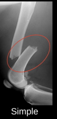
A simple fracture line is _____
1 fracture line with 2 fracture segments
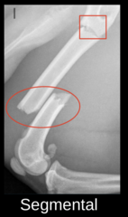
A segmental fracture line is ______
2 fracture lines that do NOT communicate.
3 fracture segments.

A Comminuted fracture is ______
> 2 fracture lines that communicate at a single point.
> 3 fracture fragments.
Butterfly fragment = Triangular fragment.
The direction of a fracture line is named based on ______
relation to the long axis of the bone;
Transverse,
Short oblique,
Long oblique,
Spiral

What is the direction of this fracture?
Transverse
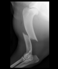
What is the direction of this fracture?
Short oblique

What is the direction of this fracture?
Long oblique

What is the direction of this fracture?
Spiral
Which bones are prone to spiral fractures?
Humerus and Tibia
What is closed fracture?
Skin is intact
What is an open fracture?
Skin is NOT intact and exposed to the environment (contaminated); High risk for infection and devitalization

How can you tell that a fracture is OPEN on radiographs?
Gas within surrounding soft tissue.
Foreign bodies.

Is this an OPEN fracture?
Yes, there is gas, soft tissue mineralization, and foreign bodies
How is displacement determined?
Position of the distal segment in relation to the proximal segment
What is required to describe ALL 3 displacement directions?
Orthogonal views
Proximal displacement = ______
Overriding
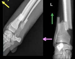
Describe the displacement of this fracture (Simple Complete, Transverse)
Cranial, Proximal, and Lateral displacement

Describe the displacement of this fracture (Simple complete, transverse)
Caudal, Proximal, and Medial displacement
How is angulation determined?
Angle of the distal segment relative to the proximal segment

Describe the angulation of this fracture (Simple complete, transverse)
Cranial and Lateral angulation

Describe an ACUTE fracture?
Sharp, well-defined margins

Describe a CHRONIC fracture?
Rounded, Tapered margins and Periosteal callus; Osteoclastic activity
Describe a TRAUMATIC fracture
Healthy bone with history of trauma of reasonable magnitude
Describe a Pathologic fracture
Secondary to weakening of bone by underlying disease; Neoplasia, Osteopenia
Animals with PATHOLOGIC fractures often have a history of _______
Minor trauma with severe lameness

Is this a traumatic or pathologic fracture?
Pathologic

Is this a traumatic or pathologic fracture?
Pathologic

What are the major descriptions for this fracture?
Major: open, comminuted, with severe soft tissue swelling

What radiographic findings are most relevant prognostically?
Subcutaneous gas and severe soft tissue swelling

What type of special fracture is this?
Avulsion fracture
Avulsion fractures occur at ______
Attachment sites of tendons, ligaments, and joint capsules
Avulsion fractures are caused by ______
Excessive pulling forces

What kind of special fracture is this?
Chip fracture
Chip fractures are _____
Small, usually corners of ONE joint (monoarticular)

What kind of special fracture is this?
Slab fracture
Slab fractures are _____
Cuboidal bones,
Biarticular,
In sagittal or dorsal plane
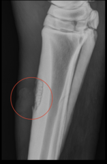
What type of special fracture is this?
Saucer fracture
Which species are saucer fracture more common in?
Horses; turn into sequestrae

What type of special fracture is this?
Depression fracture
Depression fractures are common in ______
Skulls, Sinuses, and the Nose;
Fracture fragments displaced below the normal surface.
Opaque lines instead of lucent lines.
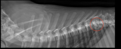
What type of special feature is this?
Compression fracture
Compression fractures are _____
From trauma that crushes the bone causing decreased dimensions in one or more areas.
Common in vertebral bodies or cubital bones.
Salter-Harris Fractures are common for ______
Young animals with OPEN physes
The prognosis ______ with increasing number on the Salter-Harris scale
worsens
Complications of Salter-Harris Fractures
Angular limb deformities,
Growth disturbances,
Joint abnormalities

A Type 1 Salter-Harris Fracture is _____
Through the growth plate

A Type 2 Salter-Harris Fracture is _____
Through the growth plate and Metaphysis

A Type 3 Salter-Harris Fracture is _____
Through the growth plate and Epiphysis

A Type 4 Salter-Harris Fracture is _____
Through all 3 elements
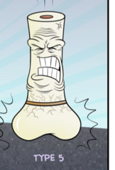
A Type 5 Salter-Harris Fracture is _____
Crush injury of growth plate
Name the layers of the PHYSIS from Epiphysis to Metaphysis
Germinal zone,
Proliferative zone,
Hypertrophic zone,
Zone of endochondral ossification
Where are chondroblasts located within the Physis layers?
Germinal zone
What is the WEAKEST layer of the Physis?
MOST Type 1 Salter-Harris fracture go through this zone!!
Hypertrophic zone

What type of Salter-Harris fracture is being shown in this image?
Type 5; Crushing of Physis

What special classification of fracture is this?
Salter-Harris Type 4; "Y" type condylar fracture

What hydrologic layers of the Physis are affected?
All of them
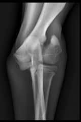
What are the most important radiographic findings regarding prognosis?
Articular involvement and Type 4

What kind of fracture is occurring here?
Smoothly marginated, Long oblique fracture extending from the lateral aspect of the mid humeral diaphysis

The metaphyseal and epiphyseal bone surrounding the fracture margins has ______
focally extensive permeative lysis with a long zone of transition and cortical destruction.

Along the medial aspect of the proximal fracture fragment there is a mild amount of ______
Amorphous Periosteal proliferation

What is the diagnosis for this fracture?
Pathologic, articular long oblique fracture of the right distal humerus; R/O neoplasia and fungal osteomyelitis