M4: Central Venous Access Devices, Blood Administration, Dialysis Catheter, Central IV Therapy Concepts
 Call Kai
 Learn
Learn Practice Test
 Spaced Repetition
 Match
Match Flashcards
 Knowt Play
1/29

There's no tags or description
Looks like no tags are added yet.
Name | Mastery | Learn | Test | Matching | Spaced | Call with Kai | Chat |
|---|
No analytics yet
Send a link to your students to track their progress
30 Terms
indications for CVAD
emergency venous access for pts requiring fluid resuscitation/hemodynamic monitoring
delivery of meds
hyperosmotic/vesicant fluids
nutrition
long-term therapy (chronic conditions)
hyperosmotic fluid examples
(>900 mOsm/L)
TPN (high glucose concentration often >10% dextrose, and electrolytes), dextrose 10%, mannitol 20%, 3% NaCl
vesicant fluid examples
chemotherapeutic agents (ex: doxorubicin, vincristine, etc.), electrolytes (ex: KCl, CaCl), diazepam, phenytoin, dopamine, norepinephrine
peripherally inserted central catheter
aka PICC
for long-term use, used to infuse high-osmolarity solutions or abx therapy
in for up to 6mos
use cephalic, basilic, brachial veins w/ sufficient diameter size
avoid in clients with end-stage renal disease who require
vein preservation for fistulas and grafts, or those with a
history of thrombosis, hypercoagulability states, or
decreased peripheral vascular flow
non-tunneled CVAD
may be used to infuse high osmolarity solutions
in for days to several wks
insertion sites may be subclavian, external/internal jugular, femoral veins
subclavian vein is favored in adult pts d/t decreased risk of catheter-related thrombosis and/or infection
external tunneled CVAD
ex: Hickman, Broviac, Groshong
long-term IV therapy (ex: chemo, hemodialysis)
may be long-term or permanent
inserted in chest area via subclavian/jugular vein, tunneled subcutaneously from proximal end of insertion site to an exit site
surgery required to tunnel catheter so that part of catheter lies in subQ tunnel → prevent organisms getting into bloodstream by separating extrance/exit pathways
implanted venous access device
aka IVAD, implanted port
long-term medication/IV therapy (ex: chemotherapy)
may be long-term or permanent
typically placed in subclavian/jugular vein and connected to small reservoir pocket → small plastic/metal chamber usually implanted just below collarbone which allows meds to be released directly into bloodstream
surgery is required, accessed using non-coring (Huber) needle and can be used immediately after placement, less visible externally, requires minimal daily care
maintenance → routine flushing w/ NS/heparin per policy, change needle/reaccess site every 7 days
non-tunneled vs. tunneled CVADs
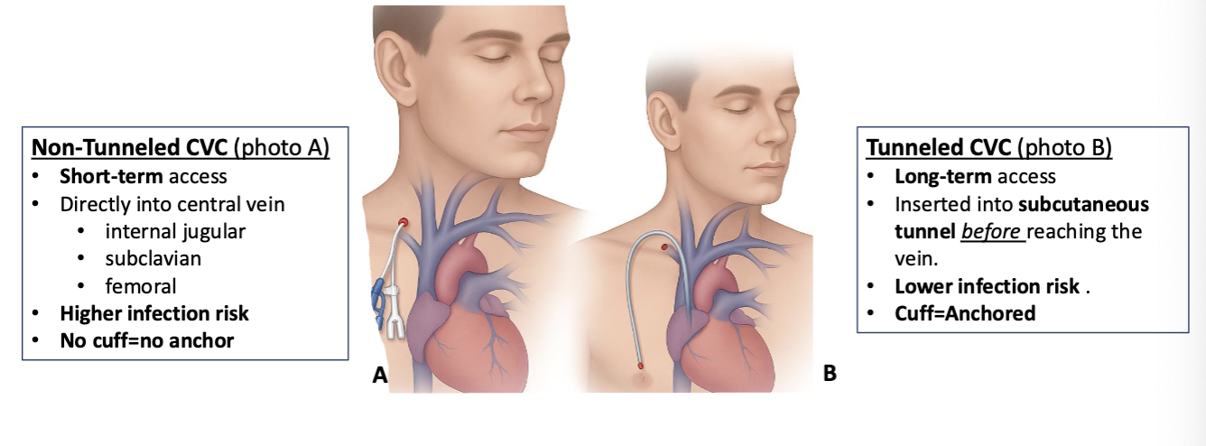
placing/using a CVAD
all CVADs require radiologic confirmation of placement prior to use
fluoroscopy during insertion of a tunneled line
x-ray after placement of a non-tunneled line
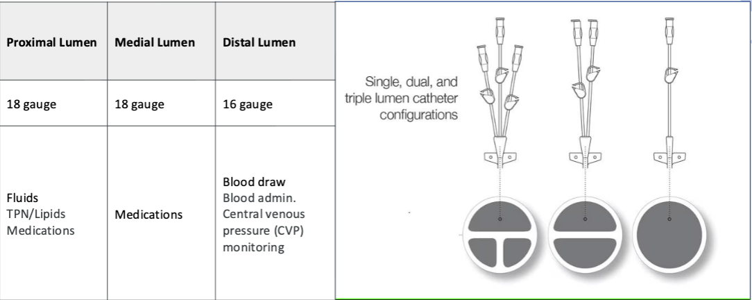
types of lumens
CVAD securement
must be stabilized/secured to pt
dislodgement/premature removal of CVAD increases complications like infections, vessel injury, treatment delays
depending on location/type of CVAD sutures or sutureless devices (Stat-lock) will be used
CVAD dressings
should always be clean, dry, occlusive
maintaining intact/occlusive dressing is essential to prevent CLABSI
if dressing becomes loose/wet/compromised in any way it must be changed immediately
A. replace dressing immediately
Which action should the nurse take first when a CVAD dressing becomes loose?
A. replace dressing immediately
B. notify provider
C. reassess in 4hrs
D. apply tape
midline catheter
used when peripheral IV access is difficult or frequent labs needed but central access NOT required
~3-8in.
same insertion site as PICC
terminates in upper arm not beyond axila, does not enter central circulation, placed at bedside w/ U/S
used for 1-4wks
used for IV fluids, abx, pain meds (NOT for vesicants, TPN, agents needing central dilution)
X-ray not required
lower risk for CLABSI
B. measure circumference
A patient with a PICC reports new arm swelling. What is your priority intervention?
A. elevate arm
B. measure circumference
C. flush line
D. warm compress
occlusion due to clot formation/malposition
assess → site, CVAD care, check equipment fxn, blood return assessment, flushing assessment
prevent → flush catheter routinely as recommended, do not flush against resistance, keep kink free, incompatible meds can cause precipitation w/in catheter
intervene → reposition pt, elevate arm overhead cough and deep breath, administer thrombolytics if appropriate, clogged CVAD may require removal
catheter damage/breakage
assess → every shift, look for leaks/tears/pinholes/drainage
prevent → 10mL syringe preferred, avoid increased P that can cause potential rupture, never flush against resistance
intervene → clamp catheter near insertion site, place sterile gauze over break/hole until repaired, CVAD may require removal
central line associated bloodstream infection
aka CLABSI
assess → catheter insertion site/surrounding area for redness, edema, drainage, tenderness; monitor pertinent lab results
prevent → maintain/utilize aseptic technique, CLABSI prevention bundle
intervene → notify HC provider, anticipate blood culture order, follow agency sepsis prevention/implementation protocol, diagnosed CLABSIs should be treated as life-threatening, abx specific to organism should be initiated
incorrect CVAD placement
assess → inadequate blood withdrawal, blood flowing back into tubing, HOTN, cardiac dysrhythmias, neck vein distention
prevent → verify catheter placement, x-ray, fluoroscopy
intervene → stop all fluid/med admin, anticipate orders for x-ray/ECG, CVAD may require removal/withdrawal to correct position
dislodgement
assess → measure/document catheter length, ID any edema at/around insertion site, palpate for coiling of catheter under skin
prevent → ensure catheter is secured at all times
intervene → cover insertion site and apply direct manual P while asking colleague to call rapid response team, pt will require monitoring for possible air embolus/reinsertion of a CVAD for critical meds
catheter migration
catheter moved from its original position
assess → patency of catheter, local irritation, swelling, inability to aspirate blood, edema of arm/hand, distended neck veins, may able to hear ‘gurgling’ sounds from catheter, possible cardiac dysrhythmias
prevent → avoid site insertion of CVAD in areas near site of a local infection, disrupted skin integrity, or scar tissue
intervene → notify provider, catheter that has migrated externally from its OG placement should not be readvanced, catheter that has migrated internally should be retracted to OG insertion length
skin erosion
assess → skin at/around CVAD insertion site, note any skin separation from catheter exit site, drainage, contusions
prevent → maintain optimal client nutritional status, avoid using same insertion ‘hole’ when accessing port multiple times
intervene → plan for removal of CVAD per order, provide effective skin care, improve nutrition as appropriate
air embolism
assess → sudden dyspnea, continuous coughing, chest pain, seizures, LOC, altered mental status, hemiparesis
prevent → catheter hubs should not be open to air, ensure all clamps are engaged appropriately
intervene → call rapid response team if suspected, admin high-flow O, place pt on L side w/ head down, begin CPR if indicated
pneumothorax
assess → sharp/stabbing chest pain (worsens when trying to inhale), SOB, cyanosis, tachypnea, dry/hacking cough
prevent → N/A
intervene → call for assistance, ask a colleague to notify rapid response team and provider, stay w/ pt, admin high-flow O, anticipate chest tube placement if pt hypoxic
infiltration/extravasation
assess → palpate over catheter insertion site dressing and surrounding area for sponginess, observe for redness/swelling, note any labored breathing/complaints of pain w/ infusions, observe IV flow rate for free-flowing fluid, aspirate for blood return
prevent → stop infusion and/or admin of vesicant soln, aspirate any remaining med from catheter in extravasation, admin antidote/therapeutic med as appropriate
intervene → discontinue IV solns, apply warm/cold compress as recommended, notify provider, anticipate order for chest x-ray
A. fever/chills, B. erythema, D. purulent drainage
Which signs indicate a possible CLABSI? (select all that apply)
A. fever/chills
B. erythema
C. clear dressing
D. purulent drainage
E. normal WBC
CVAD nrsg interventions
performing CVAD dressing changes while ensuring catheter stabilization
accessing CVADs or de-accessing inserted/implanted CVADs
performing intravenous line care/management
performing blood sampling from a CVAD
blood sampling from CVAD
main advantage → reduces pain/anxiety by avoiding repeated peripheral venipunctures
risks → infection, occlusion, inaccurate lab results if obtained improperly
steps of blood sampling from a CVAD
stop infusion of fluids/meds into the catheter’s lumens
choose appropriate CVAD lumen for obtaining samples based on largest lumen or configuration of lumen exit sites
vigorously scrub needleless connector for at least 15s w/ antiseptic scrub and let dry completely
attach prefilled 10mL syringe of preservative free NS to needleless connector using ANTT, unclamp catheter and thoroughly flush lumen w/ 10-20mL of preservative-free 0.9% NS, aspirate slowly for blood
clear dead space by using push-pull method or discarding aspirated blood
after obtaining blood sample thoroughly flush CVAD lumen w/ 10-20mL of preservative-free 0.9% NS
A. flush per protocol, C. use 10ml syringe, E. stop infusions pre-draw
Which reduce CVAD occlusion? (select all that apply)
A. flush per protocol
B. force flush
C. 10mL syringe
D. kink catheter
E. stop infusions pre-draw