exam 1 micro lecture
 Call Kai
 Learn
Learn Practice Test
 Spaced Repetition
 Match
Match Flashcards
 Knowt Play
1/78

There's no tags or description
Looks like no tags are added yet.
Name | Mastery | Learn | Test | Matching | Spaced | Call with Kai | Chat |
|---|
No analytics yet
Send a link to your students to track their progress
79 Terms
6 (7) types of microbes
bacteria, viruses, fungi, protozoa, helminths, algae and prions (newly)
existed for 3.5 billion yrs
at a micrometer or nanometer scale
immunology
study of immune system
public health micro bio and epidemiology
monitoring and control of diseases spreading
biotechnology
production of food, drugs and vaccines thru organisms
prokayrotes
prenucleus (no nucleus), simple cell, no dna, unicellular. one circular chromosome,
not in a membrane, No histones, No organelles, Peptidoglycan cell walls if Bacteria, Binary fission
eukaryotes
true nucleus, larger, complex cells, dna and multicellular. Paired chromosomes,
in nuclear membrane, Histones, Organelles, Polysaccharide cell walls, Mitosis.
viruses
neither prokaryote or eukaryote. they’re their own classification. no cells (acellular), parasitic particles composed of nucleic acid and protein.
most abundant microbe on earth
obligate intracellular parasite
bear no resemblance to cells: Lack protein-synthesizing machinery, contain only the parts needed to invade and control a host cell
Structure of Viruses
capsids- protein coats made of capsomeres
nucleocapsid- capsid together with the nucleic acid
envelope or naked
can be helical or icosahedral (20 sides)
photosynthesis
light from sun, turns to sugar. fuled by light co2, turns to organic material
decomposition
energy released in heat and broken down
bio remediation
using living organisms to remedy environment problems (ex. cleaning oil spills)
genetic engineering
manipulating genes to make new products
pasteurization
application of high heat for short time
bacilllus thuringienis
toxic crystals causing pests to have fatal infections (pesticides not harmful to humans)
normal microbiota
microbe thats normally pressent in/on humans. (not always harmful!!!) also prevents pathogen growth and produces growth factors (ex. potassium)
pathogens
ALWAYS HARMFUL microbes. 10B new infections and 12M deaths annually ☹
robert hooke
discovered cells for the first time
antonie Van Leeunwenhoek
first to obserVe Living microbes (using single lens 300x)
theory of spontaneous generation
life forms can arise from vital forces present in nonliving/decomposing matter
theory of biogenesis
came from louis pasteur, the idea that living things only come from other living things
louis pasteur
demonstrated that microorganisms are in the air
pasteurization (heated and sealed = no growth)
s shaped flask, kept microbes out but let air in
started “golden age” of micro bio
yeast causes fermentation
theory of biogenesis
rabies caused by a virus
fermentation
when sugar turns to alcohol. microbial growth causes food to spoil, bacteria that use alcohol and produce acetic acid spoil wine and turn to vinegar
joseph lister
founder of anti septic medicine, used chemical disinfectant to prevent surgical infections
robert koch
proved that a bacterium causes anthrax
identified cholera and koch’s postulates (criteria that proves a certain microorganisms cause a specific disease)
edward jenner
discovered a person w cow pox can’t get small pox
vaccination/immunity- vacca = cow
chemotherapy
treatment with chemicals
synthetic drugs used to treat disease
antibiotics
chemicals produced by bacteria/fungi that’ll kill other microbes
paul ehrlich
discovered magic bullet that can kill pathogen without hurting host
developed arsenic, salvarsan, to treat syphillis
Alexander fleming
discovered first Antibiotic, penicillium
made penicillin (kills S. aureus)
different branches of microbio
bacteriology-study of bacteria
mycology-study of fungi
virology- study of viruses
parisitology- study of protozoa and parasitic worms
George beadle and Edward tatum
showed Genes encode a cell’s Enzymes
Oswald Avery, Colin MacLeod and Maclyn McCarty
showed DNA was hereditary material
francois jacob and jacques monod
discovered role of mRNA in protein synthesis
Bruce A Beutler and Jules A Hoffmann
discovered innate immunity and molecules
ralph steinman
discovered adaptive immunity and dendritic cells
rebecca lancefield
proposed use of immunology to identify some bacteria according to serotypes
Nobel Prizes for Microbiology Research

john tyndall and ferdinand cohn
discovered present of heat resistant forms
cohn determined these forms as endospores
taxonomy
organizing, classifying, and naming living things
classification (grouping), nomenclature (naming) and identification (identifying traits for placement)
levels of classification
Domain - Archaea, Bacteria, & Eukarya
Kingdom
Phylum or Division
Class
Order
Family
Genus
Species
(dear king phillip came over for good soup)
3 domains of life
bacteria (true bacteria), archaea (odd bacteria that lives in extreme environments) and eukarya (organisms w nucleus and organelles)
binomial nomenclature
genus is capitalized and species is lowercase (ex. Staphylococcus aureus, S. aureus, Staphylococcus sp.)
specific epithet
another word for species
phylogeny
natural relatedness between groups of organisms
*know the basic idea of evolution*
shapes of bacteria
Bacillus (rod-shaped), Coccus (spherical), Spiral: Spirillum, Vibrio, Spirochete
unusual bacteria shape includes star shape and rectangular shape bacteria

arrangements
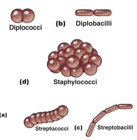
Pairs: Diplococci, diplobacilli
Clusters: Staphylococci
Chains: Streptococci, Streptobacilli
The Structure of a Prokaryotic Cell: Glycocalyx
Outside cell wall is Usually sticky
Capsule: neatlyorganized
Slime layer: unorganized and loose
Extracellular polysaccharide allows cell to attach
Capsules prevent phagocytosis
The Structure of a Prokaryotic Cell: Flagella
Outside cell wall Made of chains of flagellin
Attached to a protein hook
Anchored to the wall and membrane by the basal body
Motile Cells
Rotate flagella to run or tumble
Move toward or away from stimuli (taxis – photo, chemo)
Flagella proteins are H antigens

Axial Filaments
Also called endoflagella
In spirochetes
Anchored at one end of a cell
Rotation causes cell to move

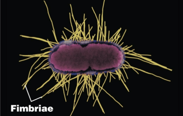
Fimbriae and Pili
Fimbriae: allow attachment
Pili: Facilitate transfer of DNA from one cell to another, Gliding motility, Twitching motility
The Cell Wall
Prevents osmotic lysis (rupturing of a cell membrane caused by excess water/osmosis)
Made of peptidoglycan (in bacteria)
Peptidoglycan
Polymer of disaccharide (NAG and NAM)
Peptidoglycan in Gram-Positive Bacteria linked by polypeptides
gram positive vs gram negative bacterial wall
g+ = Thick peptidoglycan and Teichoic acids. Lipoteichoic acid links to plasma membrane. Wall teichoic acid links to peptidoglycan. polysaccharides provide antigenic variation. killed by penicillin and detergents. 2 ring basal body
g- = Thin peptidoglycan, Outer membrane, Periplasmic space. resistant to antibiotics 4 ring basal body, endotoxin
→ outer membrane: Lipopolysaccharides, lipoproteins, phospholipids. Forms the periplasm between the outer membrane and the plasma membrane. Protection from phagocytes, complement, and antibiotics. O polysaccharide antigen, Lipid A is an endotoxin, Porins (proteins) form channels through membrane
staining slides (for contrast)
Smear: A thin film of a solution of microbes on a slide. usually fixed to attach the microbes to the slide and to kill the microbes
Stains consist of a positive and negative ion
→ basic dye: the chromophore is a cation. used once in simple stain
→ acidic dye: the chromophore is an anion
negative staining: staining background instead
mordant: used to hold the stain or coat the specimen to enlarge it
Differential Stains: Used to distinguish between bacteria. Gram stain and Acid-fast stain
gram stain
classifies bacteria into gram positive or gram negative. all gram pos are purple. gram neg decolorizing is clear, gram neg counter stain is red
gram pos: alcohol dehydrates peptidoglycan
gram neg: alcohol dissolves outer membrane and leaves holes in peptidoglycan
atypical cell walls
acid fast cell walls, like gram pos.
waxy lipid/mycolic acid bound to peptidoglycan
mycobacterium and nocardia
mycoplasmas lack cell walls. sterols in plasma membrane
archaea are wall-less or waks of pseudomurein (lack NAM and D-amino acids)
acid fast stain
stain waxy cell wall not decolorized by acid alcohol. mycobacterium and nocardia
acid fast= all red
non acid fast recolorizing is colorless, non acid fast counter stain is blue
special stains
used to distinguish part of cells:
capsule stain- cells stained, negative stain
endospore stain- primary stain is malachite green with heat. decolorizes cells (water) and counterstain is safranin
flagella stain- mordant on flagella, carbolfuchsin simple stain
damage to cell wall
lysozome digests disaccharide in peptidoglycan
penicillin inhibits peptide bridges in peptidoglycan
protoplast- spherical shape assumed by gram pos bacteria
spheroplast- spherical shaoe assumed by gram neg bacteria.
both are susceptible to osmotic lysis
L forms- wall’less cells that swing into irregular shapes
plasma membrane
has phospholipid bilayer, peripheral proteins, integral proteins and transmembrane proteins.
selective permeability allows passage of some molecules
damage to membrane by alcohol detergents etc causes leakage of cell contents
fluid mosaic model
similar consistency to olive oil
proteins move to function
phospholipids rotate and move laterally (sideways)
movement of materials across membranes
thru lipid layer and aquaporins (water channel)
active transport requires ATP and transporter protein
group translocation requires transporter protein and PEP
simple diffusion- solute moving from area of high concentration to low concentration
facilitated diffusion- solute combines with a transporter protein in membrane
osmosis- movement of water across selectively permeable membrane from area of high water concentration to area of low water concentration
→ isotonic is no movement of water. hypertonic causes shrinkage. hypotonic causes swelling or bursting
prokaryotic ribosome
protein synthesis
30s (small subunit) + 50s (large subunit) = 70s complete ribosome
inclusions
phosphate reserves: metachromatic granules (volutin)
energy reserves: polysacchardie granules, lipid inclusions and sulfer granules
endospores
resting cells resistant to desiccation, heat chemicals. ex: bacillus or clostrdium
sporulation: endospore formation
germination: return to vegitative state
history of eukaryotes
2 bill yr
evolution from prokaryotic by symbiosis
kingdom fungi
macroscopic fungi (mushroom)
microscopic (yeast): 2 morphologies. yeast ( round and asexual) and hyphae (long fungi) can be dimorphic
unicellular or colonial mostly
fungal nutrition
heterotrophic
most are harmless, some are parasites
mycoses- fungal infection
fungal organization
yeast- soft, uniform and reproduce by budding
filamentous fungi- mass of hyphae called mycelium, can be cottony/hairy. hyphae might be dividied by septate. reproductive hyphae = spores
protists
algae- eukaryotic, microscopic are unicellular, macroscopic are multicellular. all are colonial. photosynthesize w chlorophylla. cell wall, may or may not have flagella. plankton, dinoflagellaten (can cause red tides and food poisoning)
protozoa- eukaryotes that are mostly unicellular, lack tissue and cell wall. many shapes. most are harmless. feed by engulfing other things. all heterotrophs, none have chloroplasts. cytoplasm is divided into endoplasm and ectoplasm
protozoan pathogens
Pathogenic flagellates: ex Trypanosomes -T. brucei – African sleeping sickness and T. cruzi – Chaga’s disease; South America
infective amoebas: ex Entamoeba histolytica- amebic dysentery
parasitic helminths
flatworms like Cestodes (tapeworms) or Trematodes. simple excratory and nervous system
Roundworms (nematodes) – round, a complete digestive tract, a protective surface cuticle, spines and hooks on mouth; excretory and nervous systems poorly developed
nucleic acid
Viral genome – either DNA or RNA but never both
Carries genes necessary to invade host cell and redirect cell’s activity to make new viruses
Number of genes varies for each type of virus
DNA viruses Usually double stranded (ds) but may be single stranded
(ss Circular or linear
RNA viruses Usually single stranded, may be double stranded
modes of multiplication
1. Adsorption – binding of virus to specific molecules on the host cell
2. Penetration – genome enters the host cell
3. Uncoating – the viral nucleic acid is released from the capsid
4. Synthesis – viral components are produced
5. Assembly – new viral particles are constructed
6. Release – assembled viruses are released by budding (exocytosis) or cell lysis
Cytopathic effects
virus-induced damage to cells
1. Cells fuse to form multinucleated cells
2. Cell lysis
3. Alter DNA
4. Transform cells into cancerous cells
Techniques in Cultivating and Identifying Animal Viruses
Cell (tissue) cultures, Bird embryos and Live animal inoculation
prions
misfolded proteins, contain no nucleic acid. extremely resistant to usual sterilization techniques, can be fatal
Scrapie in sheep and goats
Bovine spongiform encephalopathies (BSE), a.k.a. mad cow disease
Wasting disease in elk
Humans – Creutzfeldt-Jakob Syndrome (CJS)