Airborne dz (first)
 Call Kai
 Learn
 Practice Test
 Spaced Repetition
 Match
 Flashcards
 Knowt Play
1/77

There's no tags or description
Looks like no tags are added yet.
78 Terms
What are the 3 routes of transmission?
Contact, vehicle, and vector transmission.
What are the 3 types of contact transmission?
Direct, indirect, and droplet.
How are pathogens released in airborne diseases?
When a person coughs or sneezes.
Droplet transmission is a type of ___ transmission, and airborne transmission is a type of ___ transmission.
Contact; vehicle.
How far away is the pathogen source in droplet vs airborne transmission?
Droplet: <1 m (~3 ft).
Airborne: >1 m.
Which is easier to acquire, droplet or airborne transmission?
Airborne is easy to acquire; droplet is difficult.
Give an example of a droplet-transmitted disease and an airborne-transmitted disease.
Droplet: Ebola. Airborne: measles.
What is the major portal of entry for airborne diseases?
The respiratory tract, through its mucosal membranes.
What are the 3 respiratory symptoms of airborne diseases?
Coughing, sneezing/nasal discharge, and difficulty/noisy breathing.
What non-respiratory symptom can airborne diseases cause? Give 2 examples.
Skin manifestations (rashes), such as in chickenpox and measles.
Which region is the upper respiratory tract (URT)?
The “head” region.

What structures make up the URT?
Nose, oral cavity, pharynx (throat), middle ear, and auditory tube.
Which region is the lower respiratory tract (LRT)?
The “lung” region.

What structures make up the LRT?
Larynx, trachea, bronchial tubes, and alveoli.
What are the 2 defenses of the LRT?
The ciliary escalator and respiratory mucus.
What type of barrier is the ciliary escalator?
A physical barrier.
What are the 4 host defenses against airborne pathogens?
Normal microbiota, mucus, lysozyme, and the ciliary escalator.
How does normal microbiota defend against airborne pathogens?
It competes with invading pathogens.
How does mucus defend against airborne pathogens?
It traps pathogens, which are then removed by the ciliary escalator.
How does lysozyme defend against airborne pathogens?
It’s an enzyme that destroys pathogens.
How does the ciliary escalator defend against airborne pathogens, and where is it found?
It moves mucus up and out of the body. It’s found in the lower respiratory tract.

In the ciliary escalator diagram, what does the “debris” represent?
Pathogens/antigens (Ag).

What are the 3 bacterial upper respiratory tract diseases?
Streptococcal pharyngitis
Scarlet fever
Diphtheria.
What are the 3 bacterial lower respiratory tract diseases?
Pertussis
Pneumococcal pneumonia
Tuberculosis.
What are the 7 viral airborne diseases?
Influenza, COVID-19, smallpox, chickenpox, shingles, measles, and rubella.
Streptococcal infections are caused by which genus?
Genus Streptococcus, which has many species.
What type of media is blood agar?
Differential media.
What does blood agar help differentiate?
Different streptococcal species that cause airborne diseases.
Which 2 Streptococcus species are differentiated on blood agar, and what disease(s) does each cause?
S. pyogenes: strep throat and scarlet fever.
S. pneumoniae: bacterial pneumonia.
What role do RBCs play in blood agar?
They serve as a nutrient source.
What are hemolysins?
Toxins that lyse RBCs, produced by toxigenic bacteria.
What type of toxin are hemolysins? What 2 things are associated with this type?
Type I exotoxin, associated with Gram + bacteria and fever.
What is the process of RBC lysis by hemolysins called?
Hemolysis.
What is β hemolysis, and which toxin causes it?
Complete hemolysis/RBC lysis, caused by β hemolysin.
What is α hemolysis, and which toxin causes it?
Partial hemolysis/RBC lysis, caused by α hemolysin.
What does β hemolysis do to RBCs, what zone does it make on blood agar, and which species is an example?
Completely destroys RBCs → clear zone. Ex - S. pyogenes.

What does α hemolysis do to RBCs, what zone does it make on blood agar, and which species is an example?
Partially destroys RBCs → green zone. Ex - S. pneumoniae.

Pneumonias are infections of which part of the respiratory tract?
The lower respiratory tract.
What causes typical pneumonia? Give an example.
Bacteria. Ex - S. pneumoniae → bacterial pneumonia (AKA pneumococcal pneumonia).
What causes atypical pneumonia?
Non-bacterial microbes (viruses and fungi).
Give the viral and fungal examples of atypical pneumonia.
Influenzavirus → viral pneumonia.
Pneumocystis jirovecii → fungal pneumonia.
Who gets fungal pneumonia?
Immunocompromised hosts (weak immune system), such as people with HIV/AIDS.
TB pathogenesis step 1: what happens when tubercle bacilli reach the alveoli?
They’re ingested by macrophages, but often some survive.
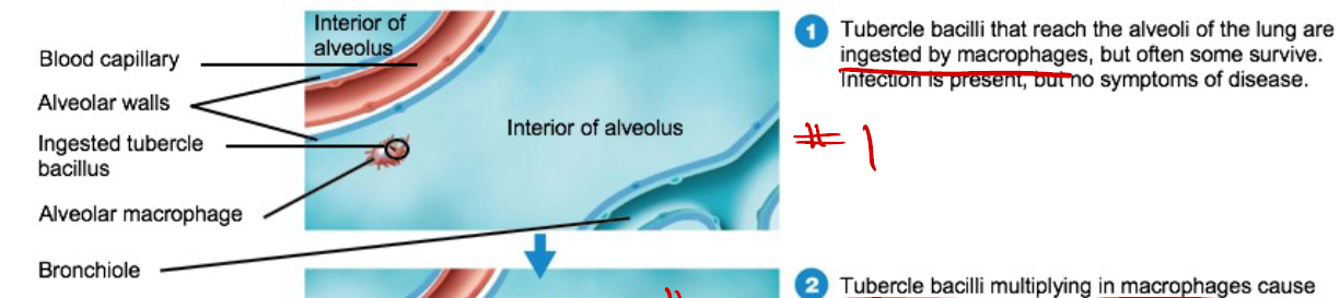
In step 1 of TB pathogenesis, are there symptoms?
No. Infection is present, but there are no symptoms.
TB pathogenesis step 2: what do tubercle bacilli do inside macrophages, and what does this cause?
They multiply in macrophages, causing a chemotactic response that brings in more macrophages and defensive cells.

In step 2, what do the recruited macrophages form?
A surrounding layer → an early tubercle.

In step 2, why does lung-damaging inflammation happen?
Most surrounding macrophages fail to destroy the bacteria and release enzymes and cytokines.
TB pathogenesis step 3: what happens to the macrophages, and what forms in the tubercle?
Many macrophages die and release bacilli, forming a caseous center.
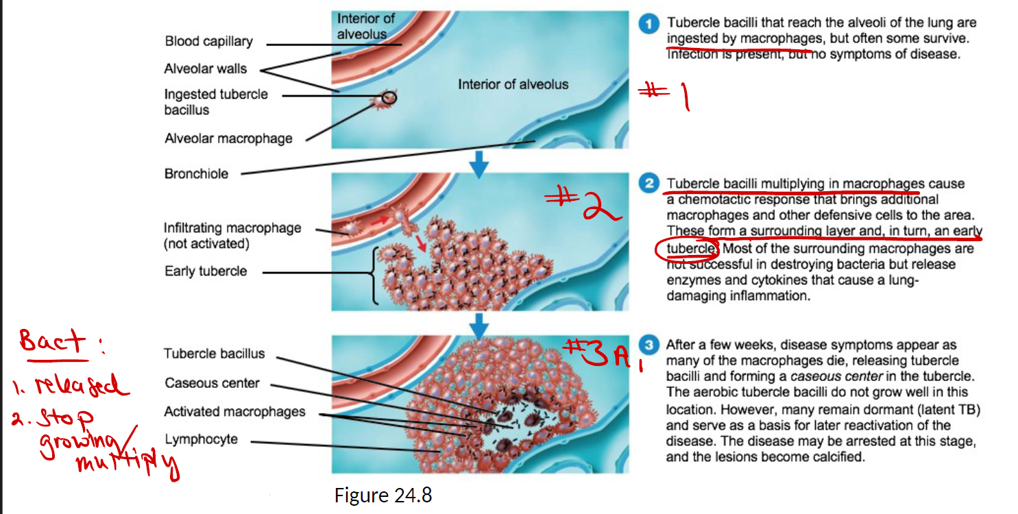
In step 3A, what 2 things happen to the bacteria?
They’re released.
They stop growing/multiplying.
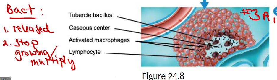
In step 3, why don’t the bacteria grow well in the caseous center, and what can they become?
They’re aerobic. Many stay dormant (latent TB) and can later reactivate.
If the disease is arrested at step 3, what happens to the lesions?
They become calcified.
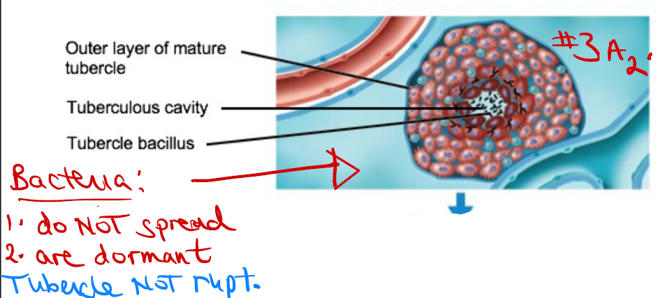
In stage 3A, what 2 things are true of the bacteria, and what’s the state of the tubercle?
They do NOT spread.
They are dormant.
The tubercle is NOT ruptured.
In stage 3B, what happens to the bacteria, and what’s the state of the tubercle?
The bacteria spread, and the tubercle is ruptured.

When the tubercle ruptures (3B), where do the bacilli spread?
Into a bronchiole, throughout the lung, and then to the circulatory and lymphatic systems.
Summarize the 3 steps of TB pathogenesis, including both outcomes of step 3
Inhaled bacteria are phagocytized by alveolar macrophages → bacteria multiply → inflammatory response.
More macrophages are recruited → they surround and isolate the bacteria inside a tubercle → tubercle lesions form.
Dying macrophages release bacteria:
a. Bacteria stop multiplying, the tubercle is NOT ruptured, lesions calcify, and bacteria stay dormant = Latent TB.
b. Bacteria grow outside macrophages → the tubercle ruptures → the pathogen spreads.
What is a tubercle?
An aggregation of activated macrophages with bacteria inside.
In step 2, what do the recruited macrophages do to the bacteria, and what forms?
They surround and isolate the bacteria inside a tubercle, and tubercle lesions form.
What is latent tuberculosis?
The tubercle is NOT ruptured, lesions heal and calcify, and bacteria remain dormant.
In step 3B, why does the tubercle rupture, and what’s the result?
Bacteria grow/multiply outside macrophages → the tubercle ruptures → bacteria are released into the lungs and cardiovascular system, and the pathogen spreads.
When a ruptured tubercle lets the pathogen spread, what 2 places can it go?
To other lung areas and to other areas of the body.
How does TB spread to other areas of the body, and what type of infection does this cause?
Through the bloodstream, causing a systemic infection.
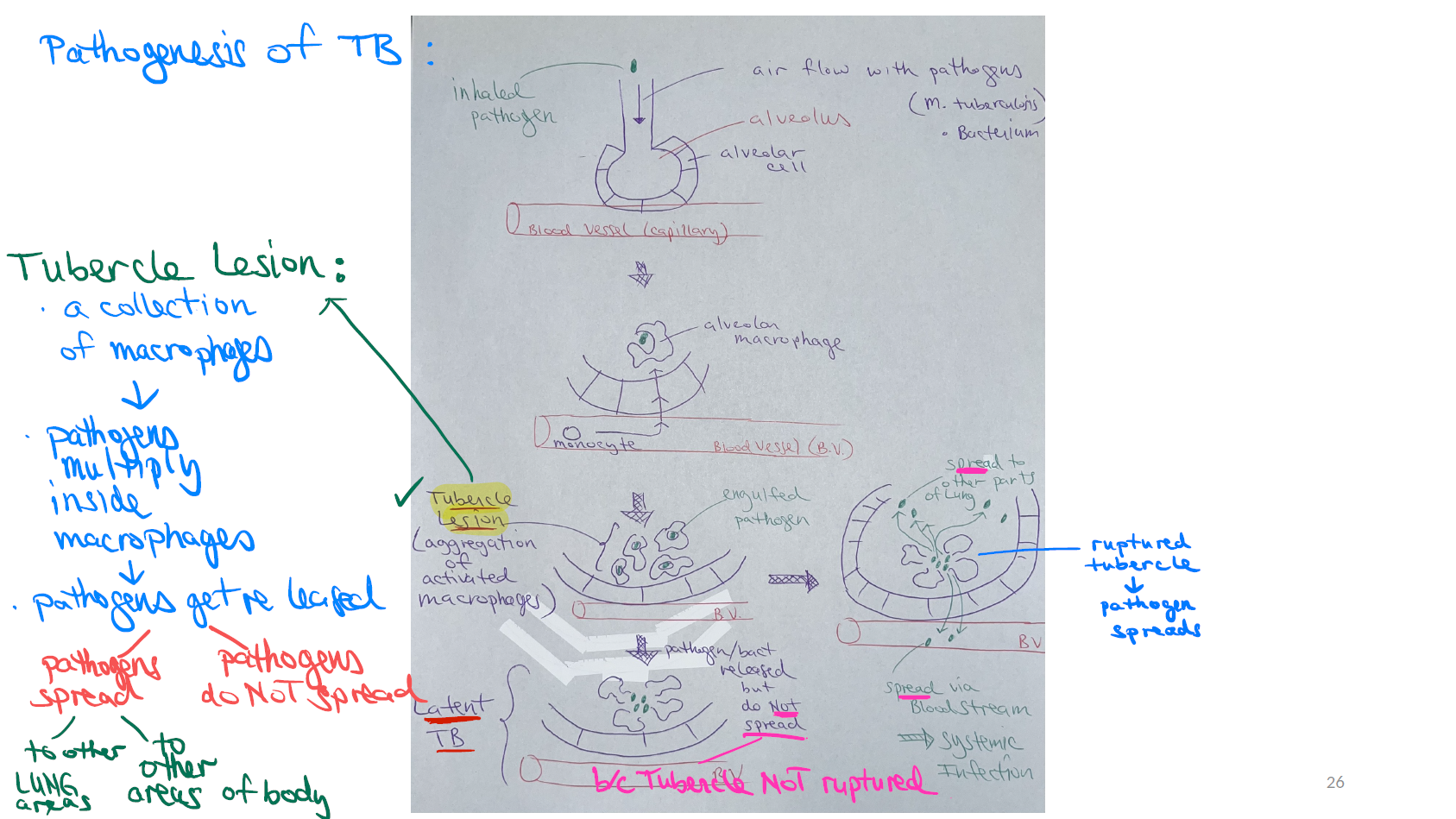
Try to explain this drawing:
TB is inhaled into the alveolus.
Macrophages engulf the bacteria.
More macrophages gather and form a tubercle lesion, where the bacteria multiply.
The macrophages die and release the bacteria.
Then one of two things happens:
Tubercle not ruptured: the bacteria don’t spread and stay dormant (latent TB).
Tubercle ruptured: the bacteria spread to other lung areas, or through the bloodstream to the rest of the body (systemic infection).
What are the 2 main types of skin lesions (rashes)?
Elevated (vesicular/papular)
Flat (macular)
What are the 2 types of elevated rashes?
Fluid-filled
Pus-filled
Which diseases cause a fluid-filled elevated rash?
Chickenpox
Shingles
Which disease causes a pus-filled elevated rash?
Smallpox.
Which diseases cause a flat (macular) rash?
Scarlet fever
Measles
What is the function of each virulence factor: β hemolysin, streptokinase, hyaluronidase, and capsules/M proteins?
β hemolysin: completely lyses RBCs.
Streptokinase: dissolves/prevents blood clots.
Hyaluronidase: dissolves the substance holding connective tissue cells together.
Capsules and M proteins: allow adherence.
What is lysogeny, and which 2 toxins are produced by it?
A bacteriophage’s toxin gene is incorporated into the bacterial chromosome (prophage), so the bacterium now makes the toxin. Examples are the erythrogenic toxin (scarlet fever) and the diphtheria toxin.
What is an antitoxin?
Antibodies (Ab) against a toxin.
What is a toxoid?
A non-toxic toxin, used in vaccines (Ex - Td).
What does “acellular” mean in a vaccine?
The entire cell (pathogen) isn’t injected, only a piece of it.
Are vaccines ever used as treatment?
No. Vaccines are prevention, NEVER treatment.
What is the difference between a monovalent and a multivalent vaccine?
Monovalent contains a single strain of a pathogen.
Multivalent contains multiple strains (Ex - flu vaccine).
What is antigenic variation, and why does it make it hard for the immune system to destroy a virus?
Spikes change through genetic recombination, forming hybrid viruses.
The immune system must start over and needs time to respond to each “new” antigenic determinant.
What does being antigenically stable mean for a vaccine?
There’s no antigenic variation, so the same vaccine can be used every time (Ex - measles).
Why is adaptive immunity not effective against latent infections?
The virus can hide in the nervous system.
What is the difference between toxemia and viremia?
Toxemia means symptoms are caused by a toxin in the blood. Viremia means the virus itself is in the bloodstream.