BISC205 Topic 1
 Call Kai
 Learn
Learn Practice Test
 Spaced Repetition
 Match
Match Flashcards
 Knowt Play
1/44

There's no tags or description
Looks like no tags are added yet.
Name | Mastery | Learn | Test | Matching | Spaced | Call with Kai | Chat |
|---|
No analytics yet
Send a link to your students to track their progress
45 Terms
Form and function of hawk moth
Inserts tongue-like proboscis to reach nectar receptacle of an orchid flower. This maximizes energy gain because it’s quick and doesn’t use much energy.

Herbivores form and function
Herbivores have longer intestines to digest hardier plants and they have larger cecums to host more bacteria that break down cellulose from plants into glucose

Hippopotamus form and function
Secrete blood sweat from their pores. It consists of several highly acidic compounds that absorb UV light to prevent sunburn. It comes out clear, turns red, and then fades to brown

Form and function of naked mole rat (three)
1) a poikilotherm (internal temperature varies considerably), so internal temperature changes with environment
2) when deprived of oxygen, they go into a suspended animation to reduce body’s energy demands. They also switch from a glucose-based metabolic system (which requires oxygen to release energy) to a fructose one that lets them make energy for cells to survive in vital organs. This can allow them to survive up to 18 minutes without oxygen.
3) has high levels of a polysaccharide called high molecular hyaluronan that accumulates in the extracellular matrix. It is involved in stimulating intracellular pathways that induce the expression of the p16 gene (tumor supressor gene). This means they do not develop cancer

Horned lizard form and function (three ways of defence)
Three ways of defence:
1) camouflage
2) scare predators by puffing body up to twice the normal size
3) scare off enemies by squirting toxic blood from their eyes. They do this by contracting muscles near their eyes to reduce blood flow out of its head, which builds up blood pressure. When the pressure is strong enough, the delicate blood vessels around their eyes rupture and shoot blood up to five feet.

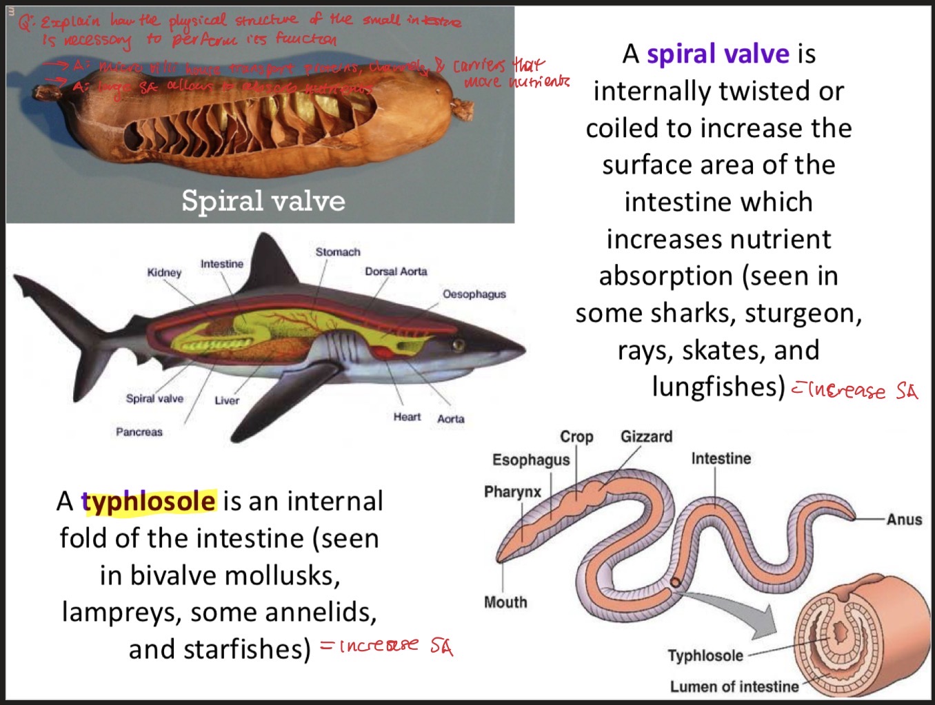
Typhlosole and spiral valve
Typhlosole: an internal fold of the intestine, which increases nutrient absorption (seen in bivalve mollusks, lampreys, annelids, and starfishes)
spiral valve: structure that is internally twisted or coiled to increase surface area of intestine, which increases nutrient absorption (seen in sharks, sturgeon, rays, skates, and lungfishes)
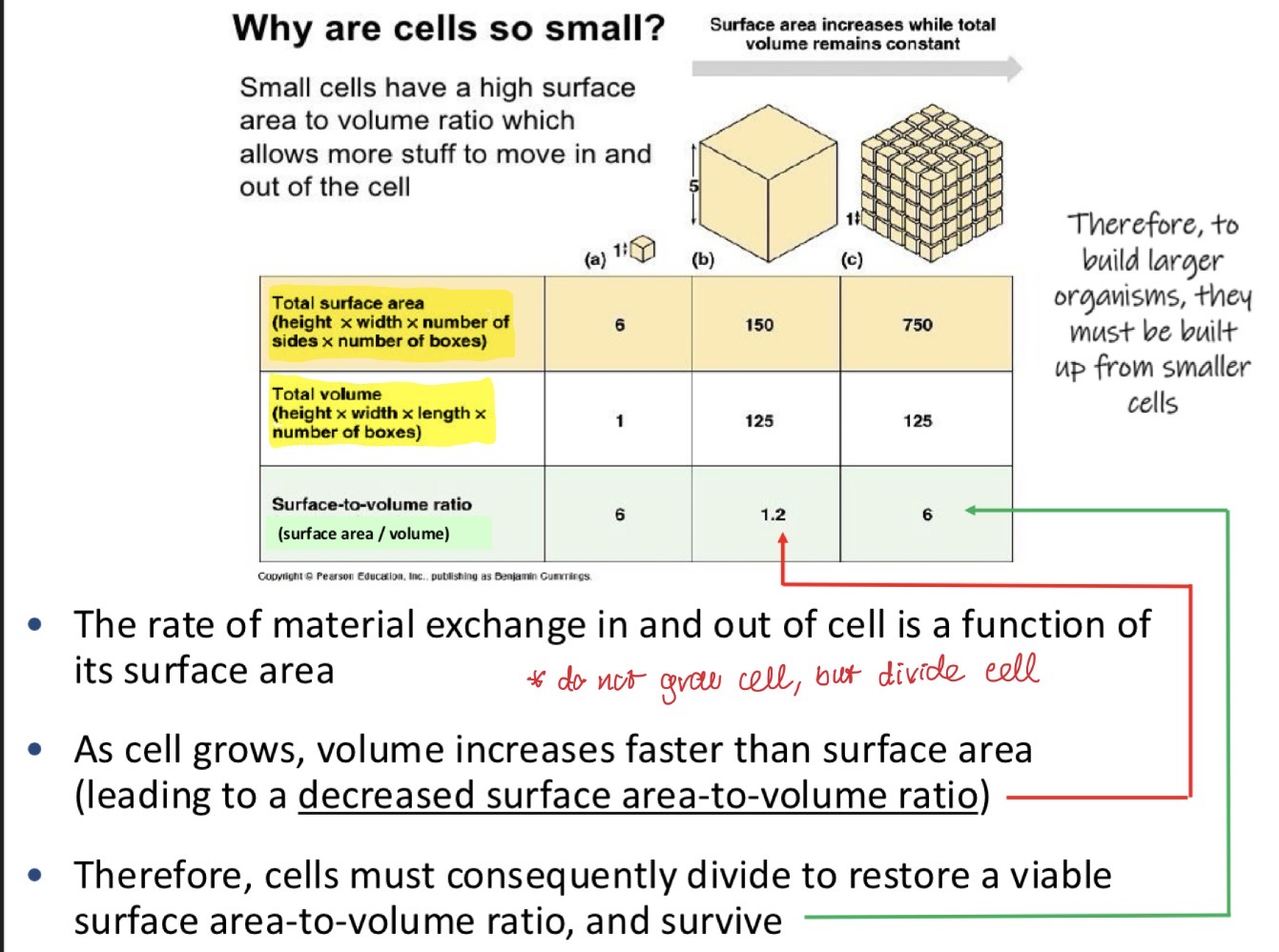
Why are cells so small?
Small cells have high surface area to volume ratio, which allows them to move more stuff in and out the cell. When cells grow, their volume increases faster than their surface area. In order to maintain this ratio, cells must divide and form organs rather than grow into really big cells.
Three adaptations that increase surface area and maintain a high surface area to volume ratio
1) flattening: thin and broad surface maximizes exposure to the environment relative to its volume (increases SA without increasing volume)
2) folding: existing surfaces are folded into complex shapes to pack more surface area into a given volume
3) branching: structures grow into intricate networks containing fine extensions
Examples of flattening
1) capillaries: small blood vessels that are highly branched to increase oxygen delivery to tissue
2) axons and dendrites: parts of neurons that have branching structures to transit and receive signals over long distances
3) plant roots: branched to increase water and nutrient uptake from the soil

Examples of folding
1) villi and micro villi: folds in the small mammal intestine for increased nutrient exchange
2) mitochondrial Cristae: folded to increase surface area for ATP production
3) brain cortex: folds increase surface area for neurons, enhancing processing capacity

Examples of flattening
1) lamellae: flattened, sheet-like structures to increase oxygen uptake
2) alveoli: thin-walled and flattened to facilitate efficient gas exchange
3) leaves: broad and flat that helps capture more sunlight for photosynthesis

Four main categories of tissues
1) epithelial
2) connective
3) muscle
4) nervous

Shapes of epithelial cells
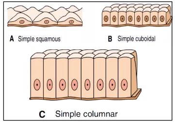
1) squamous: cells are thin and flattened, with a small centrally located nucleus
2) cuboidal: cells are cube shaped with a central nucleus
3) columnar: cells are rectangular with nucleus towards base. They usually absorb material from the digestive tract, forming inner lining of digestive organs
Layers of epithelial cells
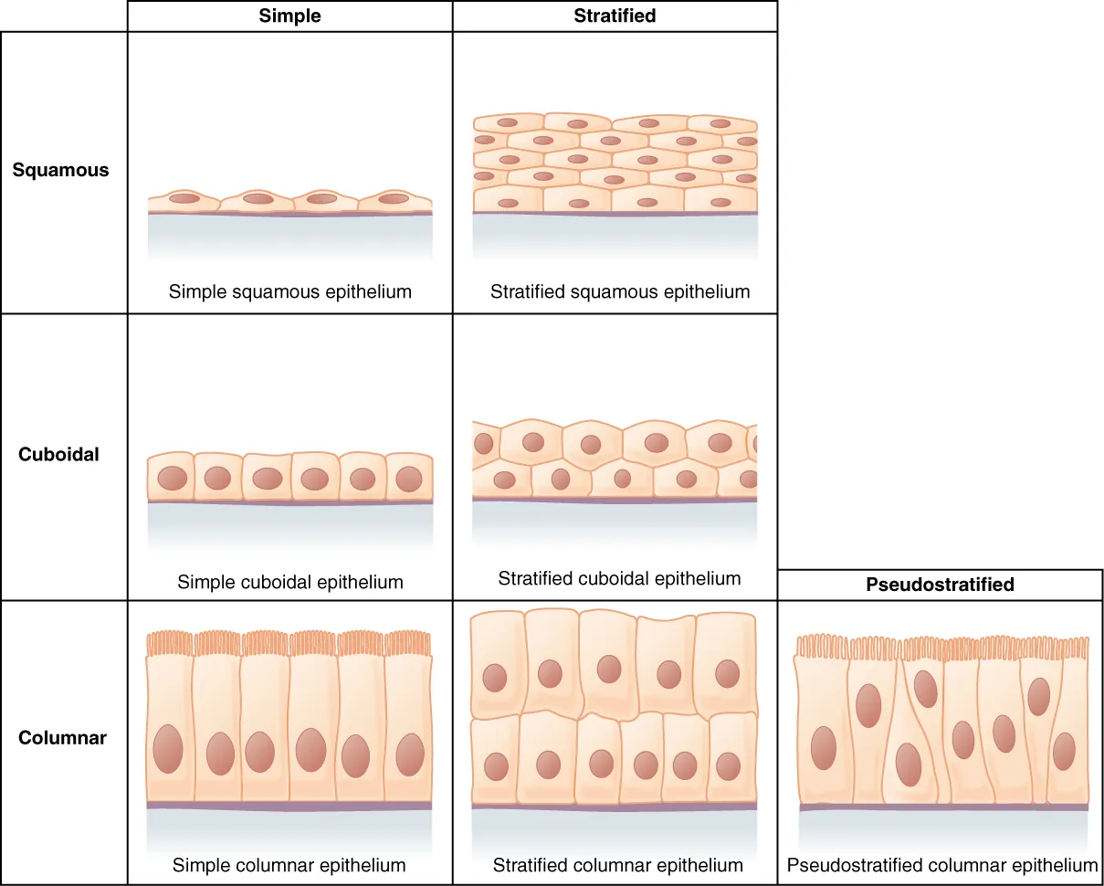
1) simple = one layer
2) stratified = two or more layers
3) pseudostratified = epithelia that appear to be stratified, but are actually simple (they look like two layers but are actually one). Can only be columnar cells because of their thicknes, so when they become crowded, the nuclei become displaced
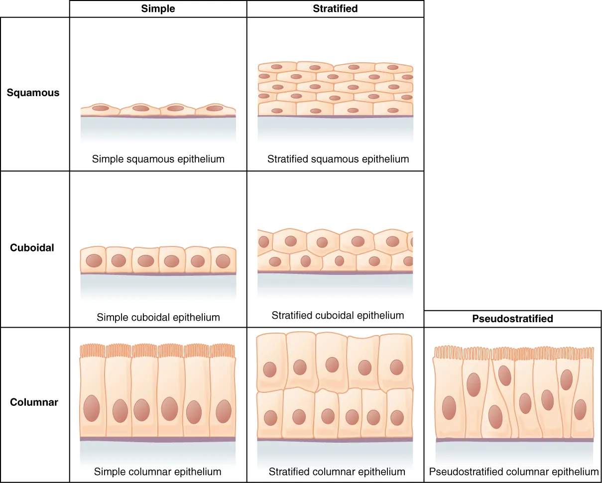
Fill in the gaps: bodily structures made of epithelial cells

The apical and basal
apical side: exposed surface of the epithelial cells
Basal side: the lower area of the epithelial cells close to the underlying body structures, like basement membrane and connective tissue
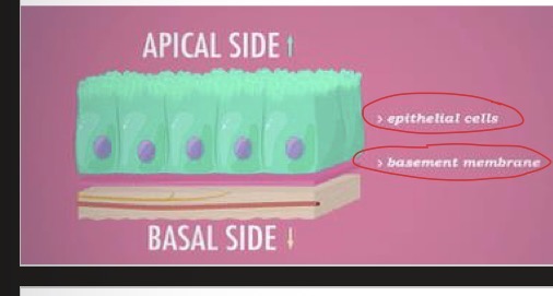
Two components that make up the basement membrane
1)basal lamina: a mixture of glycoproteins and collagen that is secreted by epithelial cells. It provides an attachment site for the epithelium, separating it from underlying connective tissues
2) reticular lamina: substance that is secreted by the underlying connective tissue. It attaches to the basal lamina

cell junction definition and the three types
Specialized intercellular connection between cell membranes
three types
1) tight junction
2) gap junction
3) anchoring junction
tight junction
separate cells into apical and basal compartments. It prevents the leakage of molecules between cells from one side of a sheet of cells to the other side

Gap junction
Intercellular passageway between membranes of adjacent cells to facilitate the movement of small molecules and ions between the neighbouring cytoplasms

Anchoring junction and the 4 types
Common on lateral and basal surfaces of cells where they provide strong and flexible connections. They do this by mechanically attaching cells and their cytoskeletons to their neighbours or to the extra cellular matrix
There are four types:
1) desmosomes
2) adherens
3) hemidesmosomes
4) focal adhesions

Adherens and focal adhesion anchoring junctions
Adheren junction: connects actin filament bundles in one cell with that in the next cell. It is a type of cell-to-cell anchoring junction
Focal adhesion junction: actin-linked cell matrix adhesion anchors actin filaments in cell to extracellular matrix. It is a type of cell-matrix anchoring junction

Desmosomes and Hemi-desmosome anchoring junctions
Desmosome junction: connects intermediate filaments in one cell to those in the next cell. It is a type of cell to cell anchoring junction
Hemi-desmosomes junction: anchors intermediate filaments in a cell to an extracellular matrix. Its is a type of cell matrix anchoring junction

connective tissue definition
The most abundant tissue type in the body, which consists of cells and the extracellular matrix

The two components that make up the extracellular matrix.
1) ground substance: a non-living, formless, viscous gel with high water content
2) protein fibers: can be elastic fibers, reticular fibers, and collagen fibers

Collagen fibers
The most abundant fiber type. They are long, thick, wavy, and have high tensile strength (resistant to stretching and tearing)

Elastic fibers
Thinner than collagen fibers. They are long, branched, and strong, but can be stretched up to 150% of the original length without breaking. They can stretch and recoil.

Reticular fibers
Thinner than collagen fibers. They are highly branches and can form mesh-like networks. This arrangement provides structural support and framework.

four examples of resident cells found in connective tissue
1) mesenchymal cells: stem cells that can differentiate into any type of connective tissue cells
2) fibroblasts: responsible for making the extracellular matrix
3) macrophage
4) adipocyte
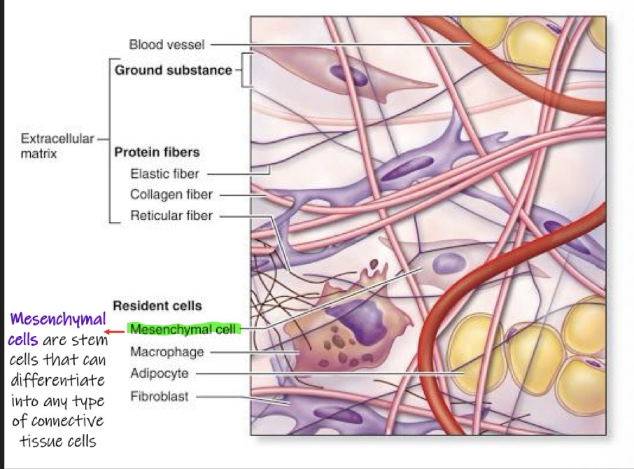
Five major types of connective tissue in vertebrates
1) loose connective
2) fibrous (or dense) connective
3) bone
4) blood
5) cartilage

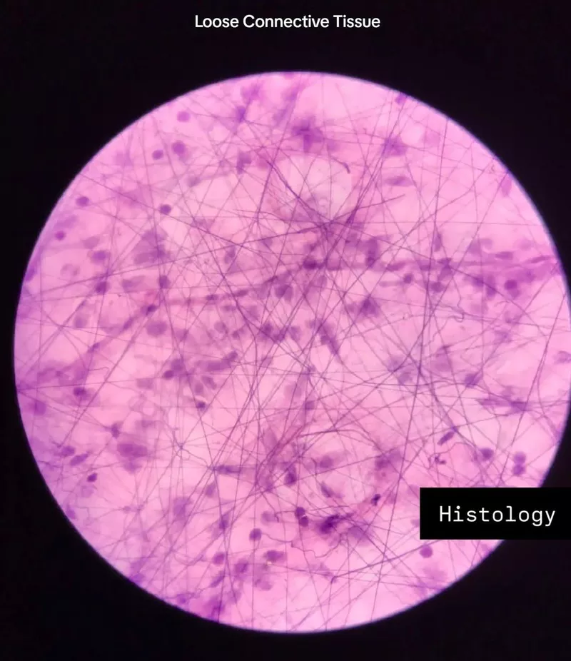
Loose connective tissue
MOST COMMON TYPE OF TISSUE IN VERTEBRATES
location: binds epithelia to underlying tissue and holds organs in place
extracellular matrix: ground substance occupies a high volume compared to the protein fibers
resident cells: contains all resident cells, with fibroblasts being the most abundant
fibrous/dense connective tissue
location: tendons and ligaments
extracellular matrix: large amounts of collagen fibres and a small amount of ground substance. this allows for higher tensile strength and a greater resistance to stretching
resident cells: contains all resident cells with fibroblasts being the most abundant

bone connective tissue
protein fiber: collagen is the most abundant
other cells:
1) osteoblasts: bone-forming cells
2) osteocytes: mature bone cells trapped in mineralized matrix
3) osteoclasts: break down and reabsorb bone

harversian system/osteons in bone tissue
microscopic structures of hard mammalian bones consists of repeating units called osteons or haversian system. Each has concentric layers of mineralized matrix deposited around a central canal. this canal contains blood vessels and nerves that service the bone.

hydroxyapatite crystals
osteoblasts produce calcium phosphate in the form of hydroxyapatite crystals, which is mainly responsible for the mineralization of bone

blood connective tissue
extracellular matrix: does not contain protein fibers. the ground substance is called plasma, which consists of water, electrolytes, nutrients, hormones, salts, and dissolved protein. More extracellular matrix in blood than cells
cells found in extracellular matrix/plasma
1) erythrocytes (red blood cells)
2) leukocytes (white blood cells)
3) thrombocytes (platelets)

two functions of cartilage
Two functions
1) strong flexible support material
2) acts as a shock absorber
Protein fibre: collagen is the most abundant, but can contain some elastic fibers depending on the type of cartilage. No reticular fibers present.

Chondroitin sulfates and chondrocytes
Chondroitin sulfates: polysaccharides that make the distinctive appearance of cartilage. They bind to ground substance proteins to form proteoglycans
Chondrocytes: cells that secrete the collagen fibers and Chondroitin sulfates

Advantages of animals with cartilaginous skeletons
Makes their Skelton more flexible and light weight (cartilage is less dense than bone). Cartilage provides support without the weight of bone, allowing these animals to be more energy efficient and buoyant in water. Examples of animals include sharks, rays, and skates

Three types of muscle tissue
1) skeletal
2) smooth
3) cardiac

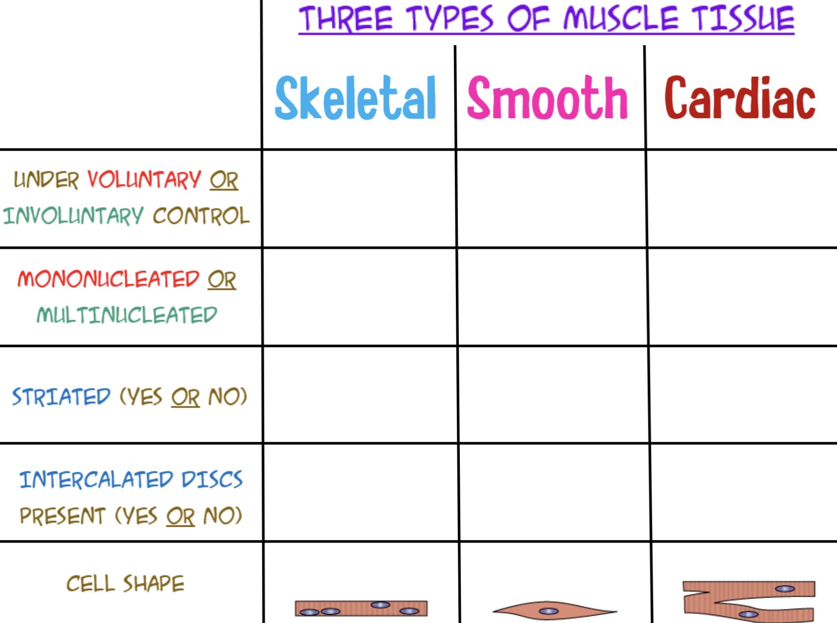
Differences between muscle tissue types
Skeletal muscle: binds on the skeleton
Smooth muscle: found in the digestive system
Cardiac muscle: found in the heart

Nervous tissues
Senses stimuli and transmits electrochemical signals in the form of nerve impulses that communicate within different regions of the animal.
Two components
1) neurons: transmits nerve impulses
2) glial cells: help nourish, insulate, and replenish neurons by providing support and protection for neurons

Two examples of glial cells
1) myelin sheath
2) Schwann cells

Axons and dendrites
Neurons have two types of projections from the cell body (where nucleus is)
1) dendrites: short branching structures which transmits electrical signals from adjacent cells to the neuronal cell body or soma
2) axons: long structures which carry electrical signals from the cell body to other cells

Cartilage connective tissue