Neuroscience Lab 1 Quiz
 Call Kai
 Learn
Learn Practice Test
 Spaced Repetition
 Match
Match Flashcards
 Knowt Play
1/32

There's no tags or description
Looks like no tags are added yet.
Name | Mastery | Learn | Test | Matching | Spaced | Call with Kai | Chat |
|---|
No analytics yet
Send a link to your students to track their progress
33 Terms
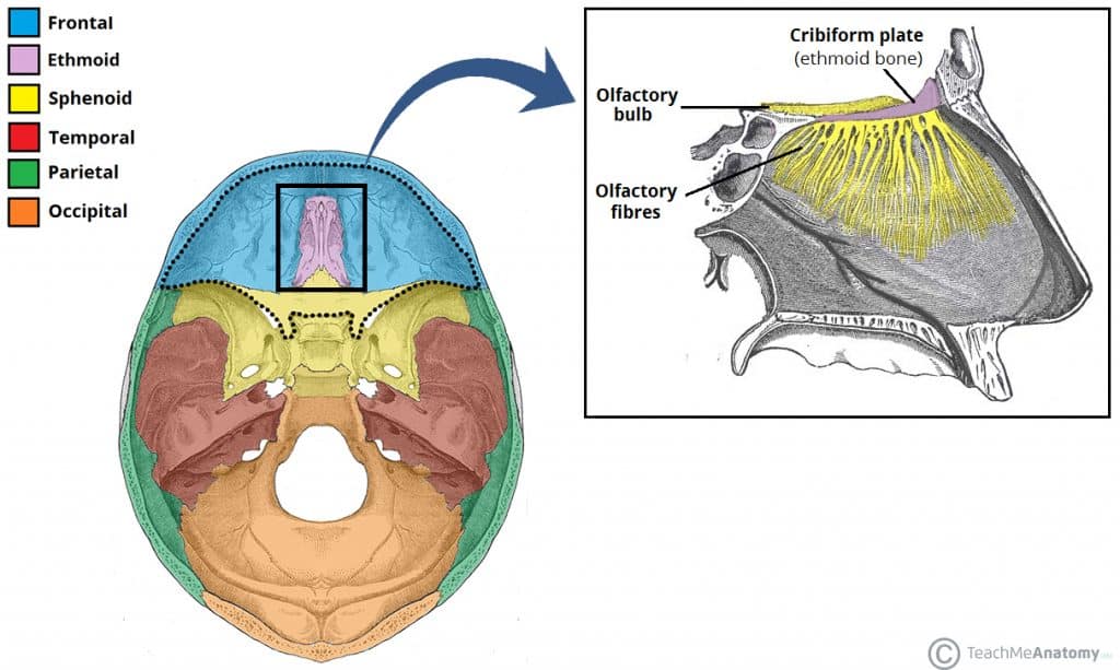
Cribiform Plate
Olfactory formina, CN1

Optic canal, CN2

Supraorbital fissure
CN3, CN4, CN6, CNV1 (opthalmic branch)

Which structure? Houses which nerve?
Foramen Rotundum - CNV2 (Maxillary branch)

Foramen Ovale - CNV3 (Mandibular branch)

Structure? Houses what?
Foramen Spinosum - middle meningeal artery

Structure? Houses what?
Foramen Lacerum - internal carotid artery

Internal auditory meatus - CN7 & CN8

Jugular Foramen

Jugular Foramen Houses…
CN9, CN10, CN11, Internal jugular vein
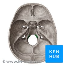
Hypoglossal canal - exit for CN12

Foramen magnum - vertebral artery, junction of brainstem + spinal cord

Falx cerebri - between cerebral hemispheres
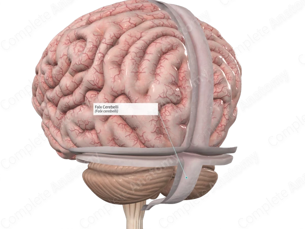
Falx cerebelli - between cerebellar hemispheres

Tentorium cerebelli - tent over cerebellum
Venous sinus path for Superior sagittal sinus
Superior sagittal sinus —> R transverse sinus —> Sigmoid sinus —> internal jugular vein
Venous sinus path for Internal sagittal sinus
Interior sagittal sinus —> straight —> L transverse sinus —> L sigmoid sinus —> internal jugular vein
Cavernous sinus path
Cavernous sinus —> petrosal sinus —> sigmoid sinus —> internal jugular vein (jugular foramen)
Ventricular system connection
Lateral ventricles —> interventricular foramina —> 3rd ventricle —> cerebral aqueduct —-> 4th ventricle

3 exits from 4th ventricle
2 lateral aperture, 1 median —> CSF enters subarachnoid space, surrounding brain and spinal cord (lumbar cistern) —> arachnoid villi —> superior sagittal sinus —> blood.
enlarged subarachnoid space at the bottom of spinal cord
lumbar cistern

3 Meningeal layers
Dura mater - arachnoid - pia mater
2 layers of Dura matter
periosteal layer, meningeal layer
Spaces in meningeal layers
Epidural, subdural, subarachnoid space
Items in subdural space, items in subarachnoid space
Subdural: bridging arteries
Subarachnoid: Arachnoid trabeculae (web like connective tissue), cerebral spinal fluid CSF

What portion of the temporal bone is this? Green region
Squamous portion of temporal bone

What part of the temporal bone is this?
Petrous portion of temporal bone
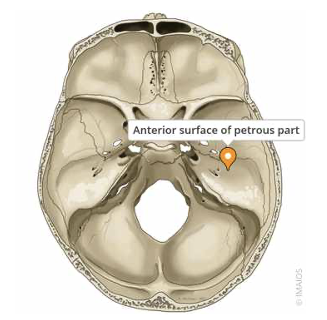

Groove on the greater wing of the sphenoid bone
Groove for middle meningeal artery

Carotid sulcus, continuation of the carotid canal, houses internal carotid artery + cavernous sinus.

Anterior fossa borders & contents
Borders:
Anterior border = frontal bone
Posterior border = anterior portion of the sphenoid lesser wing
Contents:
Frontal lobe
Middle Fossa borders and content
Borders:
Anterior border: greater wing of sphenoid
Posterior border: petrous portion (superior ridge) of temporal bone
Contents: parietal, temporal, occipital lobes
Posterior fossa borders & content
Anterior border: sella turcica posterior wall + petrous portion of temporal bone
Posterior border: back of occipital bone
Content: cerebellum