A225 - IU - Block 3
 Call Kai
 Learn
 Practice Test
 Spaced Repetition
 Match
 Flashcards
 Knowt Play
1/135

There's no tags or description
Looks like no tags are added yet.
136 Terms
Meninges
three protective membranes that surround the brain and spinal cord
Dura Mater
thick, outermost layer of the meninges surrounding and protecting the brain and spinal cord
Arachnoid Mater
weblike middle layer of the three meninges
subarachnoid space
a space in the meninges beneath the arachnoid membrane and above the pia mater that contains the cerebrospinal fluid
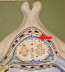
pia mater
the delicate innermost membrane enveloping the brain and spinal cord.
Cerebrum
Area of the brain responsible for all voluntary activities of the body

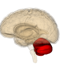
Cerebellum
A large structure of the hindbrain that controls fine motor skills.
Midbrain
A small part of the brain above the pons that integrates sensory information and relays it upward.

Diencephalon
Thalamus, Hypothalamus, Pineal Gland

Pons
A brain structure that relays information from the cerebellum to the rest of the brain

medulla oblongata
the posterior part of the brain that controls the rate of breathing and other autonomic functions

Thalamus
the brain's sensory switchboard, located on top of the brainstem; it directs messages to the sensory receiving areas in the cortex and transmits replies to the cerebellum and medulla

Hypothalamus
A neural structure lying below the thalamus; it directs several maintenance activities (eating, drinking, body temperature), helps govern the endocrine system via the pituitary gland, and is linked to emotion and reward.

pineal gland
secretes melatonin

pituitary gland
The endocrine system's most influential gland. Under the influence of the hypothalamus, regulates growth and controls other endocrine glands.

corpus callosum
the large band of neural fibers connecting the two brain hemispheres and carrying messages between them

septum pellucidum
separates lateral ventricles

optic chiasm
point at which optic nerve fibers cross in the brain

cerebral peduncles
contain fibers that carry motor output from cerebrum to other regions of CNS

middle cerebellar peduncle
connects cerebellum to pons

Pyramids
two large bulges, one left and one right, on the ventral surface of the medulla; they contain the pyramidal tracts, which carry signals for voluntary movement from the cerebral hemispheres to motor circuits of the spinal cord
lateral ventricles
A set of paired ventricles lying within the cerebral hemispheres.

third ventricle
the ventricle located in the center of the diencephalon

cerebral aqueduct
connects the third and fourth ventricles

fourth ventricle
the ventricle located between the cerebellum and the dorsal pons, in the center of the metencephalon

Cerebral Arterial Circle (Circle of Willis)
Blood vessels that surround the base of the brain, protecting the brain and providing more than one route for blood to reach brain tissue

basilar artery
An artery, formed by the fusion of the vertebral arteries, that supplies blood to the brainstem and to the posterior cerebral arteries.

vertebral arteries
Arteries that ascend the vertebrae, enter the base of the skull, and join together to form the basilar artery.

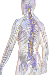
Spinal chord
Long bundles of neurons that run inside of your vertebral column. It carries impulses from all parts of the body to the brain and from the brain to all parts of your body. Reflex arcs occur here.
Anterior Horns
cell bodies of somatic motor neurons

Posterior Horns
contain axons of sensory neurons and cell bodies of interneurons

dural venous sinuses
Drain blood from the brain into the internal jugular veins.

right cerebral hemisphere
controls most of the functions on the left side of the body

left cerebral hemisphere
controls the majority of functions on the right side of the body

cortex
outermost covering of the brain consisting of densely packed neurons, responsible for higher thought processes and interpretation of sensory input

white matter
Composed of tracts of myelinated axons

frontal lobe
associated with reasoning, planning, parts of speech, movement, emotions, and problem solving

parietal lobe
Receives and interprets most somatosensory (Temperature, Touch, Pain

occipital lobe
A region of the cerebral cortex that processes visual information

temporal lobe
A region of the cerebral cortex responsible for hearing and language.

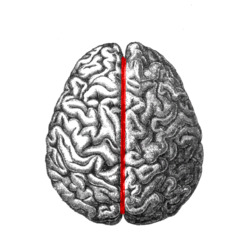
longitudinal fissure
deep furrow between left and right hemispheres
lateral sulcus
Groove that separates temporal lobe from parietal and frontal lobes

Central sulcus
separates frontal and parietal lobes

olfactory tract
the path along which the olfactory receptors send their electrical messages to the brain.

olfactory bulb
the first brain structure to pick up smell information from the nose

gray matter
Brain and spinal cord tissue that appears gray with the naked eye; consists mainly of neuronal cell bodies (nuclei) and lacks myelinated axons.
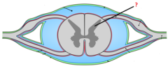
Posterior Roots
fibers carry sensory information

posterior root ganglion
contains cell bodies of sensory neurons

conus medullaris
end of spinal cord

cauda equina
collection of spinal nerves below the end of the spinal cord

central canal
A tiny channel found within the spinal cord and inferior medulla oblongata

anterior (ventral) root
Attaches on each to the anterior part of the spinal cord and contains motor (efferent) axons
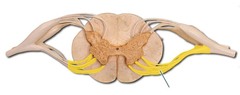
spinal nerves
Formed when a posterior nerve and an anterior nerve unite, thus contains both motor (efferent) and sensory (afferent) axons.

Soma
cell body of a neuron

Nucleolus
Found inside the nucleus and produces ribosomes

chromatophilic substance
membranous sacs in the cytoplasm of nerve cells that have ribosomes attached to their surfaces

Dendrites
Branchlike parts of a neuron that are specialized to receive information.

Axon
the extension of a neuron, ending in branching terminal fibers, through which messages pass to other neurons or to muscles or glands
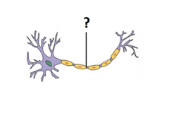
axon hillock
Cone shaped region of an axon where it joins the cell body.

Olfactory nerve (1)
the nerve that carries smell impulses from the nose to the brain

Optic Nerve (II)
sensory, vision

Oculomotor Nerve (III)
narrows pupil and focuses lens, motor, eye movement

Trochlear Nerve (IV)
motor, eye movement
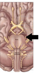
trigeminal nerve (V)
contains motor axons for the muscles of mastication
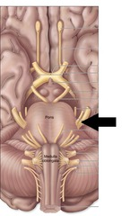
Abducens Nerve (VI)
lateral eye movement

Facial Nerve (VII)
Movement of facial expression muscles, taste (from anterior 2/3 of tongue)

vestibulocochlear nerve (VIII)
hearing and balance

Glossopharyngeal nerve (IX)
A cranial nerve that has sensory axons for taste in the posterior 1/3 of the tongue, motor axons for pharynx muscles, and some autonomic axons that control the parotid salivary gland

Vagus Nerve (X)
This nerve innervates most of the viscera in the thoracic and abdominopelvic cavities. As well as innervates voluntary structures in the neck

Accessory Nerve (XI)
Contains motor axons that go to the pharynx, larynx, trapezius, and sternocleidomastoid

Hypoglossal Nerve (XII)
Contains motor axons for the intrinsic and most extrinsic tongue muscles.

Phrenic Nerve
Provides motor innervation for the diaphragm

brachial plexus
network of interlacing nerves found in the upper arm area

radial nerve
Sensory-motor nerve that, with its branches, supplies the thumb side of the arm and back of the hand.

ulnar nerve
Sensory-motor nerve that, with its branches, affects the little-finger side of the arm and palm of the hand.

Median Nerve
Smallest of the three arm and hand nerves; runs along the mid forearm and extends into the hands

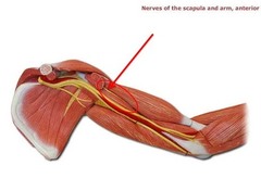
musculocutaneous nerve
arises from the brachial plexus and provides motor innervation to the muscles of the anterior compartment of the arm and sensory innervation to the lateral part of the forearm
Lumbosacral plexus
Formed of L2-S2 rami. Supplies the lower limb.

femoral nerve
innervates quadriceps and skin of anterior thigh and medial surface of leg

sciatic nerve
nerve extending from the base of the spine down the posterior side of the thigh, down the leg to the foot

tibial nerve
A division of the sciatic nerve that passes behind the knee. It subdivides and supplies impulses to the knee, the muscles of the calf, the skin of the leg, and the sole, heel, and underside of the toes.

common fibular nerve
controls muscles on the anterior and lateral leg (primarily dorsiflexors) and foot

myelin sheath
A layer of fatty tissue segmentally encasing the fibers of many neurons axon; enables vastly greater transmission speed of neural impulses as the impulse hops from one node to the next.

Neurolemmocytes
myelinate axons in the PNS

neurofibril nodes (nodes of Ranvier)
narrow gaps between myelin sheaths

medial rectus muscle
Turns the eyes inward towards the nose

Lateral rectus muscle
Turns the eyes outward away from the nose

superior rectus muscle
Turns the eyes upward

Inferior Rectus muscle
Turns the eyes downwards

Superior oblique muscle
depresses eye and turns it laterally

inferior oblique muscle
elevates eye and turns it laterally

fibrous tunic
sclera and cornea
Sclera
white of the eye

Cornea
the transparent layer forming the front of the eye.

vascular tunic
choroid, ciliary body, iris
choroid
middle, vascular layer of the eye, between the retina and the scler

ciliary body
ring of tissue behind the peripheral iris that is composed of ciliary muscle and ciliary processes

Iris
a ring of muscle tissue that forms the colored portion of the eye around the pupil and controls the size of the pupil opening

pupil
the adjustable opening in the center of the eye through which light enters

retina
the light-sensitive inner surface of the eye, containing the receptor rods and cones plus layers of neurons that begin the processing of visual information
