DSON 5014: Congenital Heart Disease Midterm
 Call Kai
 Learn
 Practice Test
 Spaced Repetition
 Match
 Flashcards
 Knowt Play
1/133

There's no tags or description
Looks like no tags are added yet.
134 Terms
Describe development of the heart: Days 18-19 (cardiogenic area)
Heart begins to form near the fetus head, in the area called the cardiogenic area
chemical signal the mesoderm to form into two strands that will become the endocardial tubes
Describe development of the heart: Days 20-21 (heart tube)
cardiogenic cords develop a lumen, and they are now called endothelial tubes
tubes migrate and fuse together to form the primitive heart tube
the tube “beats”, or rhythmically contracts to help push blood through the tube
blood enter sinus venosus to truncus arteriosus due to contractions of primitive tube
Form several distinct regions
Identify five regions of the fetal heart
truncus arteriosus
bulbus cordis
primitive ventricle
primitive atrium
sinus venosus
What does truncus arteriosus become?
will eventually divide and become the ascending aorta and pulmonary trunk
What does bulbus cordis become?
will eventually become the right ventricle
What does primitive ventricle become?
will eventually become the left ventricle
What will primitive atrium become?
will become the anterior portion of both atria and the appendages
What will sinus venosus become?
will eventually become the ends of IVC and SVC, as well as parts of the atria
Describe development of the heart: Days 23-28 (heart tube)
primitive heart tube elongates
tube is fixed at its ends and restricted by the pericardium, so as it elongates it begins to form a curvature, or a “S” shape
Describe D-looping
aka dextra-looping (latin for right)
in a normal heart, as the heart elongates and forms the S curve, it should loop ventrally and to the patient’s right
Describe development of the heart: Weeks 4-6 (cardiac septation)
results in division of the single atrio-ventricular canal into left and right sides
includes events that happen at same time:
formation of MV and TV
atrial septation
formation of IVS
septation of truncus arteriosis
formation of the semilunar valves
How are MV and TV formed?
proliferation of endocardial cushions for formation of MV and TV
How is IAS formed?
Septum primum grows inferiorly towards the endocardial cushions, space between = ostium primum
Seals with the endocardial cushions and leaving an opening = foramen secundum
Septum secundum grows besides the septum primum
The two septums fuse together to form interatrial septum
A small flap called foramen ovale remains center of the septum

How is IVS formed?
Muscular IVS grows superiorly up from the apex until it meets the ridges of the bulbus cordis
final step of IVS formation occurs when the membranous portion of the IVS forms and fuses with the endocardial cushions
How are septation of the truncus arteriosus formed ?
splitting and rotating → pulmonary stays and aortic disappears
Spiral formation at distal end, results in division of truncus into pulmonary artery and aorta
subpulmonary conus/infundibulum remains = in a small distance between TV and PV
aortic conus regresses/disappears = fibrous continuity between the AoV and MV
How are AoV/PV semilunar valves formed?
Grows from endocardial cushions in the truncus arteriosus
two of the encocardial cushions meet and fuse together to separate the AoV and PV, and the remaining endocardial cushions form the cusps of the valves
Describe development of the heart: Weeks 6 onwards
by week 6: 4 chambers of the heart are fully formed, however the heart will continue to grow and develop over the remainder of the pregnancy
muscle tissue will continue to develop and fully form by the end of week 20
What causes malformation of MV and/or TV
failure of endocardial cushions to develop
What causes ostium primum septal defect?
failure of the septum primum to fuse with endocardial cushions
What causes congenitally correct TGA or L-TGA
L- looping, causing RV/LV in the wrong spots
What causes TGA or d-TGA
failure of the spiral growth pattern results in transposition of the great vessels
aka great vessels in wrong spots
What causes persistent truncus arteriosus
failure of septation of truncus ateriosus
What causes bicuspid aortic valve AND Tetraolgy of Fallot
problem during formation of the endocardial cushions in the truncus arteriosus
Describe fetal circulation: oxygenated blood flow
Blood is oxygenated in the placenta instead of the lungs
Oxygenated blood travels through the umbilical vein and enters RA
Eustachian valve helps to push blood away from the TV and direct it towards the foramen ovale
Oxygenated blood enters the left side, and through the aorta

Describe fetal circulation: deoxygenated blood flow
Deoxygenated blood from head and upper extremities enters SVC, and then into RA
Oxygenated blood from babies lower body enters the heart via the umbilical vein, eventually into RA
In the RA, oxygenated + deoxygenated blood mix
Mixed blood will travel through the RV, PV, and PA
Due to the high resistance of the lungs in the fetus, most of the blood will travel through the ductus arteriosus and enter the aorta → aorta now have mixed oxygen content
some blood will travel through the lungs
The deoxygenated or mixed blood travels down the aorta and enters umbilical arteries
blood then travels to the placenta to become oxygenated again

Describe post natal circulation
During birth, there is compression of the chest as baby travels through birth canal
fluid is forced out of the bronchi and trachea
baby takes its first breath, air flows into lungs
pulmonary resistance drops and the umbilical cord is cut at the same time
systemic blood pressure increases → pulmonary pressure now lower than systemic pressure
Describe closure of the foramen ovale in post natal
sudden decrease in pulmonary pressure → sudden increase in blood flow into left and right pulmonary arteries
increase flow through pulmonary veins
increases LA volume and pressure
Increased LA pressure causes flap of the foramen ovale to close → no longer any communication between LA and RA
all blood entering RA must travel through the lungs
Describe closure of Ductus Arteriosus
happens a bit more delyed
right sided pressures drop and left sided pressures increase
aortic pressure rises above pressure of pulmonary artery
blood through the ductus arteriosus now changes direction, and travels from the aorta back into the pulmonary artery
should only last a few hours, as soon the pressure changes, it will signal the ductus arteriosus to close permanently → no longer any communication between aorta and right pulmonary artery
Define Congenital Heart Disease (CHD)
describe a structural abnormality of the heart and/or great vessels that you are born with
ACHD: adult congenital heart disease
What are the common latin terms to describe heart locations
situs → location
solitus → usual/common
inversus → inverted
totalis → total/entire
dextro → right
levo → left
Describe segmental approach scanning technique for assessment of CHD (5 things to prove)
do not assume chambers and valves are connected in proper order → assess every single chamber, valve, and great vessel
Cardiac position and visceral situs
Blood flow into the heart, # of IVC, SVC, etc
Blood flow through the heart
Blood flow out of the heart
Coronary artery anatomy
How to determine cardiac position and visceral situs? (step one)
apex points to the patient’s left side but is sitting at a slightly different angle, may or may not be congenital
Left → levoposition
Midline → mesoposition
Right → dextroposition
orientation of how the heart swing into a different position
What is cardiac situs
situs is the term used to describe if an organ or position of abnominal organs, is sitting normally or flipped mirror image
congenital
example: levocardia, on the left is normal and dextrocardia, on the right is “mirror image”
What is dextrocardia
congenital condition where the heart is on the right side of the chest and its apex points to the right
can be asymptomatic and often associated with other congenital heart diseases
How can you check if levocardia or dextrocardia in echo?
subcostal view, POM at 3-oclock
confirm which side is apex is pointing
Levocardia
pointing left in adult and pediatric (upside down, TEE)
Dextrocardia
pointing right in adult and pediatric (upside down, TEE)
What are the 3 types of cardia situs
Situs Solitus Totalis
Situs Inversus Totalis
Situs Ambiguous
Describe Situs Solitus Totalis
heart and abdominal organs in normal locations
Left: Stomach, spleen, pancreas, sigmoid colon
Right: Liver, cecum and appendix
Describe Situs Inversus Totalis
heart and abdominal organs are flipped
heart is dextrocardia, apex pointing to the right
liver is mainly on the left
stomach and aorta on the right
Describe Situs Ambiguous
heart and/or abdominal organs are random
the position of the vessels and the liver cannot be reliably determined
higher incidence of associated congenital cardiac defects
What are the 3 types of atrial situs?
atria determined by morphological features, not by their positions
Atrial Situs Solitus
Atrial Situs Inversus
Atrial Situs Ambiguous
Describe Atrial Situs Solitus
both atria are in their expected positions, with the RA on patient’s right and LA on the patient’s left
Describe Atrial Situs Inversus
the atria are reversed, with the LA on the patient’s right and the RA on the patient’s left
Describe Atrial Situs Ambiguous
Right atrial isomerism: patient has 2 RA’s
will likely also have two right lungs and no spleen, and associated with anomalous pulmonary venous return
Left atrial isomerism: patient has 2 LA’s
will likely also have two left lungs and is associated with anomalous systemic venous return
What are distinctive features of right and left atrium?
Right atrium:
broad based, triangular appendage positioned anteriorly
Chiari network
Left atrium:
narrow, elongated, finger-like appendage positioned posteriorly
How to determine blood flow into the heart (step 2) hint: assess systemic vs pulmonic features
Systemic:
is there an SVC and an IVC entering the RA?
are there extra connections entering the RA?
for example: persistent left SVC, patient with 2 SVCs
Pulmonic:
are there 4 pulmonary veins entering LA?
are there one or more pulmonary veins entering into different chamber?
for example: partial anomalous pulmonary vein return, total anomalous pulmonary vein return
How to determine blood flow through the heart (step three), what could go wrong (2)
flow should be RA → RV → Lungs → LA → LV → Aorta
wrong route if:
chambers in wrong location
D-Loop → normal
L-Loop → incorrect, ventricles switched sides
shunts
What are distinctive features of right and left ventricle? (4 ea)
Right ventricle:
triangular shape with coarse trabeculations
moderator band
pap muscles attached to septum and free wall
TV inserts more apically
Left ventricle
cone shaped, small trabeculations
no moderator band
superiorly inserted MV annulus with 2 pap muscles attached to LV free wall
may have false tendons
Describe atrioventricular connections (concordance vs discordance)
Atrioventricular concordance:
right atrium attaches to right ventricle
left atrium attaches to left ventricle
Atrioventricular discordance:
right atrium attaches to left ventricle
left atrium attaches to right ventricle
Where do the atrioventricular valves arise from?
arise from respective ventricles
Tricuspid valve → right ventricle no matter what side
Mitral valve → left ventricle
Describe venticulo-arterial connection (concordant vs discordant)
Concordant
pulmonary artery arises from morphological right ventricle
aorta arises from the morphological left ventricle
Discordant
pulmonary artery arises from morphological left ventricle
aorta arises from the morphological right ventricle
What is the Rule of 50% for when atrioventricular or ventriculo-arterial connections are not clear?
a chamber is a ventricle if it receives >50% of an inlet, inlet refers to AV (atrioventricular valve)
if 50% or more of a great vessel arises above a chamber, the great artery is said to be connected to that chamber
Note: if chamber does not receive 50% of an inlet valve, then it is known as rudimentary chamber
How to determine blood flow out of the heart (step four)
aortic arch patency and branching pattern
pulmonary artery bifurcation, size, and patency
presence/size of the ductus arteriosus
patency: being open and unobstructed, can be good or bad
Describe the special pediatric views: suprasternal crab view (SAX)
rotate 60 degrees clockwise from normal SSN
to evaluate pulmonary veins and SVC, able to all 4 pulmonary veins at same time

Describe the special pediatric views: suprasternal arch position
from the SSN SAX, further clockwise rotation/angulation toward opposite shoulder
Left arch, normal
first branch going to the patient’s right (left side of screen) and bifurcating suggests left arch
Right arch, abnormal
first branch going to the patient’s left (right side of screen) and bifurcating suggests mirror image of right arch

Define hypertrophic cardiomyopathy (HCM) (3 traits)
refers to LVH in absence of any underlying cause such as hypertension or aortic stenosis
LVH is most often asymmetric rather than concentric
usually interventricular septum is thicker than other walls
most common cause of death in young athletes
higher HR, long QT syndrome
coronary artery abnormalities, compressed during exercise
Describe the etiology of hypertrophic cardiomyopathy
inherited in an autosomal dominant pattern
person w/ autosomal dominant disorder has 50% chance of having affected child with mutated gene/dominant gene
gene mutations cause alterations in contractile elements of cardiac sarcomere which causes myocardial hypertrophy and fibrosis
specific gene mutations determines:
age of onset of hypertrophy
extent and pattern of hypertrophy
risk of sudden death (common heart related sudden death in <30 yro)
Describe pathophysiology of hypertrophic cardiomyopathy
characteristic features are ventricular hypertrophy and myocardial (myocyte) disarray → looks thicker than normal
25-40% HCM patients have dynamic LVOT obstruction, sometimes called hypertrophic obstructive cardiomyopathy (HOCM)
What are the types of ventricular hypertrophy (5)
asymmetric septal hypertrophy (ASH): 90% of HCM cases
only septum is big, other walls are normal
concentric hypertrophy: 4% of HCM cases
all walls thickened, should rule out normal causes of LVH, may or may not have LVOTO
apical hypertrophy: 25% of Japanese HCM patients, rare outside of Japanese population
thickened in the apical walls
mid-ventricular hypertrophy with an apical aneurysm: very rare
mid ventricle wall is thick
posterior wall hypertrophy with no septal involvement: very rare
only posterior wall
What are the types of asymmetric septal hypertrophy + 3 dynamic obstruction classifications
Resting LVOTO: gradient of >30 mmHg (2,7 m/s) at rest
Latent (provocable/dynamic) LVOTO: gradient of >30 mmHg only with provocation/Valsalva
Non-obstructive HCM: no gradient in LVOT, even during provocation/Valsava
Describe the hemodynamic consequences for ALL types of hypertrophic cardiomyopathy
Diastolic dysfunction due to thick walls
dilated LA, increased LAP, increased pulmonary pressures
Myocardial Ischemia
increased LV muscle mass → increases oxygen demand
decreased capillary network → decreases oxygen supply
elevated LV cavity pressures → compresses coronary arteries
Describe the hemodynamic consequences for SOME types of hypertrophic cardiomyopathy
LVOTO and SAM of Mitral Valve
obstructs LVOT and mimics AS
LV must contract to higher pressure to eject blood
can worsen LVH/diastolic dysfunction
can reduce cardiac output
Can cause mitral regurgitation
increased LA volume/pressure
increased pulmonary pressures
Arrhythmias
sudden death in HCM caused by lethal arrhythmia (ventricular tachycardia and ventricular fibrillation)
probably originates from disorganized heart muscle structure or from small scars
Hemodynamic consequences of HCM flow chart (4 main symptoms)

List signs and symptoms of hypertrophic cardiomyopathy (7)
majority asymptomatic
murmur
SOB
angina
syncope/pre-syncope
palpitations/arrhythmias/ sudden death
fatigue with physical activity
Role of Sonography in hypertrophic cardiomyopathy (5)
determine presence and distribution of hypertrophy
assess LV systolic function and diastolic function
determine presence and cause of LVOT obstruction
determine the exact location and magnitude of dynamic LVOT obstruction
assess the mechanism and degree of MR
How to determine presence distribution of hypertrophy
where is the hypertrophy located
HCM defined as LV hypertrophy >15 mm in absence of any underlying causes such as hypertension or AS
ASH defined as IVS to PW thickness ratio of 1.3 to 1
Assess RV bc 1/3 of patients have RV involvement
How to assess LV diastole and systole function
Systole:
most patients with HCM, the LV function with be normal or hyper-dynamic
patients with end stage HCM can develop LV dilation and decreased EF
Diastole
HCM causes increased wall stiffness, which in turn leads to diastolic dysfunction
impaired ventricular filling
increased LV filling pressures
How to determine presence and cause of LVOT obstruction (2)
Septal hypertrophy:
causing narrowed LVOT
Systolic anterior motion (SAM) of the mitral valve
venturi effect: pressure drop in LVOT → MV is sucked toward thick septum
abnormal valve apparatus → malposition of the papillary muscles, and/or longer than normal MV leaflets (drag)
assess thoroughly in 2D → Zoom, M-mode, then assess with Doppler → Colour and Spectral
Differentiate M-mode on normal mitral valve versus SAM

What are some ways to provoke or assess dynamic obstructions of LVOT?(4)
Main idea: make ventricle contract harder, or cavity smaller → bringing septum and MV closer → narrowing LVOT more
Vasodilators: amyl nitrate
increase force of LV contraction and induce LVOT obstruction (decreases afterload)
Exercise
increase force of LV contraction
Ectopic beats
increase force of LV contraction
ventricle has more time to fill, preload is increased as is the force of contraction (Frank-Starling)
Valsalva maneuver
pressure inside the chest rises, which impedes blood flow into the heart
preload and stroke volume decrease, and the LVOT is narrowed
Describe Valsalva Maneuver
Valsalva (strain phase):
Forced exhalation → ↑ intrathoracic pressure
Vein compression (IVC/SVC) → ↓ venous return
↓ Preload → ↓ LV size
Smaller LV → ↑ LVOT obstruction (esp. in HCM)
Release:
Pressure normalizes
Blood rushes back → ↑ preload
How to determine the exact location and magnitude of LVOTO
Prove where velocity is normal
Use PW to find location of stenosis, starting at apex or mid cavity and “walk down” the PW trace until you find the velocity increase
store an image just before the stenotic area and label where the PW trace
Prove stenosis location
store image of PW in the area of stenosis and label where you are
increase scale as high as possible, store the image, even if aliasing off screen
proves velocity is very high in LVOT versus aortic valve
cannot tell how high because of aliasing
Assess for gradient at rest
PW is preferred to measure the LVOT gradient, but is often not possible due to the high gradients; switch to CW only if you have to
Trace and label LVOT
determine and record LVOT peak and mean pressure gradient at rest
Assess for gradient during Valsalva
Label trace as “LVOT Valsalva”
Ask patient to perform a Valsalva maneuver
Record highest gradient → Doppler profile will often have dagger shape
Trace Doppler curve to determine LVOT mean pressure gradient with provocation
Record peak and mean pressure gradient at rest
What are the 4 minimum images to assess LVOTO
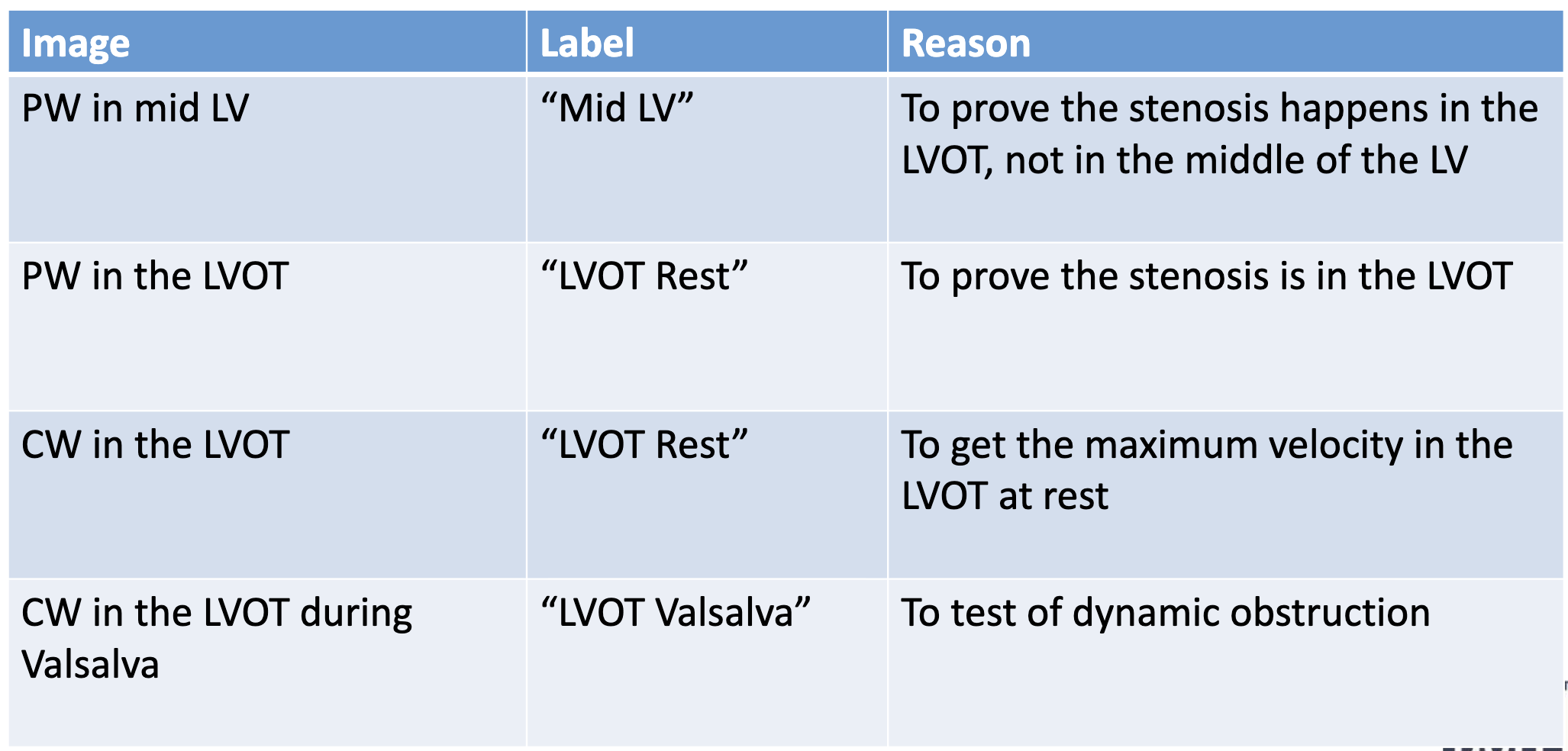
How to assess the aortic valve area in HOCM?
will not be able to use continuity equation because LVOT velocity will be elevated due to obstruction
have to visually assess AoV or perform valve planimetry from SAX
How to assess mechanism and degree of MR
SAM causes mitral regurgitation due to incomplete-coaptation/distortion of the MV leaflets in systole
mechanism for MR: Eject → Obstruct → Leak
MR jet is often posteriorly directed due to misplacement of anterior mitral valve leaflet
How to differentiate Doppler signal is MR or LVOT?
LVOT:
dagger shaped
occurs later in systole than MR
often lower velocity
MR:
round shape
occurs earlier in systole than LVOT obstruction
often higher velocity
How to diagnose HCM by definition? What are other causes of LVH (5)?
must be no known cause for hypertrophy
other causes of LVH:
Aortic stenosis
coarctation of aorta
Fabry’s disease (fatty tissue deposits in heart)
Hypertension
Infiltrative disease (amyloidosis)
What are medical treatment options for patients with hypertrophic cardiomyopathy (3)
Beta blockers
decrease oxygen demand by slowing HR and force of contraction
decrease LVOT gradient during exercise by decreasing the force of contraction
increase passive diastolic ventricular filling time due to slower HR
decrease the frequency of PVCs
Calcium Channel Antagonists
decrease stiffness and improve exercise tolerance in those who cannot tolerate beta blockers
Anti-arrhythmic Drugs
atrial fibrillation is not tolerated well in patients with HCM
What are surgical treatment options for patients with hypertrophic cardiomyopathy (3)
Septal Myectomy
surgical removal of the thickened septum, which widens the LVOT and eliminates the LVOT obstruction
Alcohol Septal Ablation
a small amount of alcohol is injected into a major septal perforator branch of the LAD, causing an MI in the basal IVS
the IVS becomes akinetic scars, and over time the wall will thin
this increases the LVOT area and decrease the severity of obstruction
Implantable Cardiac Defibrillator
an ICD is a pacemaker than can recognize dangerous heart rhythms and “shock” the patient back into normal sinus rhythm when needed
Define Arrhythmogenic Right Ventricular Cardiomyopathy (ARVC) (5)
aka arrhythmogenic right ventricular dysplasia (ARVD)
inherited in an autosomal dominant pattern
characterized by fibro fatty replacement of RV myocardium
usually right but can be left
myocytes are replaced with scar and fat tissue, leaving portions of heart with areas that do not conduct electricity or contract normally
fatty areas develop in small sections of the RV, but over time spread throughout the entire RV, and can even spread into the LV in severe cases
Describe etiology of ARVC (3)
Ventricular arrhythmias
heart failure
sudden cardiac death
Describe patient signs and symptoms of ARVC
arrhythmias ranging from PVC’s to sustained VT and ventricular fibrillation
requisitions of frequent PVCs → always assess for ARVC (esp in younger patients)
sudden cardiac death (due to arrhythmias)
patients usually present in their 20s, 30s, or 40s
Role of Sonographer in ARVC
Help with the diagnosis of ARVC
Assessment of RV function
Assessment of LV systolic function
Evaluation of TR severity
Estimate the RVSP
How to help with the diagnose of ARVC
biopsy can confirm, but sometimes false negatives due to segmental progression of disease
on echo, assess multiple views and windows and look for:
wide spectrum of RV involvement
global or regional RV dysfunction
RV structural abnormalities, wall thinning, RV aneurysm, RV dilation
highly reflective (echo-genic) moderator band
abnormal trabeculations
How to assess RV function, LV systolic function, TR severity and estimate the RVSP in ARVC
Assessment of RV function
TAPSE/TDI/Visual
more than 1 measurement for RV function should be performed
Assessment of LV systolic function
in severe cases of ARVC, fatty tissue can infiltrate LV
Evaluation of TR severity
TR occurs secondary to dilation of RV
Estimate the RVSP
in cases of “wide open” TR, TR velocity is usually low and the RVSP cannot be accurately estimated
RAP is unknown
Describe treatment options for ARVC (2)
pharmacologic therapy (anti-arrhythmic drugs)
implantable cardioverter-defibrillator (ICD)
Define LV non-compaction
“spongy myocardium” → things protruding out
characterized by 2 layered myocardium:
thin, compacted layer and an extensive non-compacted layer, with prominent trabeculations and deep recesses that communicate with left ventricular cavity but not with coronary circulation
caused by arrest of normal embryogensis of the endocardium and myocardium
Describe etiology of LV non-compaction
rare congenital abnormality
possibly due to failure of normal trabecular resorption or compaction during embryonic development, resulting in double layered LV myocardium
Describe signs and symptoms of LV non-compaction (4)
heart failure
arrhythmias (atrial fibrillation/ ventricular tachyarrhythmia)
endocardial clot with systemic embolization
sudden death
Role of Sonographer in LV non-compaction
Diagnose presence of LV non-compaction
Assess diastolic dysfunction
Assess systolic dysfunction and arrhythmia
How to determine diagnosis on LV non-compaction
more than three trabeculations protruding from LV free wall, visible in a single image plane
blood flow from the LV cavity into the intertrabecular recesses as seen with colour Doppler
ultrasound contrast may be used to help visualize trabeculations and deep recesses
two-layered myocardium, with thin outer compacted layer and a thickened inner non-compacted layer
Differential from: apical HCM, endomyocardial fibrosis, apical LV mass/thrombus
How are diastolic and systolic dysfunction caused from LV non-compaction
Diastolic dysfunction:
due to abnormal relaxation caused by the numerous prominent trabeculae
Systolic dysfunction and arrhythmias:
possibly due to sub-endocardial hypo-perfusion and micro-circulatory dysfunction
The inner heart muscle isn’t getting enough blood because the tiny vessels aren’t delivering it properly.
non compacted myocardial segments have decreased contractility, therefore LV EF is decreased
Describe treatment options for LV non-compaction (4)
pharmacologic therapy (anticoagulation, anti-arrhythmic drugs)
implantable cardioverter-defribillator (ICD)
heart rhythms
Ventricular assist device
blood circulations
cardiac transplant
Define atrial septal defects
defect in the tissue of interatrial septum that allows blood to flow directly from one atrium to the other
blood flow direction is determined by pressure gradient between the atria
What are the types of atrial septal defects (5)
Ostium secundum ASD
Ostium primum ASD
Sinus venous ASD
Unroofed coronary sinus
Patent foramen ovale (not true ASD)
Describe ostium secundum ASD (where, vs PFO, treatment)
most common, 70% of ASD
occurs in area of fossa ovalis
differentiated from a PFO by:
absence of valve/ flap of tissue of foramen ovale
presence of continuous flow (PFO is intermittent)
bigger than PFO
often closed percutaneously with closure device in cath lab, between opening
Describe ostium primum ASD (where, CHD (3), treatment)
20% of all ASD
located adjacent to atrioventricular valves (TV and MV), at the crux of heart
associated with other congenital heart diseases → atrioventricular septal defects (inlet VSD), with cleft mitral valve and Trisomy 21 (down syndrome)
surgery treatment bc cannot be treated with percutaneous device
not enough tissue to attach patch bc too close to AV valves and AV node
Describe sinus venous ASD
uncommon, 7% of ASD
located near SVC or IVC
associated with congenital diseases, with partial anomalous pulmonary vein return
best seen in bicaval view (see SVC and IVC)
Describe unroofed coronary sinus (associated w/ …)
rare, aka coronary sinus defect
not true ASD bc not IAS → but similar hemodynamic consequences
complete or partial unrooting of coronary sinus with direction communication to left atrium
because LAP > RAP → blood in LA enters CS tube and will lead to RA
will reverse if RAP > LAP
almost always associated with connection of a persistent left SVC to coronary sinus
What are some clues for unroofed coronary sinus (5), seen best in ?
better seen in TEE
unknown cardiac murmur
right sided heart enlargement
dilated coronary sinus → biggest clue
transient cyanosis or hypoxia
stroke
Describe patent foramen ovale
not true ASD bc not a hole, has a flap
25% of adults
foramen ovale should functionally close post-natally when LA P > RA P→ should seal over and remain shut
some adults, foramen does not shut and remains as “flap”
can open when RA P > LA P
no hemodynamic consequences expected with PFO, except increased risk of stroke/embolus