0. Glaucoma Exam 2, Photos
 Call Kai
 Learn
 Practice Test
 Spaced Repetition
 Match
 Flashcards
 Knowt Play
1/78

There's no tags or description
Looks like no tags are added yet.
79 Terms
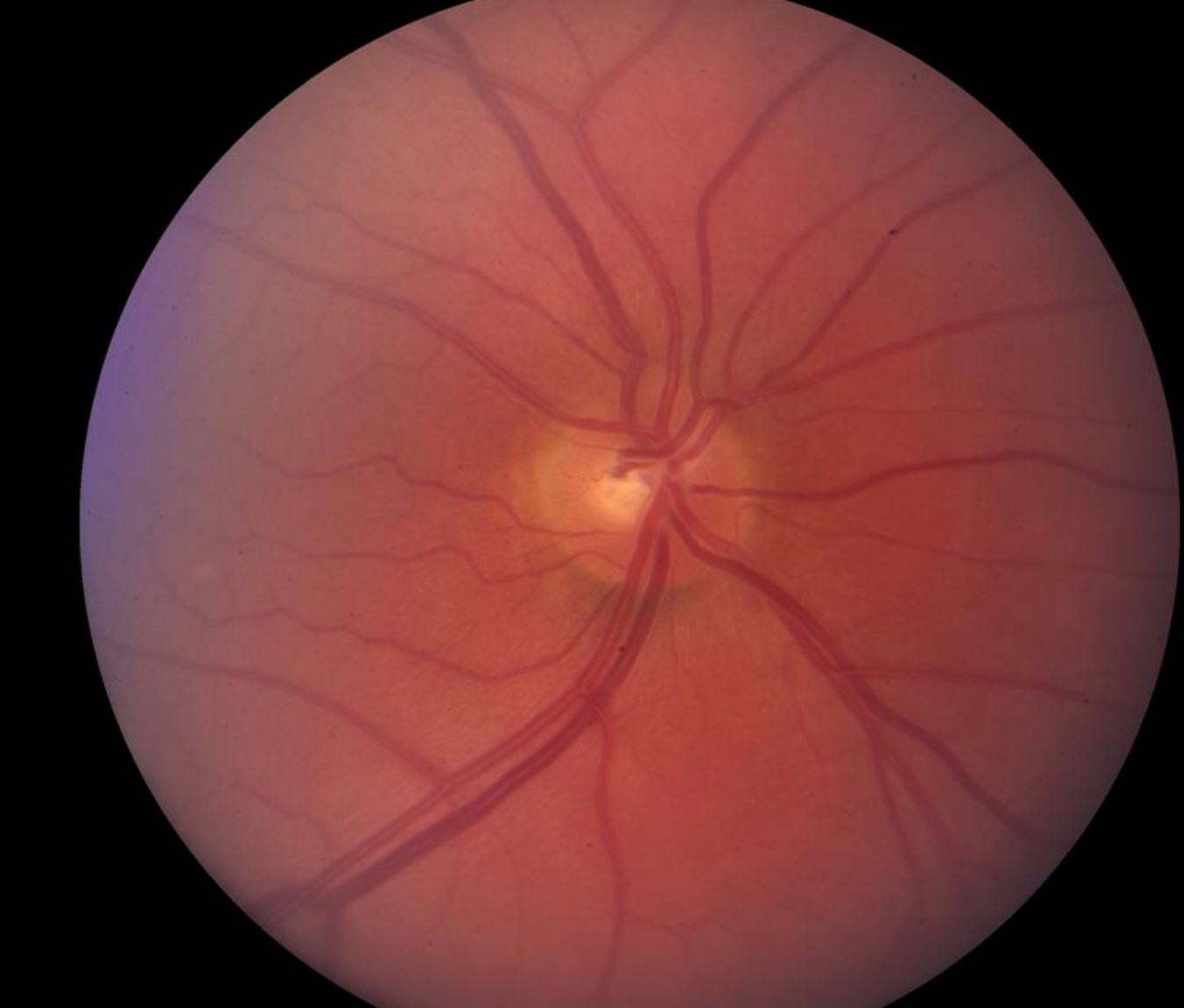
grade the C/D
0.3

grade the C/D
0.5

grade the C/D
0.5

grade the C/D
0.25

grade the C/D
0.8

describe the finding:
blurred margins


describe the finding:
missing rim tissue


finding?
RNFL / GCA Thinning

finding?
RNFL / GCA Thinning
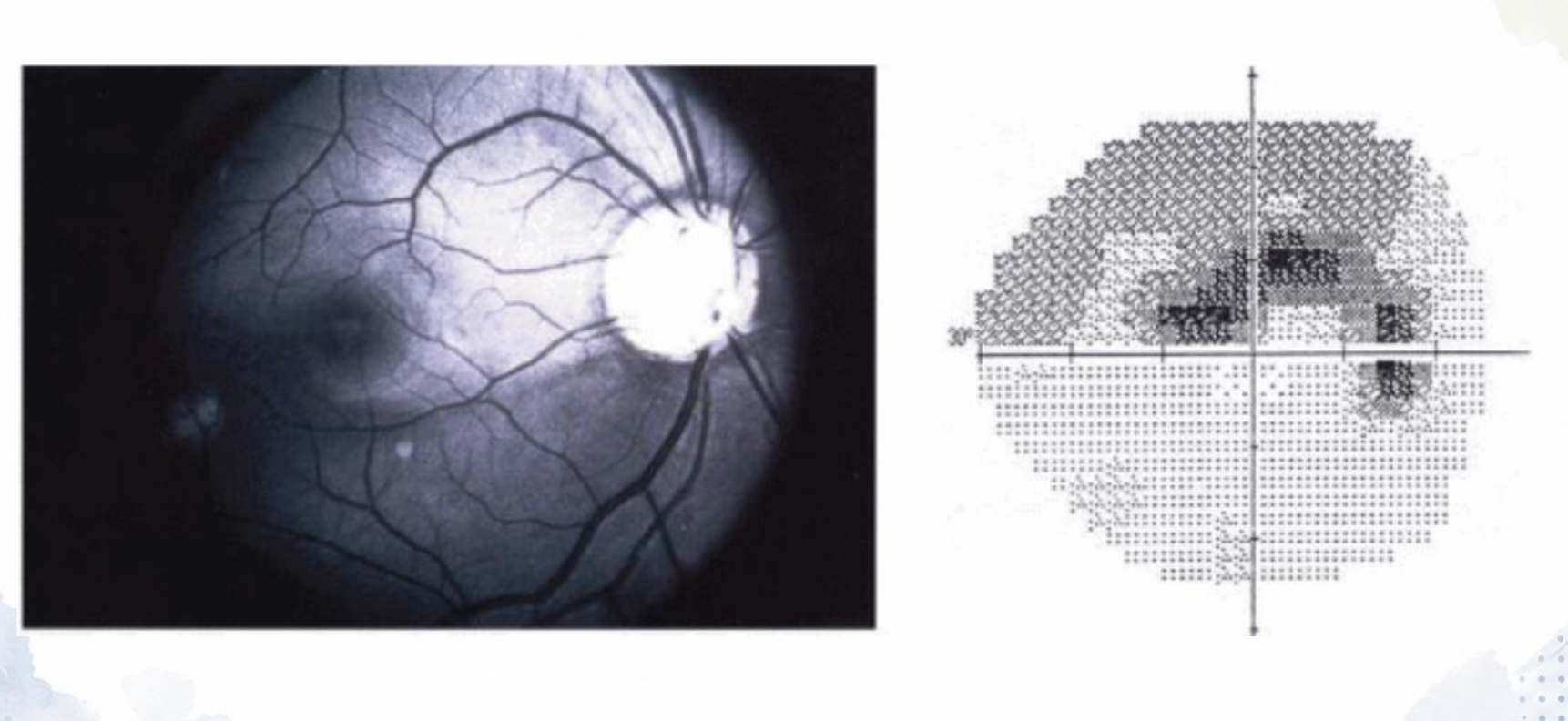
finding?
RNFL Thinning

finding?
thinning of parapapillary RNFL

finding?
thinning of parapapillary RNFL

findings? (2)
RNFL loss,
Drance

finding?
notching

finding?
notching

finding?
narrowing of optic disc rim

finding?
cupping of the optic disc

finding?
cupping of the optic disc

finding?
beta-zone peripapillary atrophy

finding?
circumlinear barging

finding?
circumlinear baring

finding?
bayonetting of vessels

finding?
bayonetting of vessels

finding?
nasalization of the vessels

finding?
nasalization of the vessels

finding?
visible lamina cribrosa

finding?
drance hemorrhage

finding?
Drance Hemorrhage

finding?
Drance Hemorrhage

Using FORGE, how would you evaluate this ON?
C/D: 0.65/0.75,
S & I rim thinning,
local RNFL defect @ 5:00,
Beta Zone PPA 3:00 - 5:00,
(-) disc heme


Using FORGE, how would you evaluate this ON?
C/D: 0.75/0.75,
Follows ISNT,
(-) RNFL defects,
(-) Beta Zone atrophy,
(-) disc heme


finding? normal or abnormal?
iris processes; normal

What structure is identified by the corneal wedge?
Schwalbe’s line

5?
Schwalbe’s Line

4?
Non-pigmented trabecular meshwork

3?
Pigmented trabecular meshwork

2?
scleral spur

1?
ciliary body

grade iris insertion using Spaeth
A

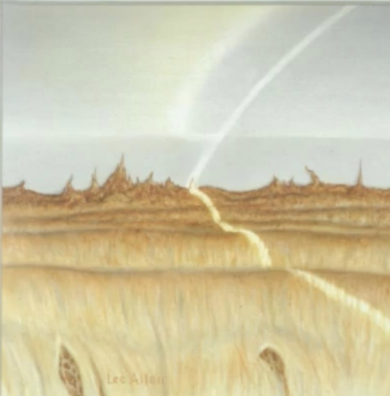
grade iris insertion using Spaeth
B


grade iris insertion using Spaeth
C


grade iris insertion using Spaeth
D


grade iris insertion using Spaeth
E


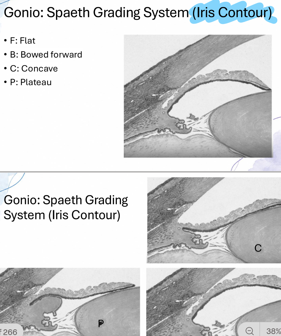
describe the iris contour using Spaeth grading
P


describe the iris contour using Spaeth grading
C


describe the iris contour using Spaeth grading
B

Grade the angle using Spaeth Grading:
D50c0

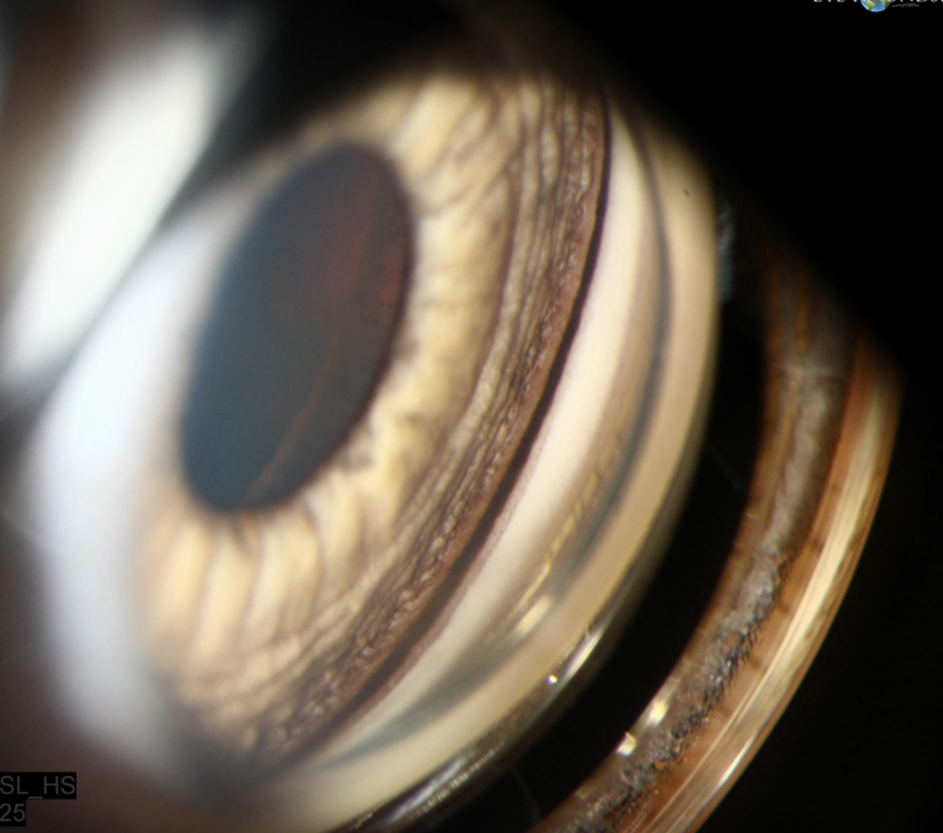
Grade the angle using Spaeth Grading:
D45f4+p


finding? normal or abnormal?
iris processes; normal
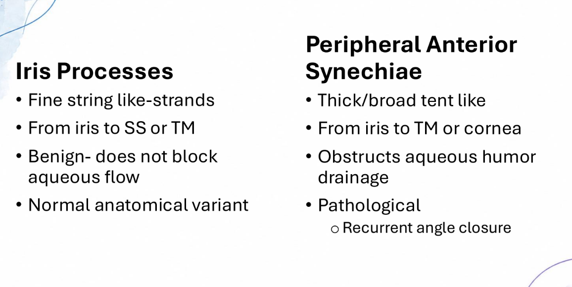

finding? normal or abnormal?
peripheral anterior synechiae; abnormal


what is the most posterior structure visible?
anterior trabecular meshwork

what is the most posterior structure visible?
Schwalbe’s Line

finding? normal or abnormal?
greater circle of the iris; normal

finding? normal or abnormal?
neovascularization of the iris
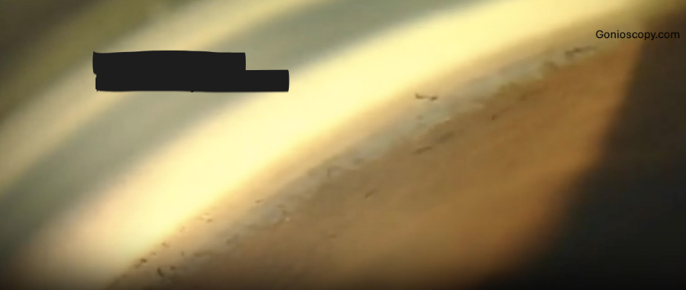
diagnosis?
angle recession

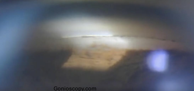
Finding? normal or abnormal?
posterior embryotoxon; normal (8-12% of population) or associated with Axenfeld Rieger syndrome

Finding? normal or abnormal?
peripheral anterior synechiae; abnormal

Finding? normal or abnormal?
pseudoexfoliation syndrome; abnormal

Where should you expect the site of damage to be located?
ONH @ 4:30


Where should you expect the site of damage to be located?
ONH @ 4:30


Where should you expect the site of damage to be located?
ONH @ 12:30


grade the severity of the glaucoma
moderate
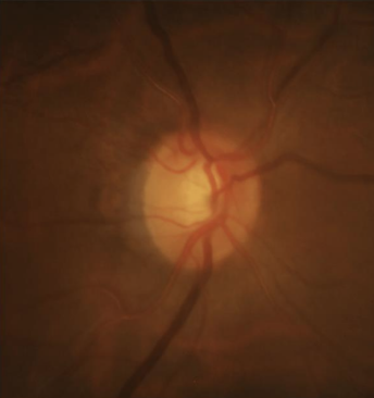
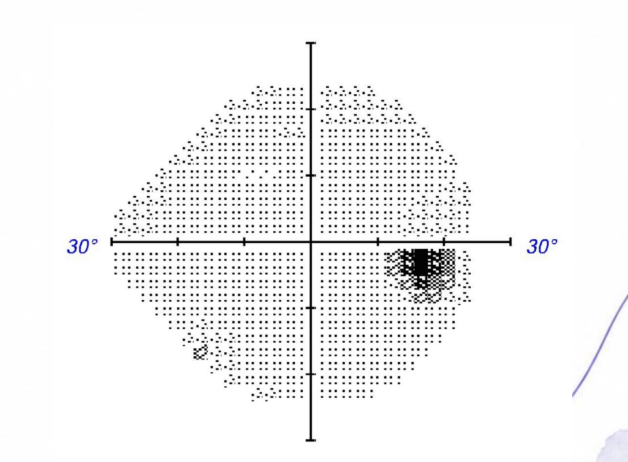
(+) RNFL defect
grade the severity of the glaucoma based on ICD-10
mild

grade the severity of the glaucoma based on ICD-10
severe
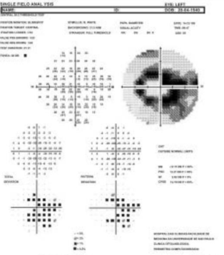
grade the severity of the glaucoma based on ICD-10
severe
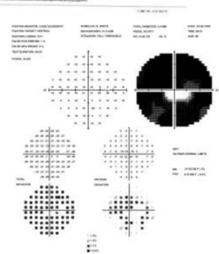
grade the severity of the glaucoma based on ICD-10
severe
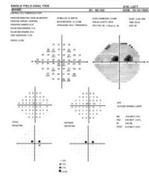
grade the severity of the glaucoma based on ICD-10
severe

class of drop?
prostaglandin analog
class of drop?
beta-blocker
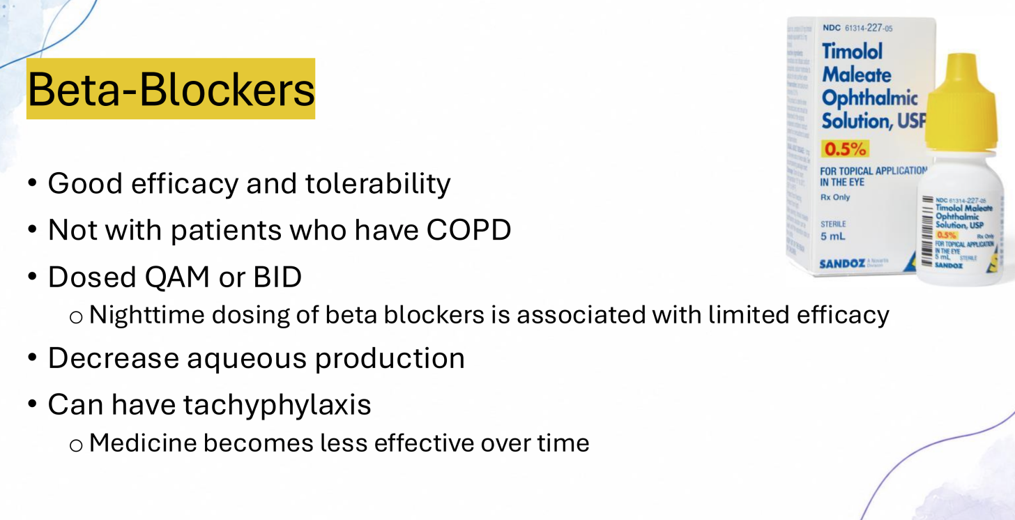

Class of drop?
alpha-2 adrenergic agonist

class of drop?
cholinergic agonist/ parasympathomimetic


class of drop?
carbonic anhydrase inhibitors


Plan?
RTC for glaucoma work up in 1 - 3 months (including OCT, VF, Pachy, Gonio, IOP)
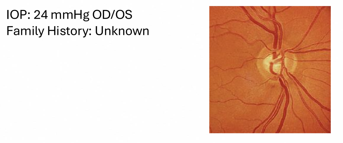

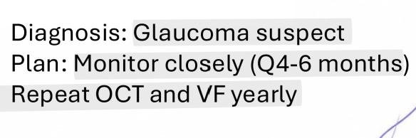
Diagnosis and Plan?
Glaucoma Suspect;
Monitor q 4-6 months + repeat OCT & field yearly


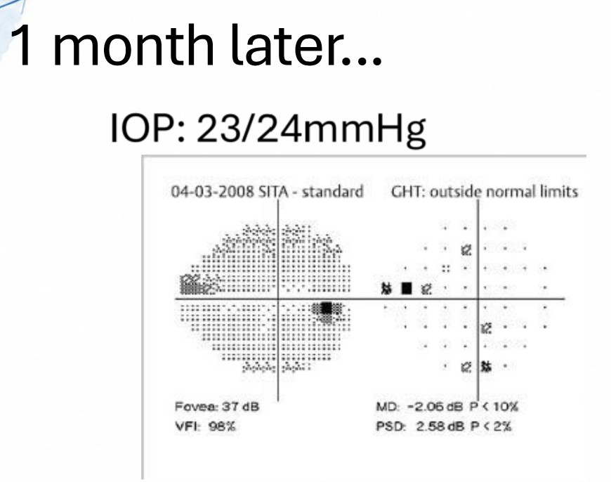
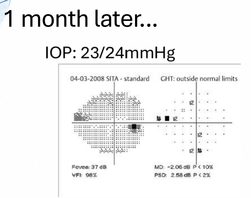
Plan?
RTC to repeat VF


Plan?
i) discuss options (SLT vs PGA)
ii) if they pick drops, start latanoprost QHS OU
iii) RTC in 6 weeks for IOP check


Plan?
continue to monitor q 4 months,
yearly VF & OCT


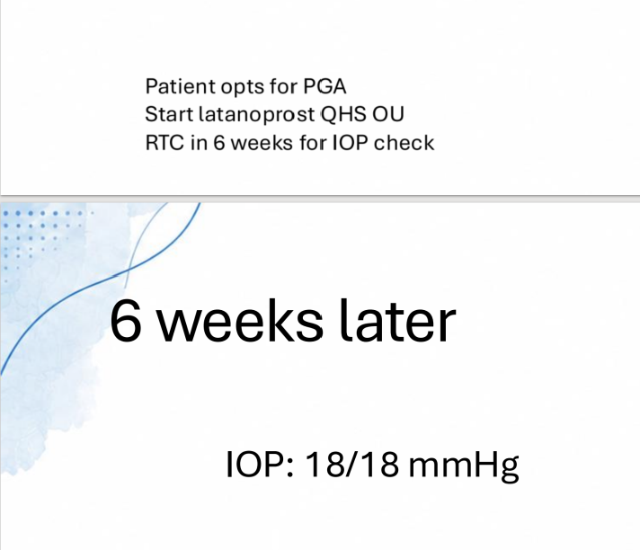

plan?
repeat VF/ confirm progression,
add additional treatment (ie: CosOpt BID OU),
RTC in 3-4 wks for IOP check

Gonio should be continued to be done how often?
yearly
