Anatomy Bone - Ex 9-11
 Call Kai
 Learn
Learn Practice Test
 Spaced Repetition
 Match
Match Flashcards
 Knowt Play
1/291

There's no tags or description
Looks like no tags are added yet.
Name | Mastery | Learn | Test | Matching | Spaced | Call with Kai | Chat |
|---|
No analytics yet
Send a link to your students to track their progress
292 Terms
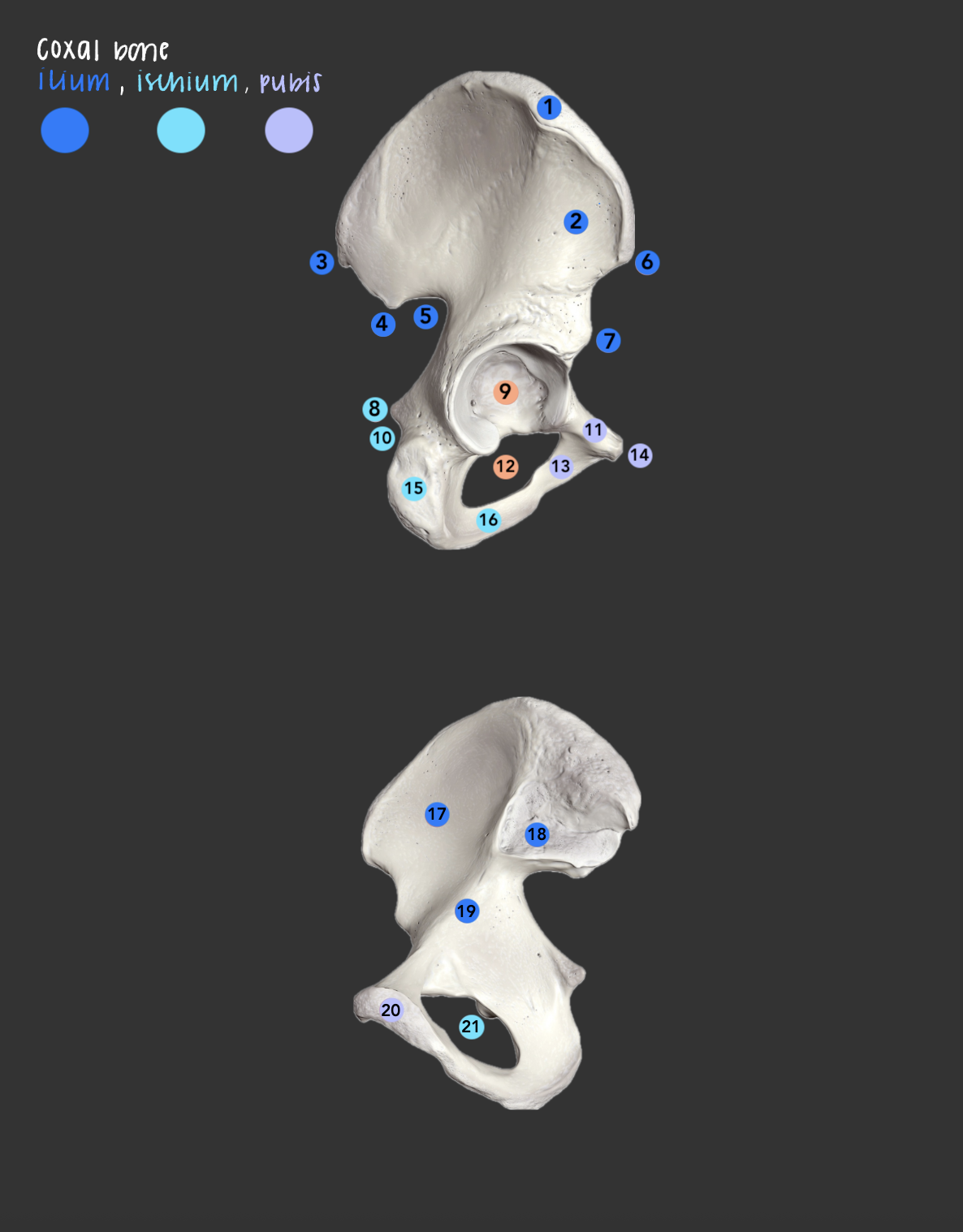
iliac crest (ilium)
what is 1?; superior ridge of the ilium; site to obtain donated bone marrow

ala (ilium)
what is 2?; (also called the wing of the ilium) is the large, upper, fan-shaped portion of the hip bone that forms the lateral wall of the false pelvis
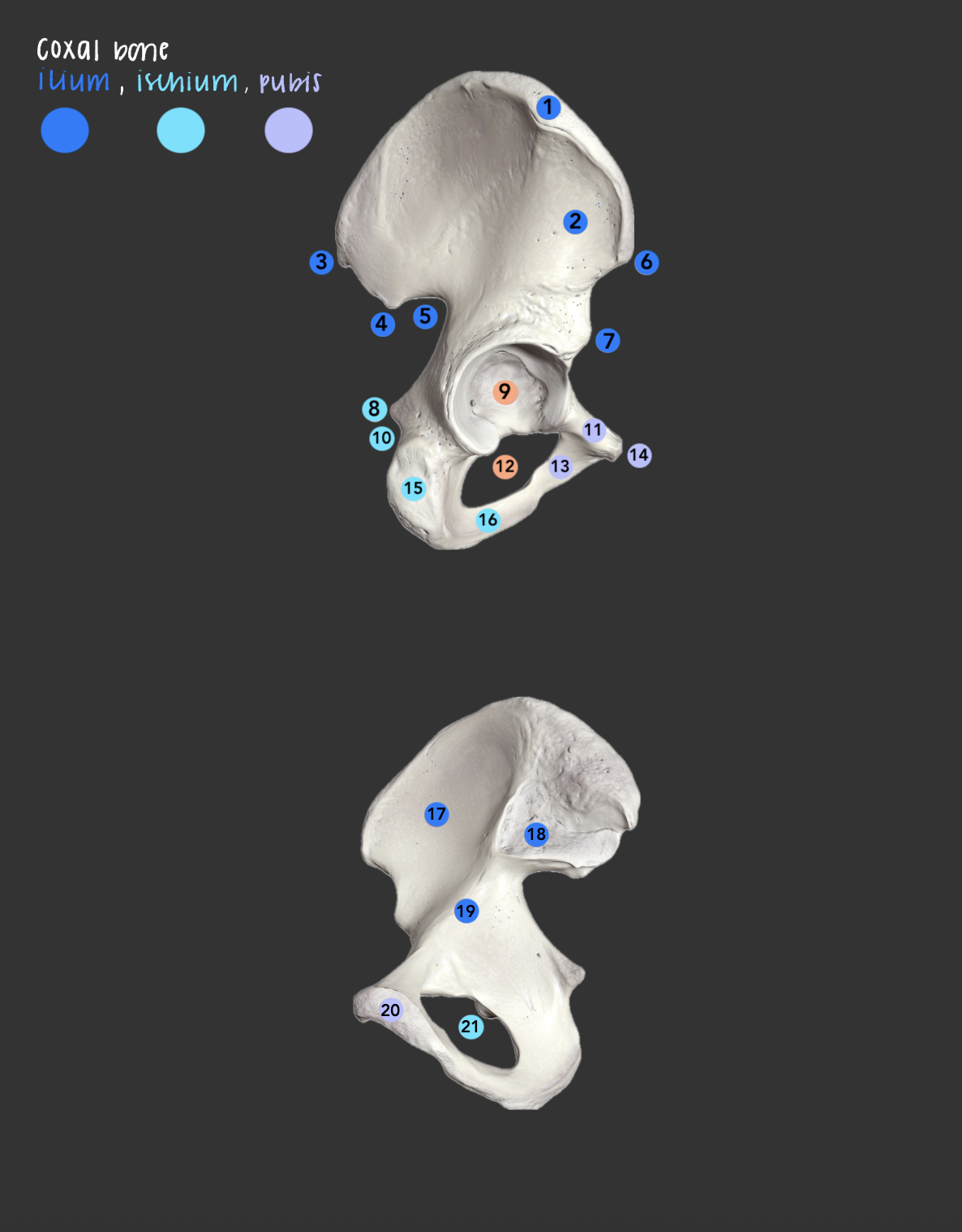
posterior superior iliac spine
what is 3?; posterior end of the iliac crest

posterior inferior iliac spine
what is 4?; projection below the PSIS on the posterior ilium
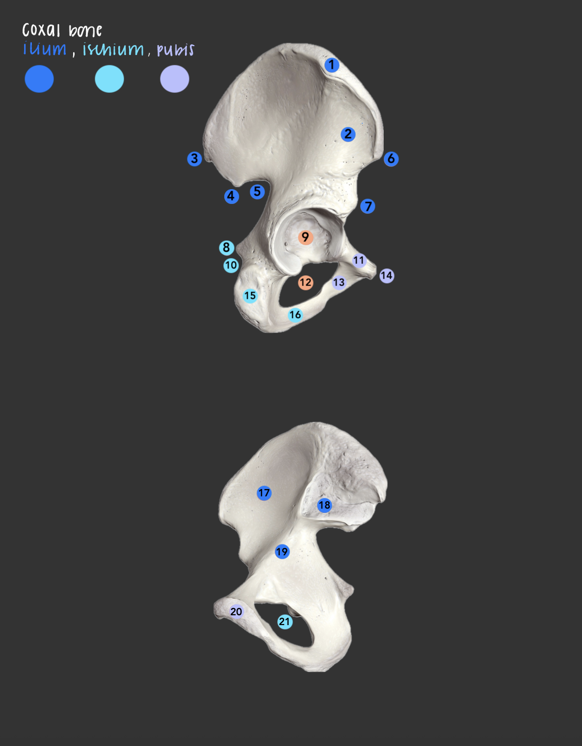
greater sciatic notch (ilium)
what is 5?; sciatic nerve passes through

anterior superior iliac spine
what is 6?; anterior end of the iliac crest

anterior inferior iliac spine
what is 7?; projection below the ASIS on the anterior ilium

ischial spine
what is 8?; a pointed bony projection on the back border of the ischium bone in the pelvis, obstetric landmark

acetabulum of coxal bone
what is 9?; socket formed by all three coxal parts; head of the femur articulates here

lesser sciatic notch (ischium)
what is 10?; notch below the ischial spine

superior ramus (pubis)
what is 11?; superior branch of pubis
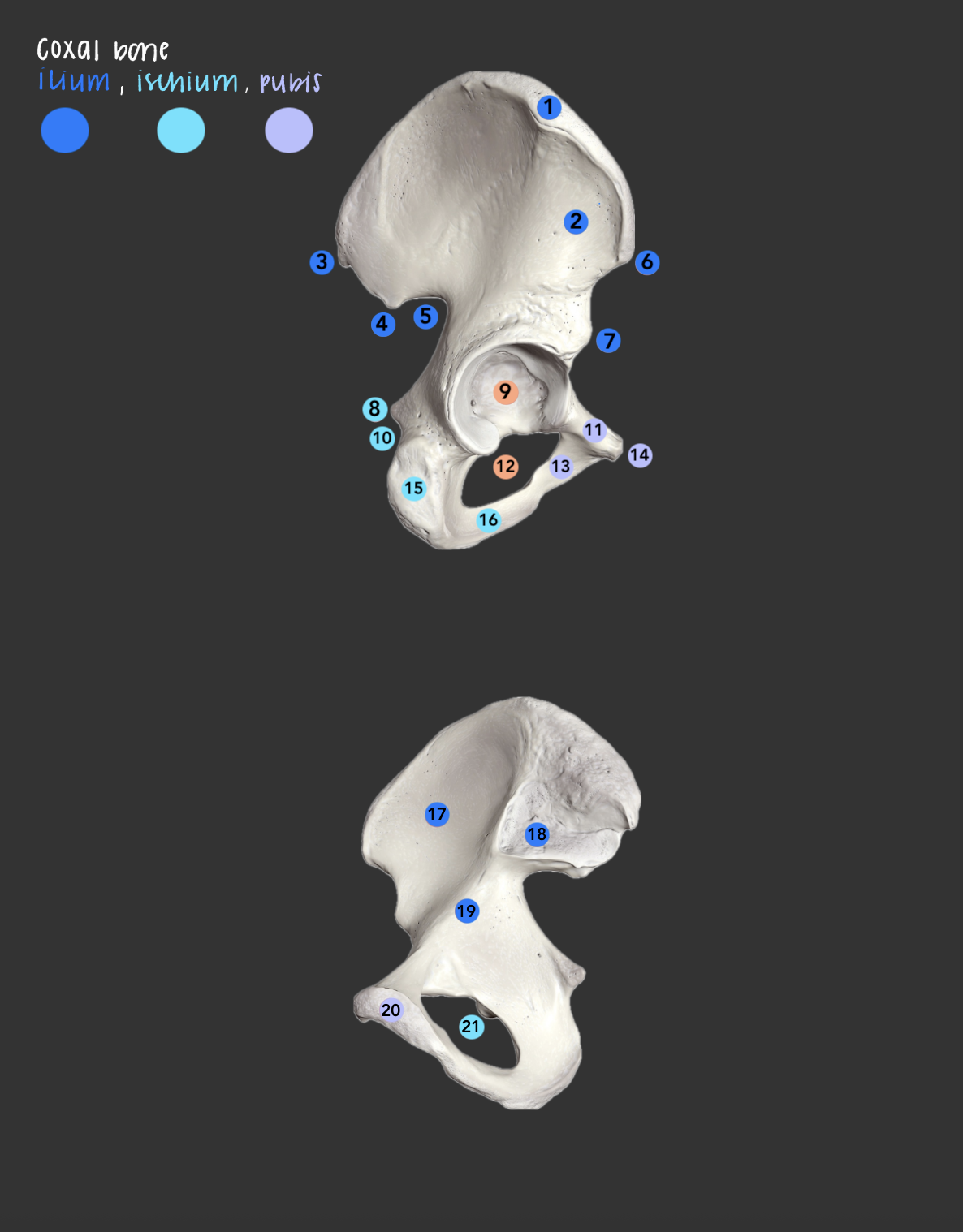
obturator foramen (ischium & pubis)
what is 12 & 21 (same structure)?; large opening formed by only the pubis and ischium
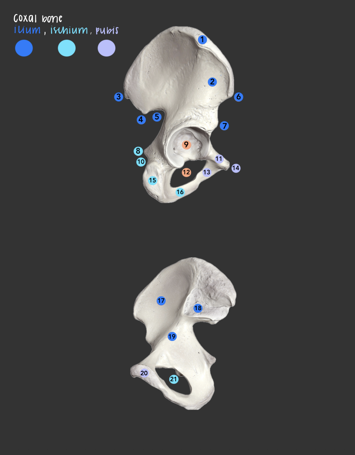
inferior ramus (pubis)
what is 13?; inferior branch of the pubis

pubic tubercle
what is 14?; small bump on the superior ramus near the pubic symphysis

ischial tuberosity
what is 15?; obstetric landmark, a pointed bony projection on the back border of the ischium bone in the pelvis

ischial ramus
what is 16?; part of the ischium that joins the inferior ramus of the pubis
iliac fossa
what is 17?; a large, smooth, and concave depression on the inner surface of the hip bone
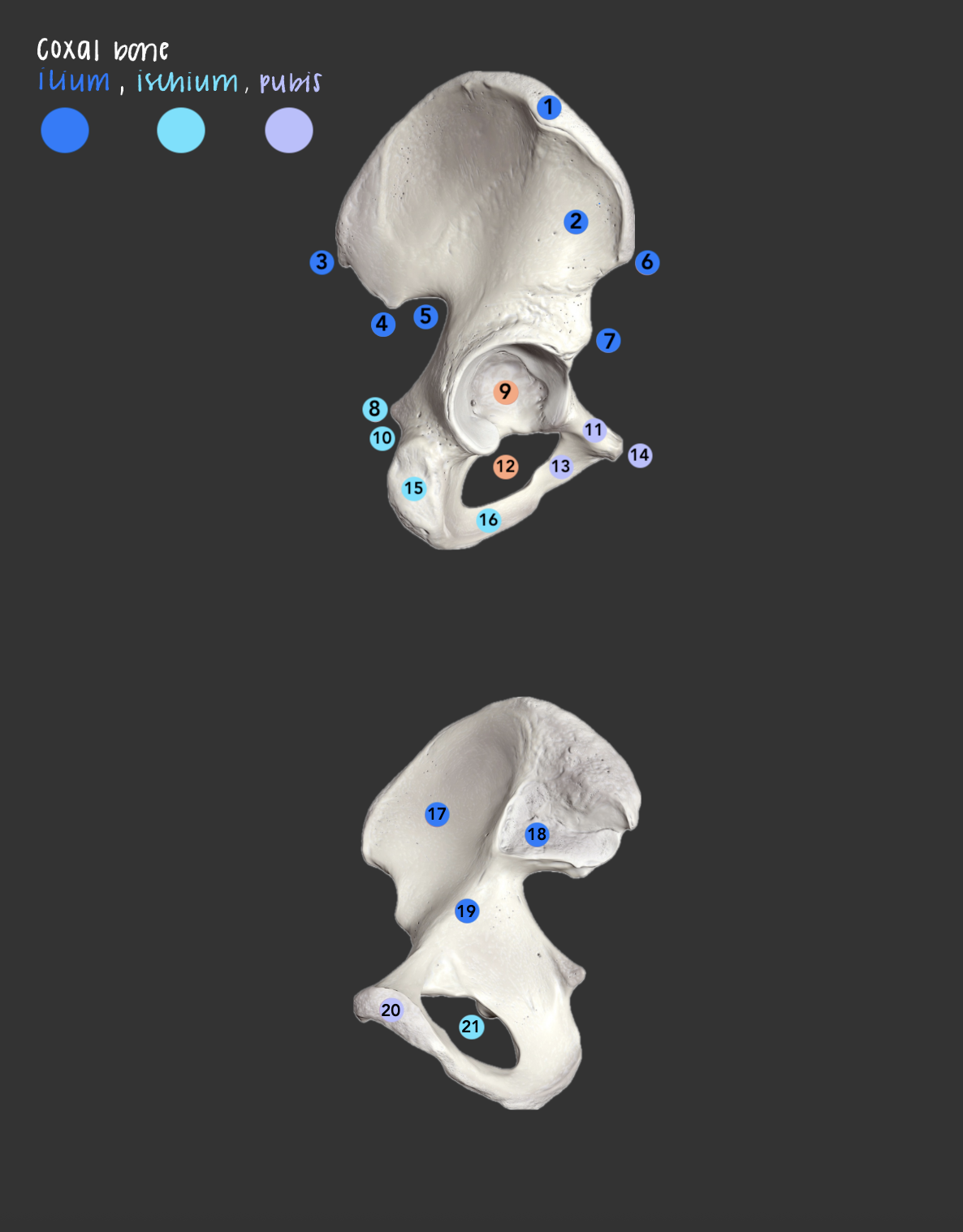
auricular surface (ilium)
what is 18?; ear-shaped articular area on the inner (medial) surface of the hip bone that connects with the sacrum to form the sacroiliac joint

arcuate line (ilium)
what is 19?; ridge on the inner ilium forming part of the pelvic brim

articular surface (pubic symphysis)
what is 20?; where the two pubic bones meet at the midline

frontal bone
makes up the forehead and upper part of eye sockets

supraorbital margin
superior rim of orbit (eyes)

supraorbital foramen (notch)
opening in the supraorbital margin for the supraorbital nerves & vessels

glabella
smooth area b/w the eyebrows

frontal sinus
What is the Blue region ? (cavity in frontal bone above eyes)

parietal bone
top of the head

coronal suture
joins frontal & parietal bones

squamous suture
joins parietal & temporal bones

lambdoid suture
joins parietal & occipital bones

sagittal suture

occipital bone
back of head

external occipital protuberance
midline bump on the back of the skull

external occipital crest
midline ridge running from protuberance toward the foramen magnum

superior nuchal line
ridge extending laterally from protuberance (attachment for neck muscle)

inferior nuchal line
ridge below the superior nuchal line (attachment for neck muscle)

occipital condyle
articulates w/ C1 (atlas)
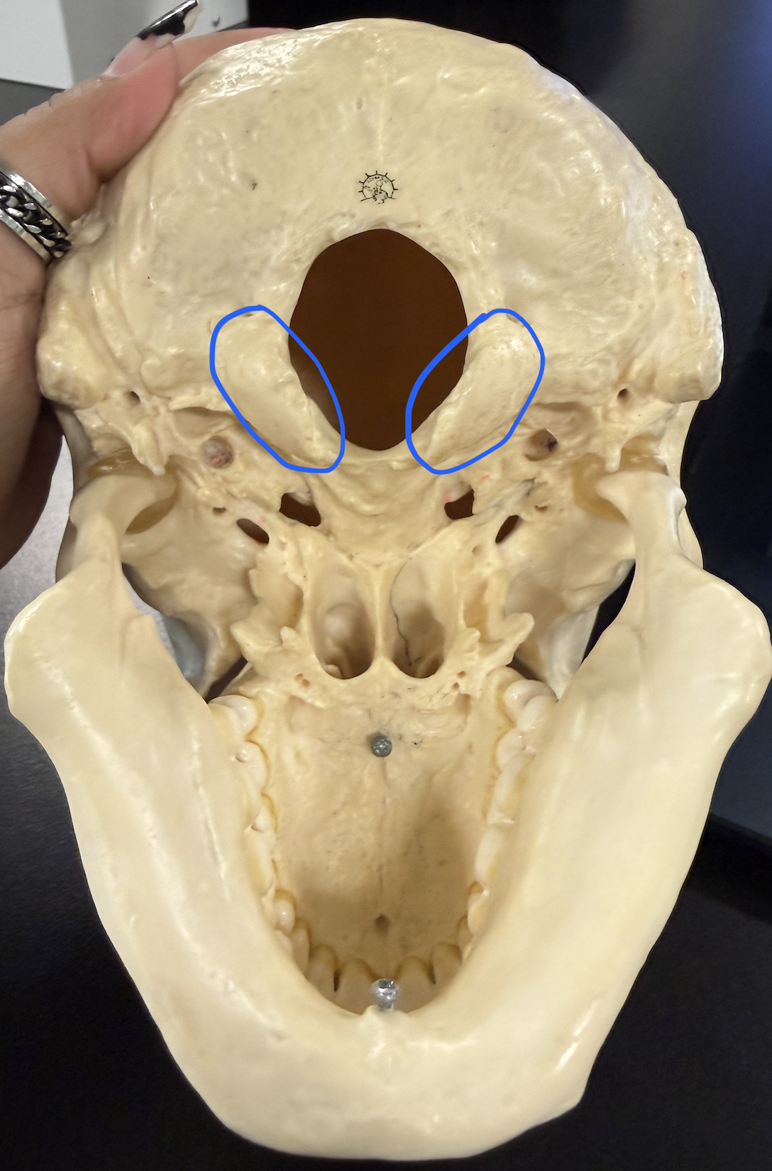
foramen magnum
large opening where the spinal cord leaves the skull

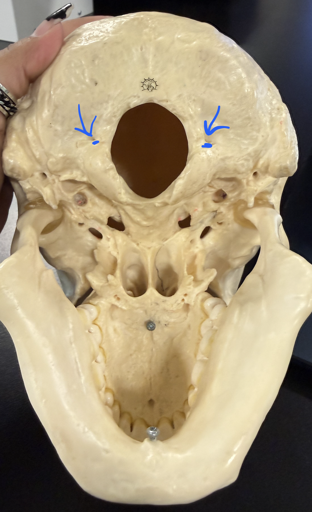
hypoglossal canal
for hypoglossal nerve; under occipital condyles (need to tilt to be visible)
temporal bone
sides of head

squamous region
thin, flat, platelike part of the temporal bone

zygomatic process
projects forward to help form zygomatic arch

mandibular fossa
articulates w/ mandibular condyle of the mandible

external acoustic meatus
ear canal opening

styloid process
slender spike below the ear canal

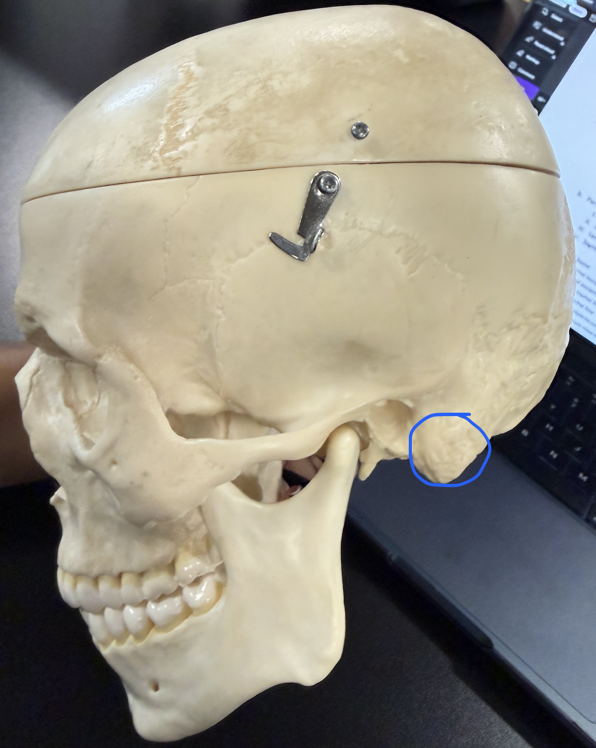
mastoid process
middle ear infection can spread here (mastoiditis), then to meninges (meningitis); sternocleidomastoid attaches here
jugular foramen
internal jugular vein, CN XI, CN X, CN IX

internal acoustic meatus
CN VIII , CN VII

foramen lacerum
jagged opening at base of the skull b/w the temporal, sphenoid and occipital bones

carotid canal
internal carotid artery

sphenoid bone
complex, butterfly-shaped bone located at the middle base of the skull

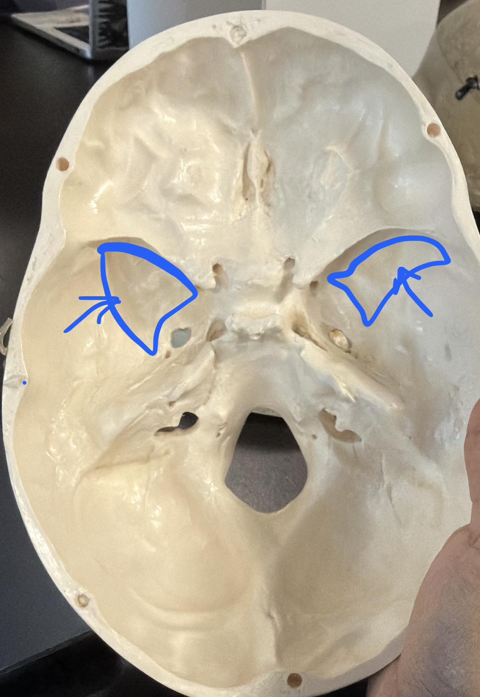
lesser wings
thin wings forming part of the anterior cranial floor and eyes

greater wings
large lateral wings forming part of the cranial floor and eyes
optic canal
optic nerve (CN II)

superior orbital fissure
CN III, CN IV, CN VI, ophthalmic branch of CN V

foramen rotundum
maxillary branch of trigeminal nerve (CN V)

foramen ovale
mandibular branch of trigeminal nerve (CN V)

foramen spinosum
smaller opening in the greater wing, posterior and lateral to foramen ovale

sella turcica
saddle-shaped depression; contains hypophyseal fossa
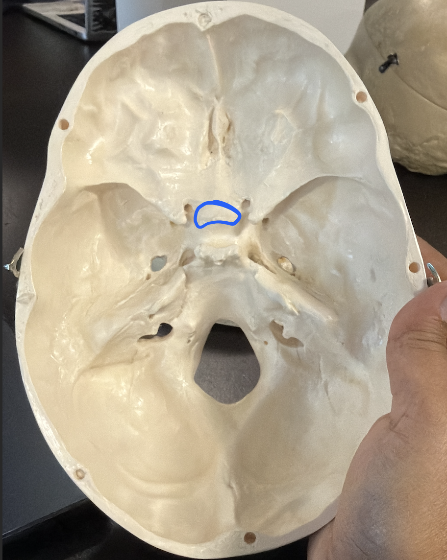
anterior clinoid processes
anterior projections of the sella turcica region
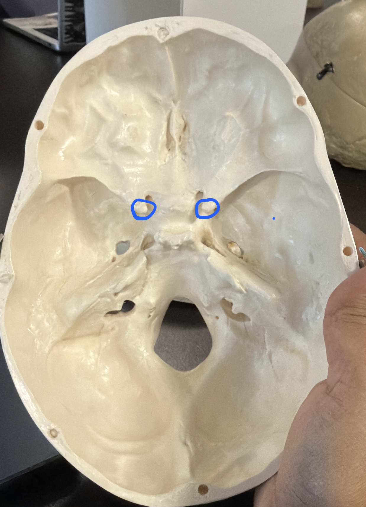
posterior clinoid processes
posterior projections flanking the sella turcica

tuberculum sellae
ridge forming the anterior edge of the sella turcica

dorsum sellae
ridge forming the posterior wall of the sella turcica

hypophyseal fossa
pituitary gland (hypophysis)

pterygoid processes
downward projections from the sphenoid; each has a lateral and medial plate

lateral plates (pterygoid)
_______ plate of the pterygoid process

medial plates (pterygoid)
______ plate of the pterygoid process

sphenoid body
central part of the sphenoid; houses the sphenoidal sinus
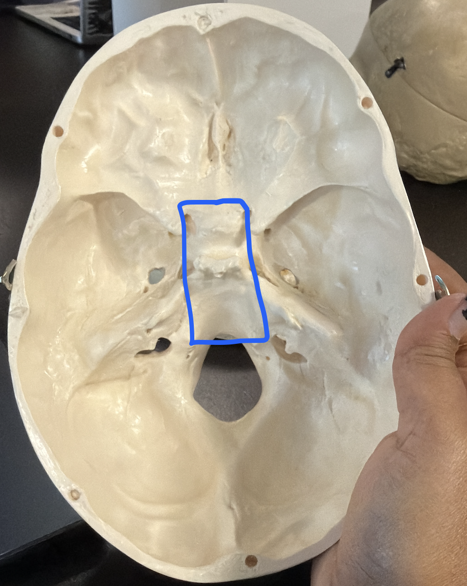
sphenoidal sinus
What is the Yellow region ? (cavity within the sphenoid body)

ethmoid bone
cube-shaped bone in the skull that separates the nasal cavity from the brain and helps form the eye sockets

crista galli
site of attachment of the dura mater (the bump that protruding out of the plate)

cribriform plate
The blue star (perforated plate forming part of the roof of the nasal cavity)

olfactory foramina
olfactory nerves (CN I)

lateral masses
side portions of the ethmoid; contain the ethmoidal sinuses

ethmoidal sinus
What is the Green region ? (air cells within the lateral masses)

superior nasal concha
upper scroll-like projection in the nasal cavity

middle nasal concha
middle scroll-like projection in the nasal cavity

perpendicular plate
forms the upper half of the bony nasal septum

mandibular condyle
articulates w/ the mandibular fossa of the temporal bone

mandibular notch
concave notch b/w the condylar and coronoid processes

coronoid process
anterior projection of the ramus

mandibular ramus
masseter attaches here; spastic masseter is an early sign of tetanus; parotid gland sits superficial to the masseter (mumps = parotid infection)

mandibular angle
corner where the ramus meets the body

mandibular body
horizontal, curved part of the mandible holding the teeth

mental foramen
opening on the anterior body of the mandible (chin)

alveolar margin
ridge of the mandible containing the tooth sockets

mandibular foramen
mandibular branch of CN V; where dentists inject Novocain for lower teeth

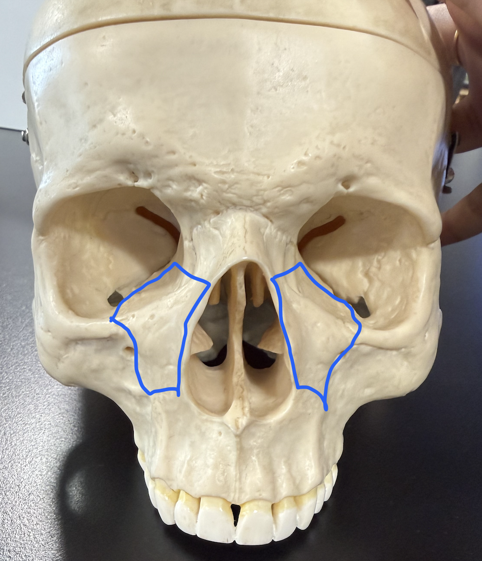
frontal process (of maxillary)
w/ lacrimal bone, forms lacrimal fossa (lacrimal sac sits here)

inferomedial orbital margin
lower medial rim of the orbit
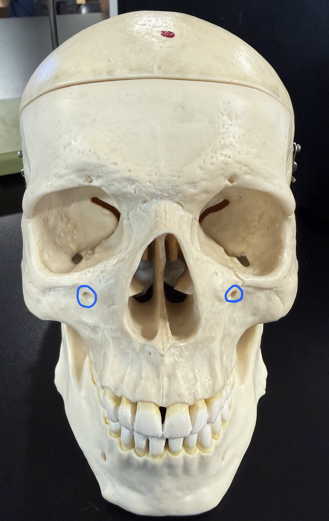
infraorbital foramen
opening below the orbit
zygomatic process
articulates w/ zygomatic bone

alveolar margin
ridge containing the upper tooth sockets

palatine process
forms the anterior 2/3 of the hard plate

incisive fossa
depression behind the incisors on the hard palate

maxillary sinus
What is the Pink region ? (largest paranasal sinus, within the maxilla)

inferolateral orbital margin
lower lateral rim of the orbit

temporal process
joins the zygomatic process of the temporal bone (zygomatic arch)

frontal process (of zygomatic)
articulates w/ the frontal bone

maxillary process (of zygomatic)
articulates w/ the maxilla

nasal bones
small paired bones forming the bridge of the nose

lacrimal fossa
with the frontal process of the maxilla, seats the lacrimal sac
