Neuroanatomy: structure and function
 Call Kai
 Learn
Learn Practice Test
 Spaced Repetition
 Match
Match Flashcards
 Knowt Play
1/84

There's no tags or description
Looks like no tags are added yet.
Name | Mastery | Learn | Test | Matching | Spaced | Call with Kai | Chat |
|---|
No analytics yet
Send a link to your students to track their progress
85 Terms
Rostral
toward the beak or nose; points to the front (anterior) of the brain
ex) the frontal lobe is rostral (anterior) compared to the occipital lobe
Caudal
toward the tail; points to the back (posterior) of the brain
ex) the occipital lobe is caudal (and posterior) to compared to the frontal lobe
Dorsal
toward the back; points to the top of the human brain
ex) the cerebral cortex sits dorsal (superior) to the brainstem
Ventral
toward the belly; points to the bottom (base) of the human brain
ex) the hypothalamus sits ventral (inferior) to the thalamus
Anterior/Posterior
synonymous with front and back of the head/brain in standard orientation
Medial/lateral
toward the middle (midline) or toward the outer edges
Why the meaning shifts between the brain and spinal cord?
Because of the upright posture of humans and the 90-degree ben (cephalic flexure) that occurs at the junction of the brainstem and forebrain
Central Nervous System (what it includes/functions)
consists of the brain and spinal cord; integrates and processes information, generates thoughts emotions and memories, initiates motor commands (processing)
Peripheral Nervous System (PNS)
cranial nerves, spinal nerves, ganglia; connects CNS to body, carries sensory input to CNS (afferent), carries motor output to muscles/glands (efferent) (detection)
Somatic
largely under voluntary control and involves a single neuron pathway from spinal cord to muscle; somatic reflex arc forces you to jump or flinch (subdivision of PNS)
Autonomic
controls involuntary, automatic functions (subdivision of PNS)
Sympathetic
(“fight or flight”) - mobilizes the body for action/stress (branch of autonomic nervous system)
Parasympathetic
(rest and digest) - conserves energy, promotes recovery
Explain how the central, peripheral, somatic, and autonomic divisions work together during an everyday event, such as reacting to a sudden loud noise
Sound waves hit your ears, activating sensory neurons within the PNS, the neurons instantly rush an electrical signal up toward your spine and brain, the auditory cortex in the CNS processes the sound, while the amygdala (brains alarm system) evaluates it as a threat, CNS sends split second orders back down through the PNS to initiate two types of responses (voluntary/internal survival tactics), before your brain fully comprehends what the noise was, a somatic reflex arc forces you to jump or flinch, as soon as the CNS processes the threat, your somatic system powers your muscles so u can turn your head toward the noise, duck or cover your ears, autonomic division (sympathetic branch - heart rate goes up, pupils widen —> parasympathetic branch - rest and digest, lowers heart rate, slows breathing)
Frontal lobe
movement, planning, decision-making, personality, executive functions
Parietal
touch, spatial awareness, integrating sensory information
Temporal
hearing, language comprehension, memory, spatial navigation
Occipital lobe
vision and perception
Damage to frontal lobe?
Will impair voluntary motor function, high-level executive cognition abilities, sensory and perceptual abilities will remain largely unaffected
Gray matter (in the brain)
made of neuronal cell bodies (somas), dendrites, synapses, unmyelinated axons, and glial cells
Function: processing and integration
Location: forms the outer surface layer, cortex
White matter (in the brain)
myelinated axons, forms the inner core, connects regions to each other
Function: transmission and connection
Limbic system
includes structures important for learning and memory, cognitive functions, emotional regulation, sense of smell
Components: hippocampus, cingulate gyrus, thalamus, hypothalamus, amygdala, mammillary bodies
Hippocampus
involved in learning, memory encoding, memory consolidation, and spatial navigation (not skill) —> Patient H.M.
Patient H.M.
lost some key cognitive functioning, kept the rest
the anterior temporal lobes on both sides were removed
patient could remember things for a very short time, which suggested that there is indeed a difference between short and long term memory (anterograde amnesia: inability to form new memories after onset of a disorder)
Amygdala
emotional processing, fear conditioning, modulation of memory, autonomic regulation
Thalamus
helps with integrating and relaying sensory information (central hub of the brain)
Hypothalamus
a small region located directly above the brainstem, helps maintain homeostasis
Cingulate gyrus
for emotional procession, pain perception, processing speed, memory, attention, etc
Mammillary bodies
important for memory and sense of direction; connected to the hippocampus via the fornix
Olfactory bulbs
receive, process, and relay neural signals representing odors from the nasal cavity to higher brain centers
Basal ganglia
includes the substantia nigra, caudate nucleus, globus pallidus, and putamen
looping neural system that is key in motor control
Substantia Nigra
located in the midbrain, “black substance”, produces chemical dopamine to help control movement, reward, and coordination
Parkinson’s disease affects nerve cells here
Brain stem
receives sensory information from the skin and muscles of the head and provides the motor control for the head’s musculature; conveys information from the spinal cord, and regulates levels of arousal and awareness through reticular formation
Midbrain
most superior of the subdivisions of the brain stem; aids in vision, hearing, motor control, sleep/wake, arousal (alertness), and temperature regulation
Superior and Inferior Colliculi
there are two of each on the midbrain, important for vision and hearing
Medulla Oblongata
directly rostral to the spinal cord; Includes several centers responsible for vital autonomic functions such as digestion, breathing, and the control of heart rate
Pons
rostral to the medulla; conveys information about movement from the cerebral hemispheres to the cerebellum
taking drugs can over or under activate these functions
Diencephalon
lies rostral to the midbrain and contains two structures - the thalamus processes most of the information reaching the cerebral cortex from the rest of the central nervous system; the hypothalamus - regulates autonomic, endocrine, and visceral functions
Cerebrum
comprises two cerebral hemispheres, each consisting of a heavily wrinkled outer layer (the cerebral cortex) and three deep-lying structures (components of the basal ganglia the hippocampus, and amygdaloid nuclei); divided into two hemispheres, seperated by the longitudinal fissure
Spinal Cord
Receives and processes sensory information from the skin, joints, and muscles of the limbs and trunk; control movement of the limbs and the trunk
Dorsal horn (spinal cord)
processess afferent sensory information
Afferent neurons
carry sensory information from receptors in skin/other regions
Central nervous system
goes TO the brain
ventral horn (spinal cord)
processes efferent motor information
Efferent Neurons
exiting the brain
Dura mater
protective membrane that protects the brain; the outermost sheet; surrounds the CNS
Pia mater
delicate innermost layer
Arachnoid
substance between the dura mater and pia mater that cushions the brain in cerebrospinal fluid (CSF)
Gyri and sulci
the brain’s surface is folded into ridges (gyri) and grooves (sulci); increases the brain’s surface area and houses various functional regions
Dendrites'/if it breaks
Receives messages from other neurons
If it breaks: the neuron never gets the message in the first place
Order signals travel
Dendrites, cell body (soma), axon hillock, axon, axon terminals
Cell body (soma)/if it breaks
adds up the messages and decides whether to fire
If it breaks: the neuron can’t make new proteins or keep its parts supplied, so over time the whole cell stops working
Axon/if it breaks
carries the signal over a long distance
If it breaks: the signal starts but never reaches the end
Axon terminals
passes the message to the next neuron
If it breaks: the signal reaches the end, but the next neuron never hears it
Axon hillock (axon initial segment)
junction between soma and axon; decision point where summation of inputs occurs; if threshold is reached, action potential starts here
Arbor Vitae
the tree-like pattern of white matter in the cerebellum responsible for transmitting information within this brain structure
Superior colliculi
paired structures in the midbrain that play a key role in visual processing; resembles a butt
Lateral ventricle
fluid-filled cavity in the brain that plays a role in cerebrospinal fluid circulation
Optic chiasma
the point where optic nerve fibers partially cross, allowing us to process visual information from both eyes
Pineal gland
Small structure in the brain that produces melatonin, a hormone involved in sleep-wake cycles; above the superior colliculus
Thalamus
A central relay station that processes sensory information to the cortex
Cerebellum
responsible for maintaining balance, and fine-tuning motor skills
Fornix
A bundle of nerve fibers that plays a role in memory and connects the hippocampus to other brain regions
Cerebellum
walnut little brain in the back; helps to coordinate muscle movement and fine motor control; behind the pons, functionally connected to the three main organs of the brain stem (medulla oblongata, the pons, the midbrain)
Corpus callosum
connects the left and right hemispheres
Explain how multiple brain structures contribute to a complex behavior catching a ball describing the sensory, motor, cognitive, or emotional role of each.
Catching a ball:
1) Occipital lobe - sensory processing
Receives raw visual data from the eyes to detect the ball’s shape, color, and initial movement
2) Parietal Lobe - sensory and cognitive
tracks the spatial location and trajectory of the ball. It calculates where the ball is in relation to your own body
3) Prefrontal cortex - cognitive
focuses attention on the ball, filters out background distractions, and makes the decision to initiate the catch
4) Motor cortex - motor execution
Sends specific electrical signals down the spinal cord to trigger muscle contractions in your arms, hands, fingers
5) basal ganglia - motor control
Prepares and smooths the motor plan ensuring you don’t overshoot or move too abruptly
6) Cerebellum - motor precision
Acts as the real-time error-corrector; constantly updates your movements based on the balls changing speed and wind adjustments
Explain how multiple brain structures contribute to a complex behavior feeling fear describing the sensory, motor, cognitive, or emotional role of each.
1) thalamus (sensory relay): receives the visual image of the snake
2) Amygdala: Processes the data; it instantly identifies the threat and triggers the survival instinct before you even consciously realize what you are looking at
3) Hypothalamus (autonomic control): receives the alarm from the amygdala and activates the fight-or-flight response, spiking your heart rate, blood pressure, and adrenaline
4) Prefrontal cortex (cognitive evaluation): decides if the snake for instance is real or just a rubber toy, ultimately signaling the amygdala to stand down if there is no actual danger
Myelination
dramatically increases both the speed and energy efficiency of action potential propagation by acting as an electrical insulator around the axon
Demyelination: slowed or blocked signaling, sensory and motor deficits, neurological diseases like MS
Dendritic Spine Loss
impaired learning and memory formation bc spine remodeling is essential for encoding new information, losing spines prevents the brain from strengthening new neural pathways, cognitive decline, synaptic failure
Sagittal
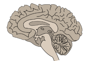
Coronal

Horizontal

Precentral Gyrus
located immediately anterior to the central sulcus, acts as the primary motor cortex, important for voluntary movement
Inferior Frontal Gyrus
the lower part of this gyrus houses Broca’s area; damage to it results in Broca’s aphasia where people can understand things normally but they can’t put a sentence together
Postcentral gyrus
located immediately posterior to the central sulcus; serves as the Primary Somatosensory cortex
Superior Temporal Gyrus
contains the Primary auditory cortex and wernicke’s area (in the dominant (left) hemisphere where language is)
Fusiform gyrus
located on the inferior/ventral surface, it is highly specialized for visual processing , particularly object and facial recognition
Damage: people cannot put together a pattern and recognize faces, even their own
Lingual gyrus
located on the medial surface below the calcarine sulcus, it is involved in vision and color perception
Damage: lose a quarter of their vision
Contralateral Communication
pertaining to the other side; most of our senses decussate to the contralateral hemisphere
Ipsilateral communication
belonging to or occurring on the same side of the body
Cerebrospinal spinal fluid (CSF)
bathes, cushions, and supports the brain and spinal cord within the central nervous system