Muscles and Joints
 Call Kai
 Learn
 Practice Test
 Spaced Repetition
 Match
 Flashcards
 Knowt Play
1/86

There's no tags or description
Looks like no tags are added yet.
87 Terms
Joints
Articulations of bones
Functions of joints
Hold bones together
Allow for mobility
Ways joints are classified
Functionally
Structurally
Synarthroses
immovable joints
Amphiarthroses
slightly moveable
joints
Diarthroses
freely moveable joints

Fibrous/ immovable joints
The bones at an immoveable joint cannot move - they
overlap or interlock, and are held together by a tough
fibre, e.g. the skull.
Generally immovable

Cartilaginous/slightly moveable joints
The bones at a slightly moveable joint can only move
a little - they are held together by strong straps
called ligaments and are joined by protective pads
known as cartilage, e.g. the ribs.
Immovable or slightly moveable

Synovial joint/freely moveable
At a freely moveable joint the bones move freely.
They are also known as synovial joints, and
are the largest group of joints found in the body,
e.g. the hips, shoulders and knees.
Freely moveable
Examples of Fibrous Joints
Bones united by fibrous tissue
Examples
Sutures
Syndesmoses
Allows more
movement than
sutures
Example: distal
end of tibia and
fibula
Cartilaginous join example
Bones connected by cartilage
Examples
Pubic
symphysis
Intervertebral
joints
Synovial Joint example
Articulating
bones are
separated by a
joint cavity
Synovial fluid
is found in the
joint cavity
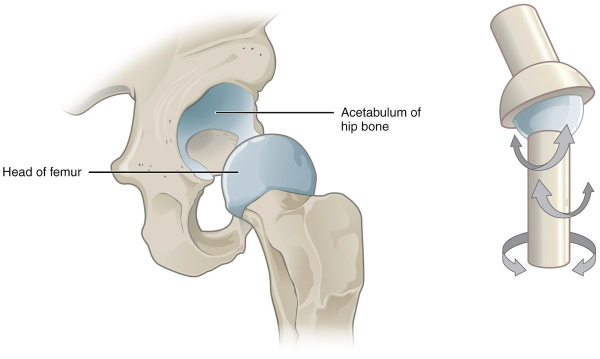
Ball and Socket Joints
The ball has a head shaped of one bone which articulates
with a cuplike socket.
MultiAxial
Allows the greatest range of movement
Examples
HIP
SHOULDER

Hinge Joints
They are cylindrical shaped of 1 bone articulates with
a depression of an adjacent bone
Movement is restricted to bending and straightening
UniAxial
EXAMPLES
ELBOW
KNEE

Pivot Joints
These are rounded, pointed or concave of one bone
which articulates which a ring shaped bone.
UniAxial
Movement is restricted to 1 bone rotating around
the longitudinal axis.
EXAMPLE
VERTEBRAE OF
THE NECK

Gliding Joints
The articulating surfaces are flat.
It allows limited movement
Non-Axial (sometimes Uniaxial)
EXAMPLES
HAND BETWEEN THE CARPELS

Saddle Joints
The articulating surfaces are shaped like a saddle
Biaxial
It allows very limited movement
EXAMPLE
THUMB JOINT
Muscles
Effectors which enable movement to be carried out
responsible for almost all the movements in animals
Cardiac and Smooth muscle
controlled involuntarily by the autonomic nervous system
Skeletal Muscle
Voluntarily controlled by the somatic nervous system
cause the muscles to move at the joints
Tendon
Attach muscle to bone
Tendons transmit muscle force to the bone.
Made of collagen fibres & are very strong & stiff
Antagonistic Muscle Action
Muscles are either contracted or relaxed
When contracted the muscle exerts a pulling force, causing it to shorten
Since muscles can only pull (not push), they work in pairs called antagonistic muscles
The muscle that bends the joint is called the flexor muscle
The muscle that straightens the joint is called the extensor muscle

Elbow Joint
The best known example of antagonistic muscles are the bicep & triceps muscles
The extensor is the tricep
the flexor is the bicep

Muscle Structure
single muscle e.g. biceps contains approx 1000 muscle fibres.
These fibres run the whole length of the muscle
Muscle fibres are joined together at the tendons
Each muscle fibre is actually a single muscle cell
This cell is approx 100 in diameter & a few cm long
These giant cells have many nuclei
Their cytoplasm is packed full of myofibrils
These are bundles of protein filaments that cause contraction
Sarcoplasm (muscle cytoplasm) also contains mitochondria to provide energy for contraction
Dark and Light
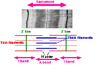
The E.M shows that each myofibril is made up of repeating dark & light bands
In the middle of the dark band is the M-line
In the middle of the light band is the Z-line
The repeating unit from one Z-line to the next is called the sarcomere

Cross Bridges
A very high resolution E.M reveals that each myofibril is made up of parallel filaments.
There are 2 kinds of filament called thick & thin filaments.
These 2 filaments are linked at intervals called cross bridges, which actually stick out from the thick filaments

The Thick Filament (Myosin)
Consists of the protein called myosin.
A myosin molecule is shaped a bit like a golf club, but with 2 heads.
The heads stick out to form the cross bridge
Many of these myosin molecules stick together to form a thick filament

Thin Filament (Actin)
The thin filament consists of a protein called actin.
The thin filament also contains tropomyosin.
This protein is involved in the control of muscle contraction
The Sliding Filament Theory
So, when the muscle contracts, sarcomeres become smaller
However the filaments do not change in length.
Instead they slide past each other (overlap)
So actin filaments slide between myosin filaments
and the zone of overlap is larger
Repetition of the cycle
One ATP molecule is split by each cross bridge in each cycle.
This takes only a few milliseconds
During a contraction 1000’s of cross bridges in each sarcomere go through this cycle.
However the cross bridges are all out of synch, so there are always many cross bridges attached at any one time to maintain force.

Irritability
ability to receive and respond to a stimulus

Contractility
Ability to shorten when an adequate stimulus is received
Nerve Stimulus to Muscles
Skeletal muscles must be stimulated by a nerve to contract
Motor unit
One neuron
Muscle cells stimulated by that neuron
A motor neuron and all the muscle fibers it activates

Neuromuscular junctions
association site of nerve and muscle

Synaptic cleft
gap between nerve and muscle
Nerve and muscle do not make contact
Area between nerve and muscle is filled with interstitial fluid
Neurotransmitter and Transmition
Neurotransmitter-chemical released by nerve upon arrival of nerve impulse
The neurotransmitter for skeletal muscle is acetylcholine
Neurotransmitter attaches to receptors on the sarcolemma
Sarcolemma becomes permeable to sodium (Na+)
Sodium rushing into the cell generates an action potential
Once started, muscle contraction cannot be stopped (all or nothing)
Contraction of a Skeletal Muscle
Muscle fiber contraction is “all or none”
Within a skeletal muscle, not all fibers may be stimulated during the same interval
Different combinations of muscle fiber contractions may give differing responses
Graded responses – different degrees of skeletal muscle shortening
Muscle Response to Strong Stimuli
Muscle force depends upon the number of fibers stimulated
The grouping of one motor neuron and the muscle fibers it innervates is known as a motor unit. A single motor neuron usually innervates more than one muscle fiber, since the motor neuron’s axon can have multiple terminals at its end. Motor units can be as large as a few thousand muscle fibers to one neuron, but motor units of this size usually only occur in parts of the body that don’t engage in fine, precise movements. For example, your leg might contain large motor units but your hands wouldn’t.
More fibers contracting results in greater muscle tension
Muscles can continue to contract unless they run out of energy

Isotonic contractions
Myofilaments are able to slide past each other during contractions
The muscle shortens

Isometric contractions
Tension in the muscles increases
The muscle is unable to shorten
Unsuccessful movement of Lift

Excitation: Step 1
Action potential (AP) travels to nerve ending called the AXON Terminal.
ACh released at the neuromuscular junction
Activation of Sarcolemma and t-tubules by Ach (Ach bonds with receptors)
AP is generated in the muscle fiber
Depolarization of sarcolemma and t-tubules
Sarcoplasmic reticulum releases Ca++ into Sarcomere
Clean Up of the Synapse
3 ways
Re-uptake
only small molecule chemical neurotransmitters can be taken back up. only small molecule chemical neurotransmitters can be taken back up. Advantage = Neurotransmitter can be recycled
Enzymatic Destruction
The neurotransmitter can be destroyed directly either in the cleft or in the pre-synaptic terminal using certain enzymes. Two major enzymes are involved in the destruction of the neurotransmitter. {Monoamine Oxidases (MAO) and Catechol-O-Methyltransferase (COMT)}
Diffusion
In the simplest form of termination, the neurotransmitter can simply diffuse out of the synaptic cleft and away from the receptors and into nearby blood vessels
The Sliding Filament Theory of Muscle Contraction
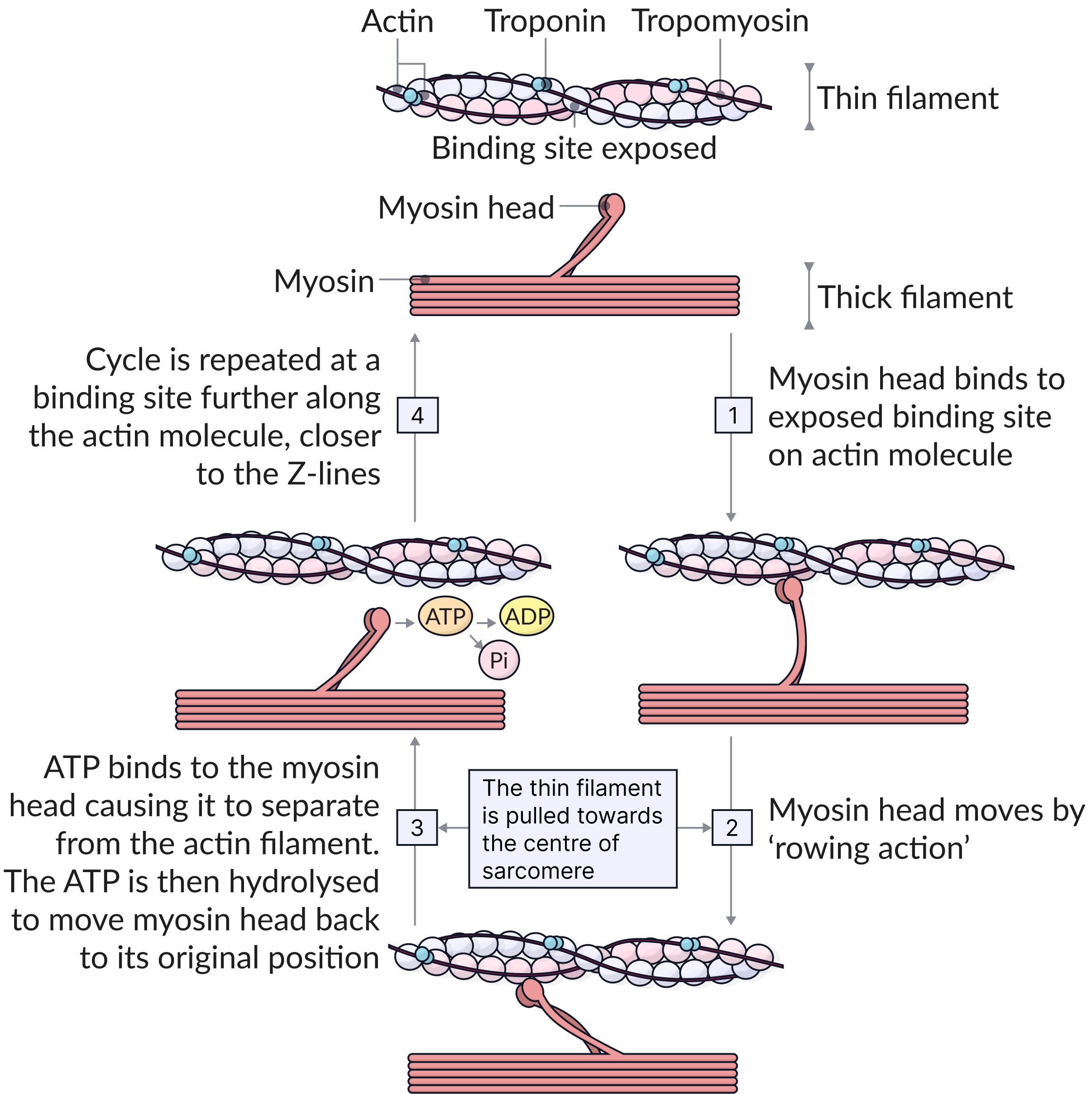
Activation by nerve causes myosin heads (crossbridges) to attach to binding (active) sites on the thin filament
Myosin heads then bind to the next site of the thin filament
Energy for Muscle Contraction
Initially, muscles used stored ATP for energy
Bonds of ATP are broken to release energy
Only 4-6 seconds worth of ATP is stored by muscles
After this initial time, other pathways must be utilized to produce ATP
The Sliding Filament Theory of Muscle Contraction
This continued action causes a sliding of the myosin along the actin
The result is that the muscle is shortened (contracted)
Contraction: Step II
Ca++ binds to troponin on Tropomyosin
Tropomyosin Twists causing Active actin sites to be exposed
Cross bridges or MH bind to actin
Rachet Effect = Cross bridges or MH rotate (kicks out ADP & P)
Myosin heads disengage when ATP attaches (see next slide)
Cycle repeats (Ca++ and ATP needed) OVER and OVER called the Power Stroke
Excitation-Contraction Coupling
Energy Production needed for the contraction stage
Action potential reaches muscle fibers
In MH’s ATPase is activated
ATP is cleaved by ATPase
MH + ATP → ADP+ P ion+ Energy
This process disengages the cross bridge or MH during the power stroke
Relaxation Step III
Ca++ is pumped back to sarcoplasmic reticulum by ion pumps
Active pump utilizing ATP
Actin sites are covered by troponin and tropomyosin
Ca++ remains stored in the reticulum

Muscle fatigue
a transient and recoverable reduction in the force of muscle contraction which occurs during exercise.
is muscle being Stimulated
Muscle does not contract or as much
A condition resulting from prolonged and strong contraction of a muscle
product accumulation or substrate depletion are the two causes
Three categories of fatigue
Central, Peripheral and Neural
Central
weakness manifests as an overall, bodily or systemic, sense of energy deprivation
Ex. = Running a Marathon
Peripheral
weakness manifests as a local, muscle-specific incapacity to do work
Ex. = Weight Training
Neural
weakness can be both central and peripheral
Muscle Fatigue and Oxygen Debt
When a muscle is fatigued, it is unable to contract (still receives stimulus)
The common reason for muscle fatigue is oxygen debt
Oxygen must be “repaid” to tissue to remove oxygen debt
Oxygen is required to get rid of accumulated lactic acid
Increasing acidity (from lactic acid) and lack of ATP causes the muscle to contract less
Oxygen debt
During long periods of vigorous activity:
lactate levels accumulate/build up
glycogen reserves in the muscles become low as more glucose is used for respiration, and additional glucose is transported from the liver.
This build-up of lactate produces an oxygen debt.
As body stores of glycogen become low, the person suffers from muscle fatigue.
After exercise
The body's tolerance of lactate is limited so when a period of exercise is over, lactate acid must be removed.
Lactate is taken to the liver by the blood, and either:
oxidised to carbon dioxide and water
converted to pyruvate and glucose, then glycogen - glycogen levels in the liver and muscles can then be restored
These processes require oxygen. This is why, when the period of activity is over, a person’s breathing rate and heart rate do not return to normal straight away.
Taking in the amount of oxygen required to remove the lactate, and replace the body's reserves of oxygen, is called repaying oxygen debt.
When someone who has been exercising pays back an oxygen debt, it can take from a few hours for normal exercise, to several days after a marathon.
Metabolites
the substances (generally waste products) produced as a result of muscular contraction.
Include: ADP, Mg2+Include: ADP, Mg2+, reactive oxygen species and inorganic phosphate.
Accumulation of metabolites can directly or indirectly produce metabolic fatigue within muscle fibers through interference with the release of calcium or reduction of the sensitivity of contractile molecules actinAccumulation of metabolites can directly or indirectly produce metabolic fatigue within muscle fibers through interference with the release of calcium or reduction of the sensitivity of contractile molecules actin and myosin to calcium.
Chloride
Intracellular chloride inhibits the contraction of muscles
due to “false alarms”= small stimuli which may cause them to contract
This natural brake helps muscles respond solely to the conscious control but also has the effect of reducing the force of conscious contractions.
Fatigue mechanisms
Factors that help determine when performance will deteriorate include:
Exercise type, duration and intensity
Types of muscular contractions
Physical fitness or conditioning
Age
Diet
Environmental factors
Mental state of the performer VCE Physical Education
Isotonic Contractions
-occur when a muscle contracts and changes length.
-muscle shortens as it generates force, such as lifting a weight
Isometric Contractions
occur when a muscle contracts without changing length
muscle fibers generate force against an immovable object, such as holding a heavy weight or pushing against a wall

concentric
muscle shortens as it contracts to meet resistance.
-This type of contraction requires more oxygen and burns more calories.
-An example of a concentric contraction is flexing your leg to climb stairs.
-Bicep Curl moving the weight in Flexion
Increased muscle mass and size
Improved overall strength
Easier to perform for beginners
eccentric
muscle lengthens as the resistance becomes greater than the force the muscle is producing.
-This type of contraction is easier than concentric, but it can break parts of the muscle and cause soreness.
-An example of an eccentric contraction is slowly lowering a weight from a curled position.
Greater muscle damage and hypertrophy
Increased strength and power
Improved tendon and ligament strength
Reduced risk of injury (when performed correctly)
Skeletal Muscle Hypertrophy
-increasing the size/mass of muscle
Myofibrillar hypertrophy
increase in protein subunits (contractile proteins)
increase in sarcomeres
resistance training
Connective Tissue Hypertophy
build up of connective tissue (fascia)
Improves integrity of muscle (Stiffens muscles)
Heavy loading, explosive stretching, or high-volume plyometrics
Sarcoplasmic Hypertrophy
non contractile proteins
increase sarcoplasm
increases fuel resevoir
Mechanical Tension
eccentric & Concentric
Metabolic Stress
increases supply of ATP
Increase production of proteins (translation increases)
Muscle Damage
produces lactaid causing muscle to swell
trauma to the actin & myosin

Single Twitches
Muscles relaxes completely between stimuli

Summation
Stimuli closer together do not allow muscle to relax fully

Summation leading to unfused tetanus
Stimuli are far enough apart to allow muscle to relax slightly between stimuli
Summation leading to complete tetanus
Muscle reaches steady tension
Latent period
begins at stimulation and typically lasts about 2 msec
the action potential sweeps across the sarcolemma and the sarcoplasmic reticulum releases calcium ions
contraction phase
tension rises to a peak
calcium ions are binding to troponin,
active sites on thin filaments are being exposed,
cross-bridge interactions are occurring
The contraction phase ends roughly 15 msec after stimulation
Relaxation Period
The period after a contraction when the tension in the muscle fiber returns to resting levels
continues for about another 25 msec
calcium levels are falling
active sites are being covered by tropomyosin
number of active cross-bridges decline
Twitch
ingle stimulus–contraction–relaxation sequence in a muscle fiber.
Twitches vary in duration.
Twitches in one eye muscle fiber can be as brief as 7.5 msec, but a twitch in a muscle fiber from the soleus, a small calf muscle, lasts about 100 msec
twitches in a skeletal muscle do not accomplish anything useful. All normal activities involve sustained muscle contractions.