exam 2
 Call Kai
 Learn
Learn Practice Test
 Spaced Repetition
 Match
Match Flashcards
 Knowt Play
 Podcast
1/494

There's no tags or description
Looks like no tags are added yet.
Name | Mastery | Learn | Test | Matching | Spaced | Call with Kai | Chat |
|---|
No analytics yet
Send a link to your students to track their progress
495 Terms
Perichondrium (perimeter of the cartilage)
Layer of dense connective tissue surrounds it.
Helps resist outward
Blood vessel for nutrient delivery
Appositional growth
cartilage - forming cells in perichondrium secrete matrix expand from outside
Interstitial growth
Chondrocytes in lacunae divide and secrete new matrix, expanding from within
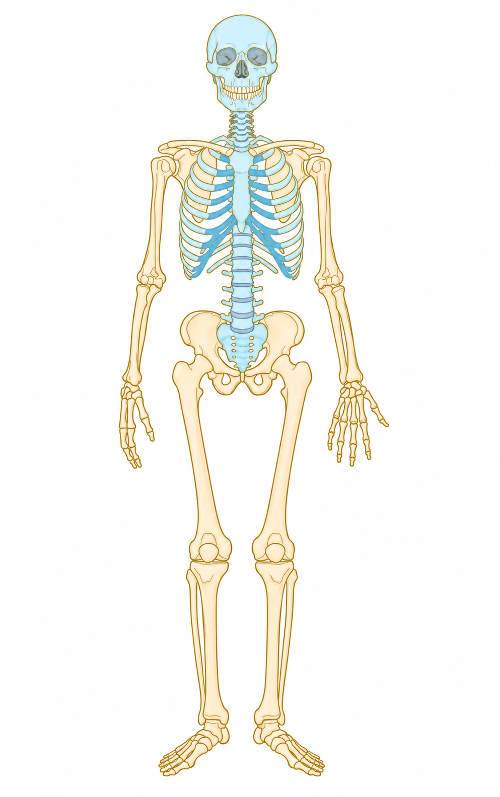
Axial skeleton
Long axis of body
Skull, vertebral column, rib cage

What’s this
Axial skeleton

Appendicular skeleton
upper and lower limbs
girdles attaching limbs
What’s this?
Appendicular skeleton

Spongy bone
Honeycomb of flat spines

What’s this
Spongy bone

Compact bone
Dense, smooth, outer layer
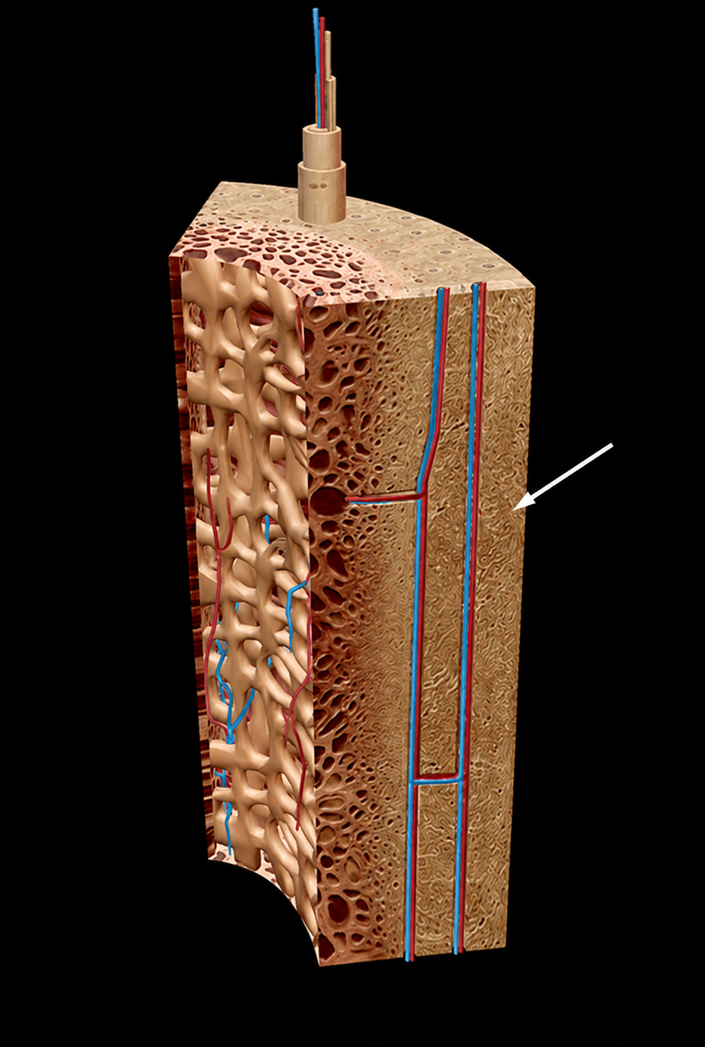
What’s this
Compact bone

Periosteum
Outside of compact bone
dense irregular connective tissue
Osteogenic inner layer
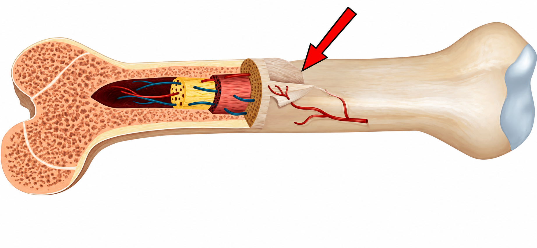
What’s this
Periosteum

Endosteum
Inside of compact bone
Areolar connective tissue
Delicate, covers trabeculae, canal lining, also has osteogenic cells
Hematopoiesis occurs here

What’s this
Endosteum
Hematopoiesis
Creation of all cell types in red marrow
trabecular cavities of spongy bone and trabeculae of flat bones
Newborns: red marrow found in medullary cavities (long bones) and all spongy bone (Babies have more blood cells)
Adults: red marrow in beads of femur and humerus, but mostly active in flat bone trabeculae and some irregulars

Epiphyseal line
Remnant of childhood epiphyseal plate where bone growth occurs. (Stop stretching vertically)

What’s this
Epiphyseal line

Trabeculae
Thin plates/ struts of bone
Forms 3D network inside spongy bone
Spaces between them contain bone marrow
Helps stay strong while remaining relatively lightweight

What’s this
Trabeculae
Osteogenic cells
Stem cells, responsible for building bones, can develop into osteoblasts
actively dividing in membrane
Differentiate into osteoblasts or bone lining cells
Some remain as stem cells
Osteoblasts
Bone formers
secrete mineralized matrix: steroid
Collagen and calcium binding protein
Collagen makes up 90% of bone protein
osteoblasts: actively dividing

Hyaline cartilage
Covers bone regions at a movable joint

Diaphysis

What’s this
Diaphysis

Epiphysis
Bone ends , compact bone, wrapping around spongy bone

What’s this
Epiphysis
Osteocytes
Mature, live in lacunae, no longer divide
maintain matrix, sense stress or strain
Communicate information to other cells for bone remodeling

Identify
Frontal bone: Located on the front of the skull

What is this?
Frontal sinuses: hollow spaces within the frontal bone

What’s this
Suture

Suture
Immovable fibrous joint
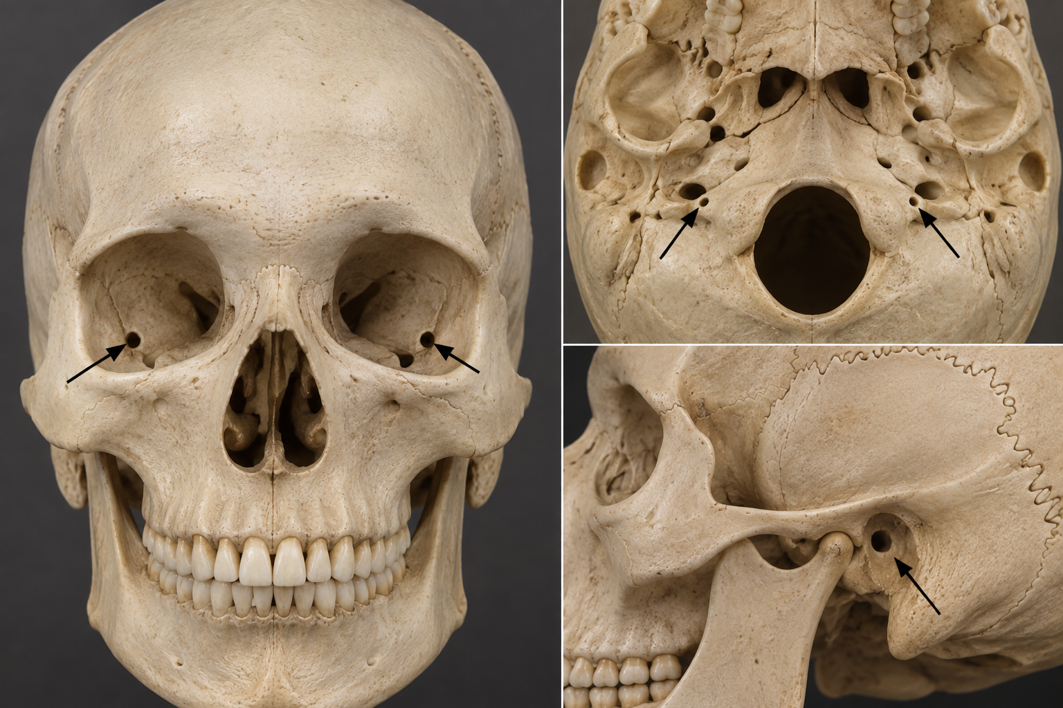
Whats this
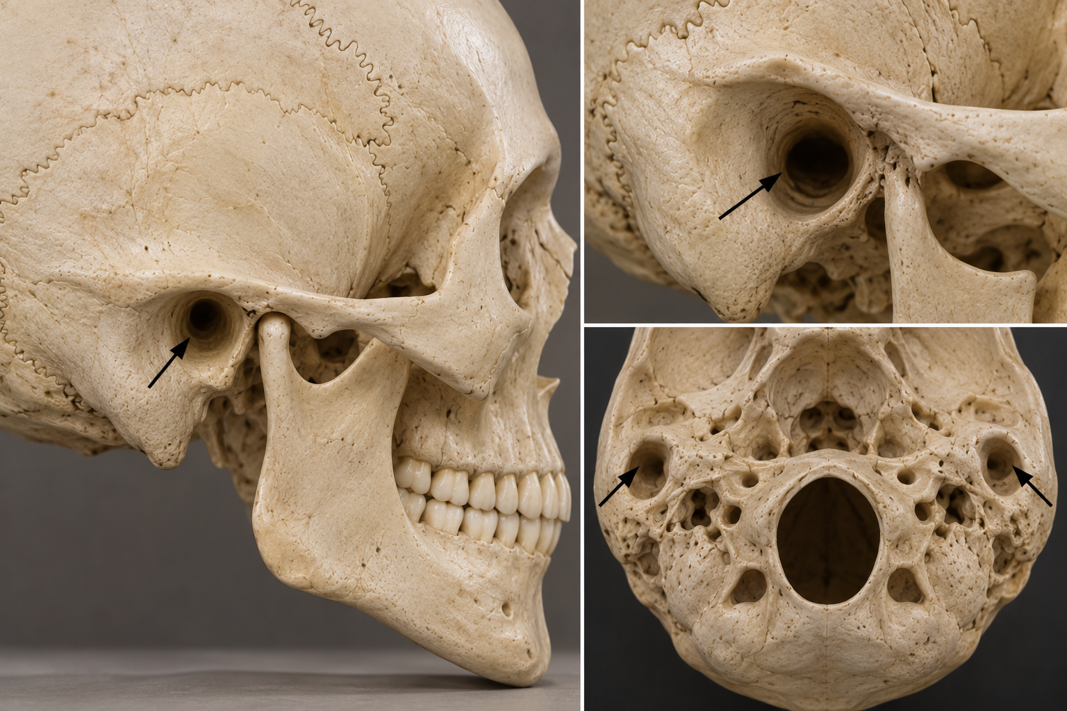
Foramen

Foramen
Round / oval opening
What’s this
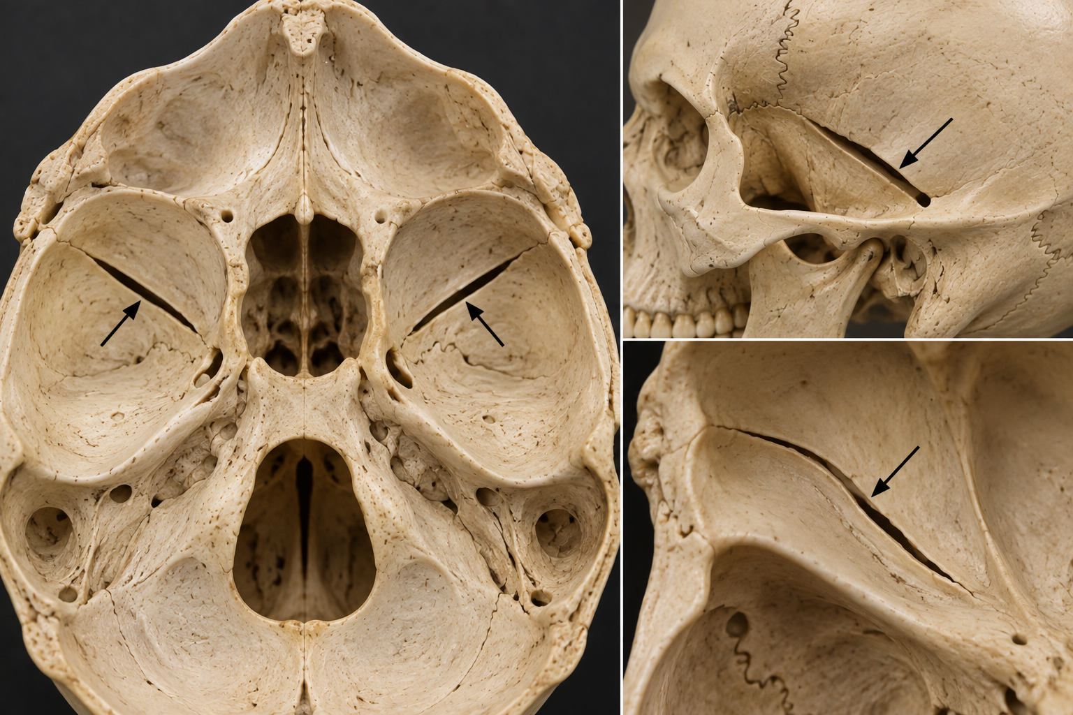
fissure

Fissure
Slit like opening or groove in a bone that allows nerves and blood vessels to pass through
What’s this
Meatus

Meatus
Canal like passageway

What’s this
Spine

Spine
Pointed projection of bone

What’s this
Process

Process
Bong prominence, projection of or bump sticking out from bone

What’s this
Tubercle
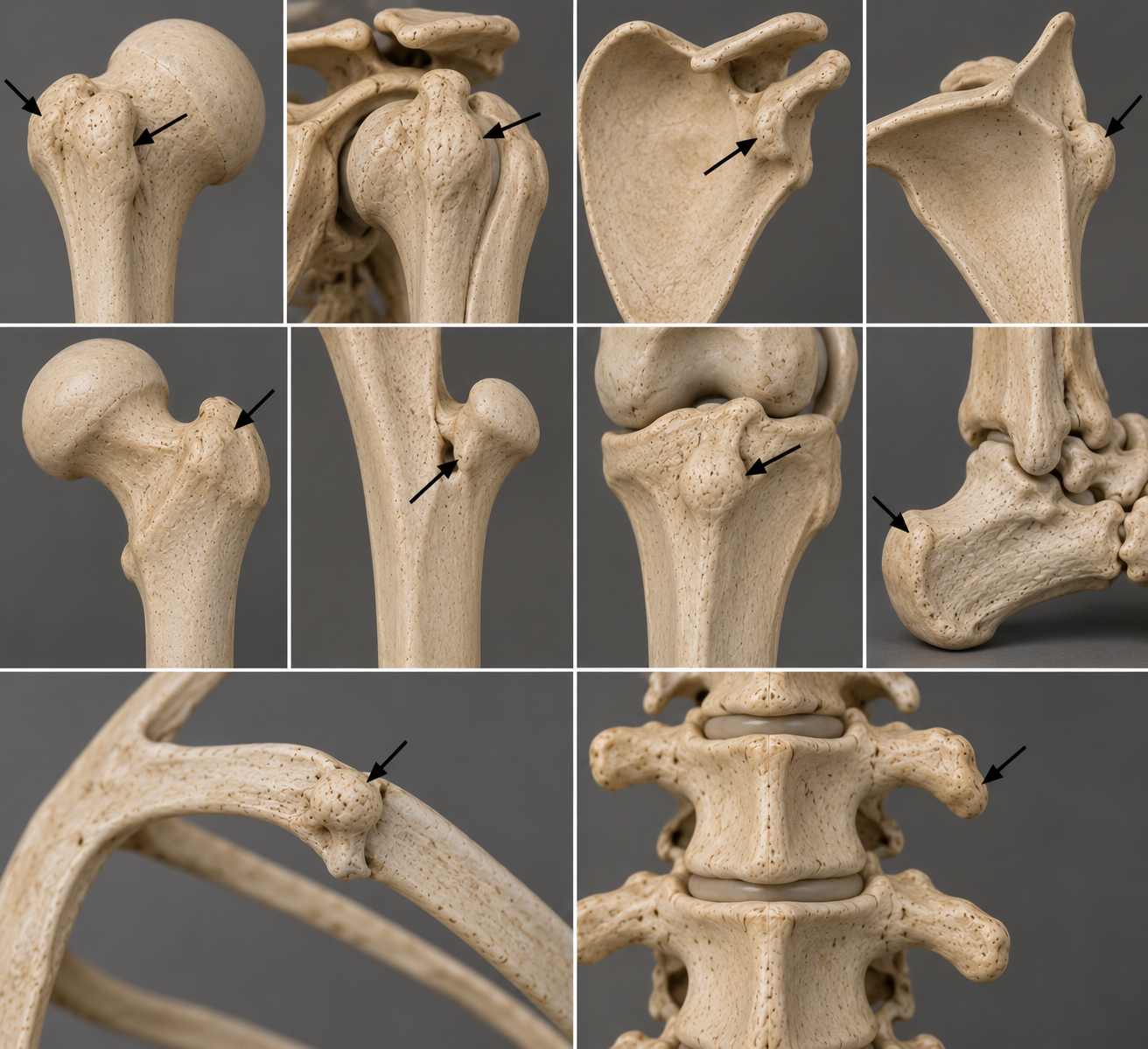
Tubercle
Small, rounded projection or bump

What’s this
Tuberosity

Tuberosity
A large rounded roughened projection of bone that usually serves as an attachment site for muscles or a ligaments

What’s this
Fossa

Fossa
Shallow depression, Hollow, or basin like area in a bone

Whats this
Groove

Groove
Long narrow depression or channel in the surface of a bone

What’s this
Condyle

Condyle
A rounded smooth projection at the end of a bone that forms part of a joint

What’s this
Epicondyle

Epicondyle
Raised area on Condyle located above or adjacent to Condyle
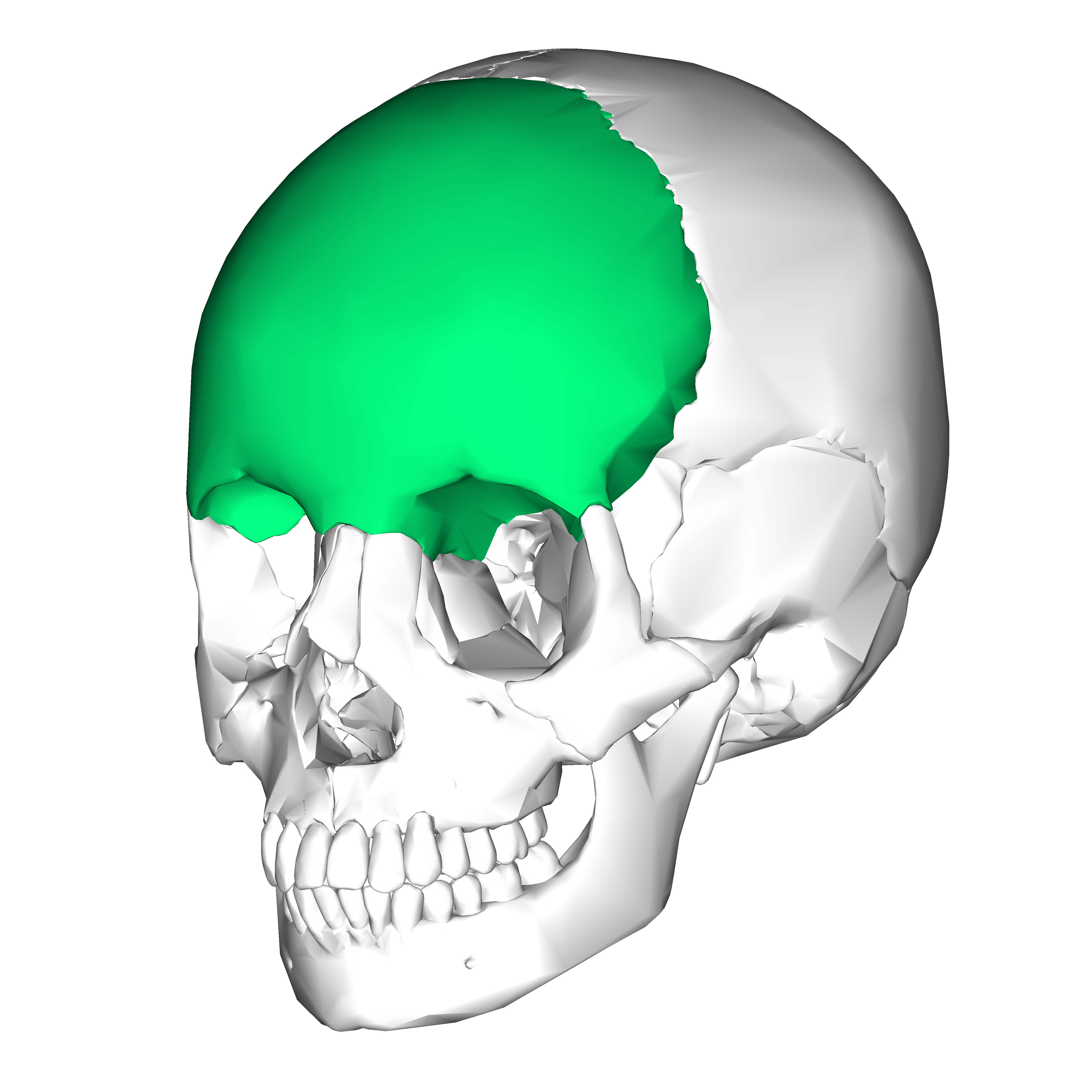
Frontal bone and landmarks. (5)
Located on the front of the skull
Frontal sinuses
Coronal suture
Glabella
Supraorbital foramen
Supraciliary arches

What’s this ?
Frontal sinuses
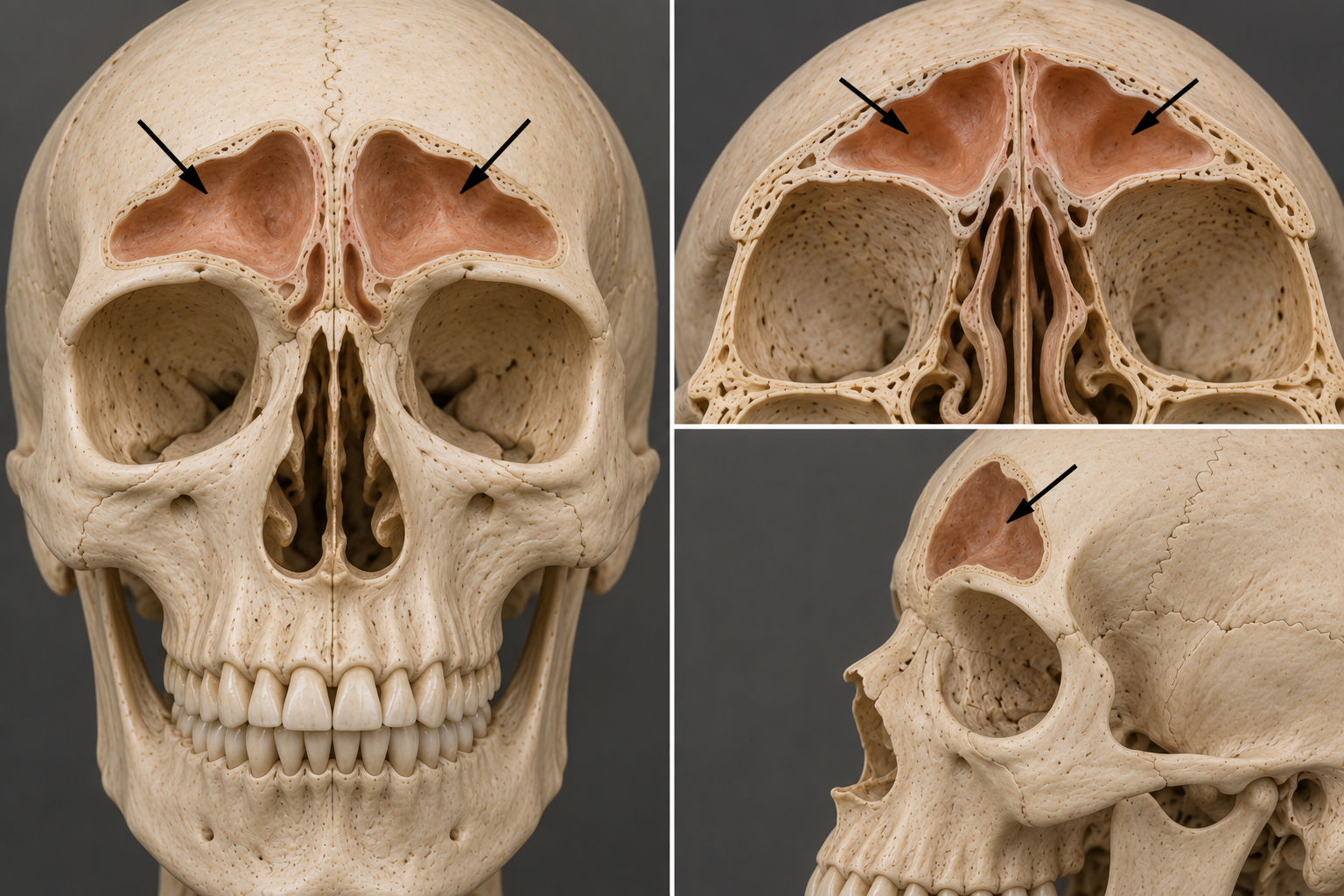
Frontal sinuses
Hollow spaces within the frontal bone

What’s this
Coronal suture

What’s this?
Glabella

Coronal suture
Articulates with the parietal bones

Glabella
Smooth surface between the supraorbital Ridge

Whats this ?
Supraorbital foramen

Supraorbital foramen
Opening on the superior aspect of the orbit that transmit the supraorbital nerve, artery and vein

Whats this
Supraciliary arches
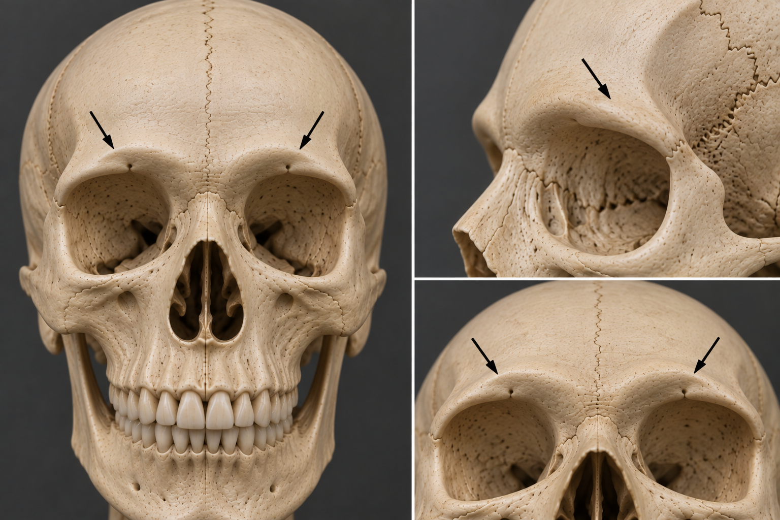
Supraciliary arches
Bony prominence on the supraorbital margin

Whats this
Parietal bones

Parietal bones and landmarks (3)
Per idle bones are paired bones that formed the lateral walls of the skull
Sagittal suture
Lambdoid suture
Squamous suture

Whats this?
Sagittal suture
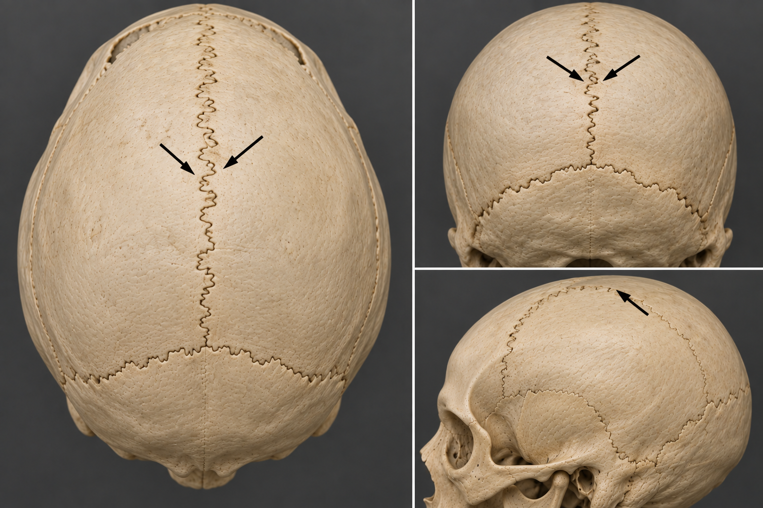
Sagittal suture
Articulates with the parietal bone.

What’s this ?
Lamboid suture

Lamboid suture
Articulates with the occipital bone

Whats this ?
Squamous suture

Squamous suture
Articulates with the temporal bone

What’s this ?
Temporal bone
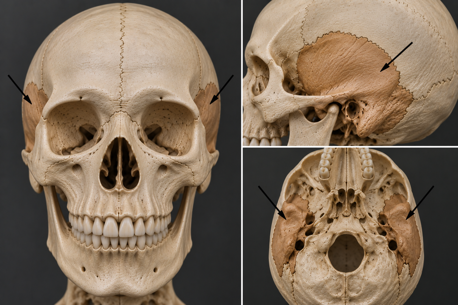
Temporal bones and landmarks (7)
temporal bones are paired bones that form the lateral walls of the skull
internal acoustic meatus
External acoustic meatus
Petrous region
Mastoid process
Styloid process
Zygomatic process
Stylomastoid Foraman

What’s this ?
Internal acoustic meatus

Internal acoustic meatus
Transmit nerves for hearing, balance, and facial expressions

What’s this ?
External acoustic meatus

External acoustic meatus
Passageway for sound waves to reach the Tympanic membrane (eardrum)

What’s this?
Petrous region

Petrous region
The dense portion of the school that houses the cochlea (Hearing) And vestibular apparatus (Balance)

What’s this ?
Mastoid process
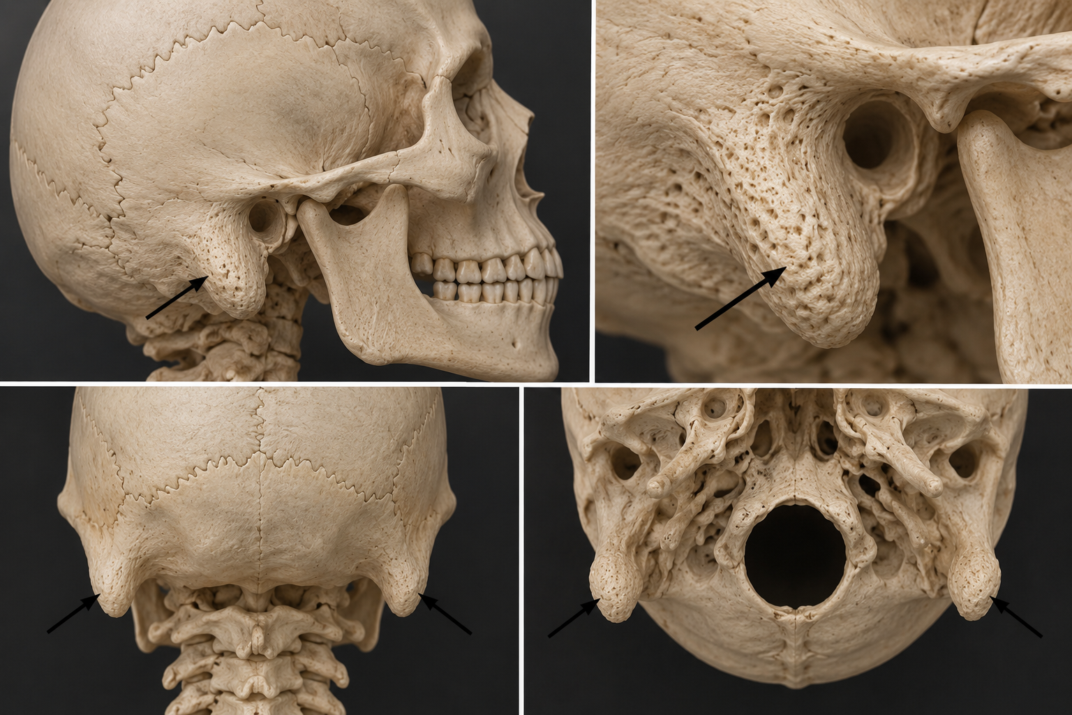
Mastoid process
Rounding Protuberance on the Postero inferior temporal bone

What’s this
Styloid process
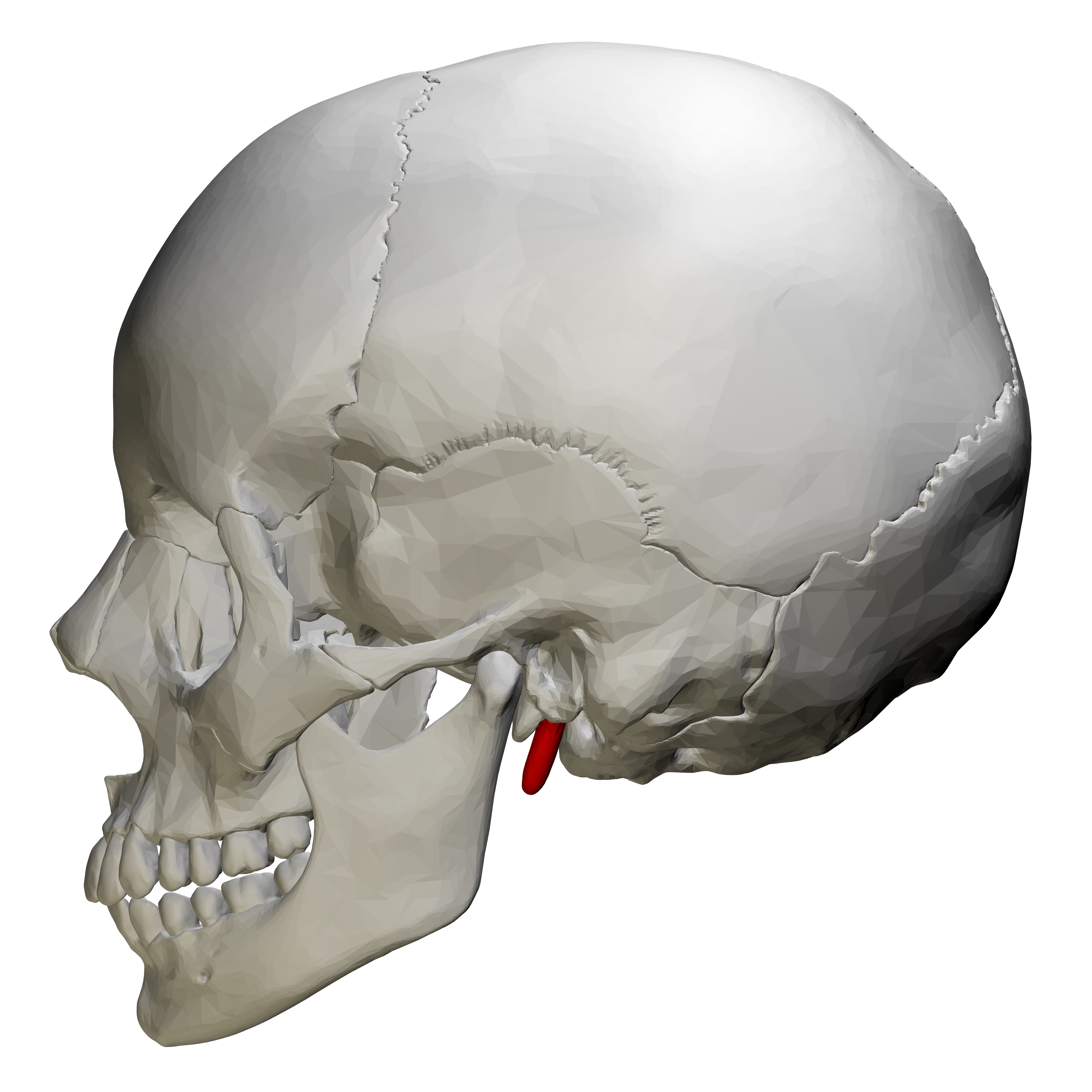
Styloid process
Pointy projection of temporal bone for muscle and ligament attachment.

What’s this
Zygomatic process

Zygomatic process
Projection that articulates with the zygomatic bone creating zygomatic arch

What’s this?
Stylomastoid foramen

Stylomastoid foramen
Opening between the styloid and mastoid process that transmit the facial nerve

what’s this?
Occipital bone
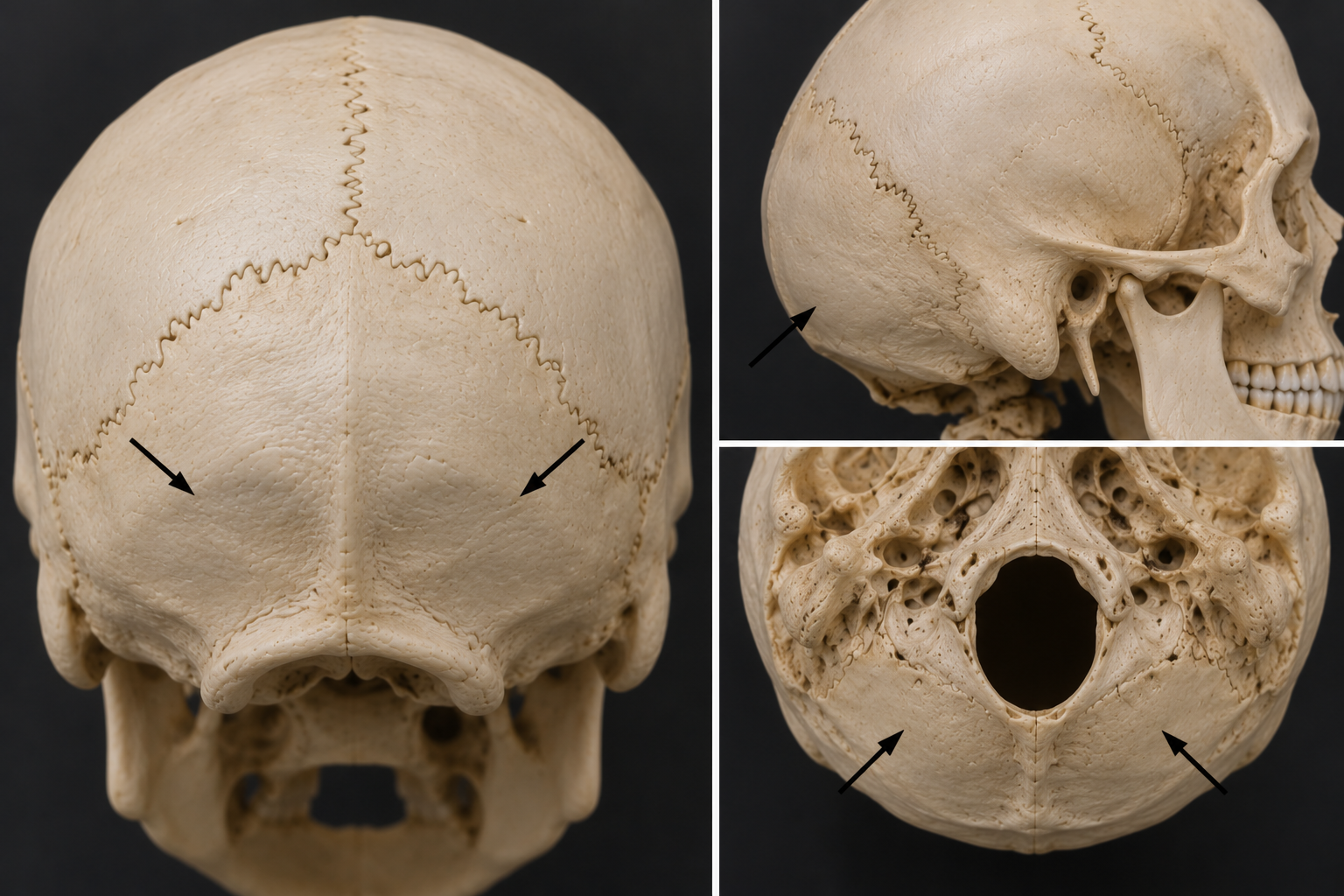
Occipital bone and landmarks (5)
Forms the posterior region of the skull
Foraman Magnum
External occipital protuberance
Occipital condyles
Hypoglossal Canal
Jugular Foraman

Whats this ?
Foramen magnum

Foramen magnum
Large opening at the base of the school, which transmits the spinal cord, vertebral arteries, and spinal accessory nerve

What’s this?
External occipital protuberance

External occipital protuberance
A prominent bump on the posterior aspect of the skull

What’s this?
Occipital condyles

Occipital condyles
Articulates with C1 vertebra (Atlas) to make the Atlanta occipital occipital joint.

What’s this?
Hypoglossal canal

Hypoglossal Canal
Located adjacent, the occipital condyles, and transmits the hypoglossal nerve

What’s this?
Jugular foramen