L10: Ankle and Foot Bones
 Call Kai
 Learn
 Practice Test
 Spaced Repetition
 Match
 Flashcards
 Knowt Play
1/39

There's no tags or description
Looks like no tags are added yet.
40 Terms
How many bones in the foot? How is body weight transferred?
What are the bones?
There are 26 bones in each foot.
Body weight is transferred from the tibia into the talus and out the calcaneus.
Rear and midfoot tarsal bones (5): Navicular, cuboid, and 3 cuneiforms.
5 metatarsals
5 phalanges
3 phalanxes (2-5)
2 sesamoid bones beneath 1st metatarsophalangeal joint
What does the lateral and medial malleoli of the fibula and tibia provide?
They are enlarged and protrude horizontally and medially.
1) Provides general medial and lateral stability
2) Serves as a pulley for tendons that wrap around posteriorly, increasing the MA of muscles.
Increased L/T relationship and lengthening the moment/force arm.
Primarily in inversion and eversion actions
Pulley: MA greatly increases for which evertors? Invertors?
Peroneus brevis and longus wraps around posteriorly on the lateral malleoli, greatly increasing the MA of eversion
Tibialis posterior, flexor digitorum longus, and flexor hallucis longus wrap around posteriorly on medial malleolus, greatly increasing the MA of inversion
Distal tibiofemoral joint
Is it the ankle joint?
What is its classification?
Are injuries common? How are they classified?
Not the ankle joint, but it is close, so it is often associated.
Is a syndesmotic, amphiarthrodial joint.
Often gets injured in high contact sports, and is classified as a “High Ankle Sprain,” when the anterior inferior tibiofibular, posterior tibiofibular, interosseus ligament, and interosseus membrane get injured.
Ankle/ Talocrural Joint.
Consists of?
Classified
What are the degrees
Greater range of dorsiflexion when
why?
Consists of the distal tibia, distal fibula, and the talus
It is classified as a hinge joint, so only plantar/dorsi -flexion
PF: 50 degrees
DF: 15-20 degrees
Greater range of dorsiflexion when the knee is flexed
With the gastroc shorter at the knee, tension reduces, allowing it to be longer at the ankle.
What is the motion potential of the distal tibiofibular joint?
During dorsiflexion, the talus moves up towards the tibia, and the fibula externally rotates (3-5 degrees), creating space and allowing a greater range of dorsiflexion.
During plantarflexion, the talus moves down. The fibula internally rotates (3-5 degrees) and moves medially back into place, closing the space.
Opens and closes by 1-2mm

Subtalar joint and transverse tarsal joints
Created by…
What movement occurs?
Classified as
Subtalar: talus and calcaneus
Transverse tarsal: Between the talus & navicular + calcaneus & cuboid
Where inversion (20-30) and eversion ( 5-15) occurs
Classified as gliding joints

Intertarsal and tarsometatarsal joints are classified as..
Gliding joints.
Metatarsophalangeal joints
Are between?
Classified as
Are between phalanges and metatarsals
Condyloid classification
Ankle proper and foot joint injuries
Ankle sprains are common, which consists of stretching or tearing a ligament(s)
What are the type of sprains?
Low ankle excessive inversion ankle sprain
injury to ligaments on low, lateral side of foot
Low ankle excessive eversion ankle sprain
injury to ligaments on low, medial side of foot.
How many arches does the foot have?
There are three arches.
Most people have one or all, but some people may also be flat-footed.

What are the arches of the foot?
Longitudinal arches (2)
1) Medial longitudinal arch: from the calcaneus to the talus, 3 cuneiforms, and proximal ends of metatarsals 1-3.
is the most common arch
2) lateral longitudinal arch: from the calcaneus to the cuboid, and proximal ends of 4th and 5th metatarsals
Transverse arch
extends across foot from 1st to 5th metatarsal.
What are the movements of ankle?foot?
Dorsi- and plantar- flexion
Eversion: decrease in angle laterally
Inversion: decrease in angle medially
Toe flexion/extension
Supination: combo of ankle doing plantarflexion, subtalar inversion, and forefoot adduction
Pronation: combo of ankle dorsiflexion, subtalar eversion, and forefoot abduction
How are the muscles divided?
Muscles are divided into four compartments by dense fascia. This facilitates venous return and prevents swelling of muscles during exercise.
Anterior, lateral, superficial posterior, and deep posterior compartment.
Anterior compartment
Dorsiflexor group: tibialis anterior, peroneus tertius, extensor digitorum longus, and extensor hallucis longus.
Lateral compartment
Peroneus longus and brevis, the two most powerful evertors
Superficial posterior compartment
Gastrocnemius, soleus, and plantaris
Plantarflexors
Deep posterior compartment
Flexor digitorum longus, flexor hallucis longus, popliteus, and tibialis posterior.
All are plantarflexors and invertors.
What injuries are common
Acute and chronic injuries are common
Shin splints, achilles tendon, and cramps
What are shin splints
most often involves
may be partially prevented by
Shin splints is a common term describing pain in the shank. It is a non specific diagnosis of muscle-tendon pain.
Most often involves the tibialis posterior, medial soleus, anterior tibialis, and/or extensor digitorum longus. Occurs by getting microtears and strain from running and jumping with poor mechanics.
May be partially prevented by strengthening and stretching plantar- and dorsi- flexors.
Rupture of Achilles tendon
occurs
It is a very disabling injury that often happens when someone jumps and lands wrong.
Painful cramps/Charly Horses
they are
may be due to
may be relieved by
They are acute muscle spasms in the gastrocnemius and soleus muscles (mostly)
May be due to random AP, dehydration, K+ deficiency, overworked muscles, etc.
May be relieved through active and passive dorsiflexion.
Muscle grouping according to location and function
Anterior ankle and foot: dorsiflexors
Posterior: plantarflexors
Lateral: Evertors
Medial: Invertors

Gastrocnemius
A
Knee flexor and plantarflexor
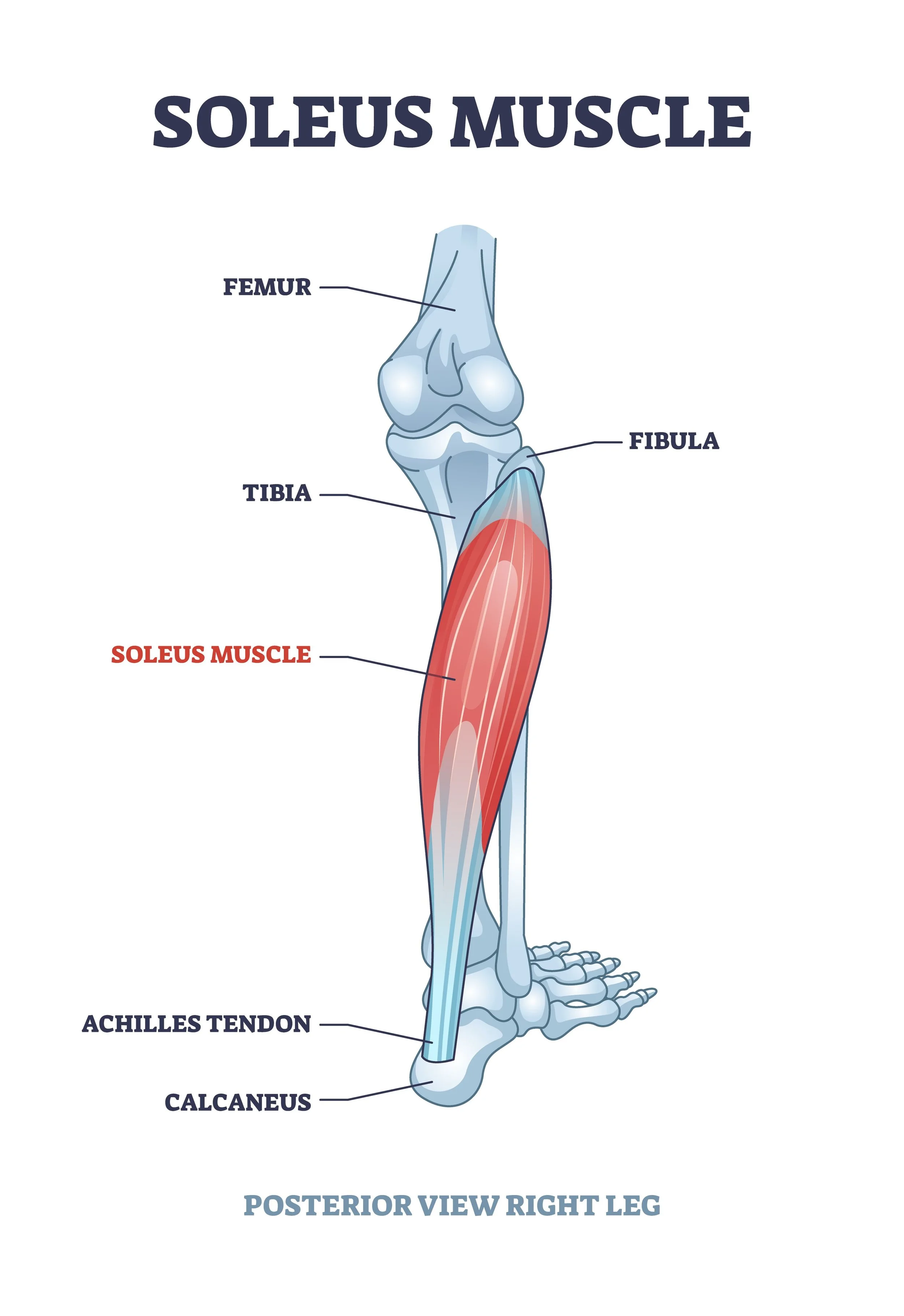
Soleus
A
Plantar flexor

Peroneus longus
A
Eversion of foot (gets MA)
Plantarflexor

Peroneus brevis
A
A: eversion of foot (gets MA)
plantarflexor

Peroneus tertius
Eversion of foot
Dorsiflexion of ankle (no MA)

Extensor digitorum longus
A
Eversion of foot
Dorsiflexion of ankle
Extensor of four lesser toes

Extensor hallucis longus
Dorsiflexion of ankle
Weak invertor of foot
Extension of hallux
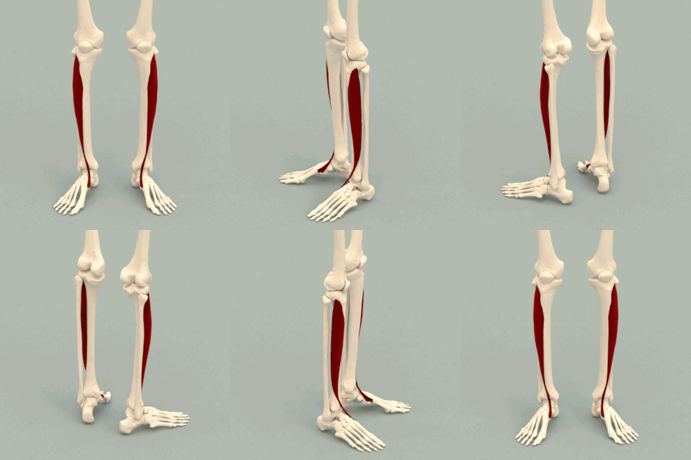
Tibialis anterior
A
Dorsiflexor and Invertor

Tibialis posterior
A
Plantarflexor and invertor

Flexor digitorum longus
A
Inversion of foot
Plantarflexion of ankle
Flexion of four lesser toes

Flexor hallucis longus
a
Inversion of foot
plantarflexion of ankle
flexion of hallux
Agonists: Dorsiflexors
Tibialis anterior
Extensor digitorum longus
Peroneus tertius
Extensor hallucis longus
Agonists: Plantarflexors
Gastrocnemius
Soleus
Tibialis posterior
peroneus longus
peroneus brevis
Flexor digitorum longus
Flexor hallucis longus
Agonists: Invertors
Tibialis posterior
Tibialis anterior
flexor digitorum longus
flexor hallucis longus
Agonists: Evertors
Peroneus longus
Peroneus brevis
Peroneus tertius
Extensor digitorum longus
Segment moved
close chain
open chain
CC: shank
OP: foot