Anatomy Exam 2
 Call Kai
 Learn
Learn Practice Test
 Spaced Repetition
 Match
Match Flashcards
 Knowt Play
1/106

Earn XP
Description and Tags
Muscular System
Name | Mastery | Learn | Test | Matching | Spaced | Call with Kai | Chat |
|---|
No analytics yet
Send a link to your students to track their progress
107 Terms
Muscle Tissue Types
Smooth (involuntary)
non-striated fibers
cardiac (involuntary)
striated fibers
skeletal (voluntary)
striated fibers
How do muscles move?
Muscles pull equally on both ends
they create force by contracting
must oppose gravity to produce movement or maintain a position
Extrinsic Muscles
muscles originating outside of the part of the body they act on
Intrinsic Muscles
muscles originating in the part of the body they act on, being contained within the corresponding body part
Muscle Belly
central part of muscle
Tendon
Macro:
connects muscle to bone
concentrates pull of muscle to small area on bone
very strong
Aponeurosis
sheet like elastic tendon acting as insertion site for muscle fibers
Fascia
band/sheet of connective tissue separating muscles and other internal organs
attaches, stabilizes, encloses, and separates muscles and other internal organs.
Micro Muscle: Muscle Belly
covered in Epimysium
comprised of bundles of muscle fascicles
Muscle Fasicle
Perimysium divides muscle fascicles into compartments
comprised of bundles of muscle fibers
Muscle Fiber
Endomysium divides muscle fibers into compartments
comprised of bundles of myofibrils
Type I: slow twitch, resistant to fatigue and associated with more endurance activities
Type ll: fast twitch, easily fatigues, and associate with power based activities

Sarcomere
basic contractile unit of a muscle fiber
compromise of Myosin Filaments (thick) and Actin Filaments (thin)
Sliding Filament Theory
overlapiing myofilaments slide past each other when muscle cell is stimulated by nerve impulse, shortening and causing muscle contraction
shortening of the sarcomere associated with
muscle contraction
Penate Muscle (Uni, Bi, or Multi)
fibers angulated
produces greater tension
reduced speed and range of motion secondary to ineffective shortening

Parallel Muscle
fibers are parallel

Fusiform Muscle
larger central belly and tapered ends
poor force production due to fewer fibers
can shorten up to 70% of original length, contributing to improved speed and ROM

Convergent Muscle
fibers widespread over sizeable area, converging to single attachment point

Circular Muscle
fibers arranged concentrically

Muscle Structure

Epimysium
connective tissue surrounding the entire muscle body
Perimysium
connective tissue sheath that surrounds the muscle fasicle
Endomysium
connective tissue that surrounds each muscle fiber
Type ll Muscle Fibers
Type llx
highest intensity contractions
short periods of time
Type lla
medium amount of power for a medium amount of time
Isometric Contraction
force of muscle equals that of resistance
no change in joint angle or length of muscle
Isotonic contraction
concentric
force of muscle is greater than that of resistance. fibers shorten
eccentric
force of muscle is less than that of resistance. fibers lengthen
Agonist
muscle contracting to provide main force
antagonist
muscle positioned opposite of agonist and release/lengthens to allow agonist to contract
prime mover
muscle responsible for largest force production for given join action.
primary driving force for an action
synergist
assists prime mover to provide more fluid/accurate motion and to increase strength/power output
stabilizer
muscle contracting isometrically to provide support or steady a body part against forces
provide balance, control movement, and prevent injury
neutralizer
muscle that contracts to prevent secondary motion caused by prime mover or synergists
Irritability
muscles ability to recieve and respond to stimulus from associated nerve
elasticity
muscles ability to return to its resting length after being stretched
viscous
plastic properties of tissue
extensibility
ability of muscle to be stretched or increase in length beyond resting length
stretching application
Elongation occurs for both the muscle and its connective tissue
when stretching.
The elastic elongation recovers and the plastic elongation remains.
Elements are influenced by temperature, and rate/duration of applied
forces
Central Nervous System
Main processing center of the nervous system and control all of the body's functions
cerebrum
cerebellum
brain stem
spinal cord
Concussion Symptoms
headache
dizziness/nausea
balance impariments
imparied cognitive function
light sensitivity
Peripheral Nervous System
Responds To Signals/commands from your cerebral cortex.
Relays Information From
Areas of your body back to cerebral cortex.
Peripheral Nervous System Structures
1. Nerve Roots
2. Brachial & Lumbar Plexus
3. Peripheral Nerves
Afferent Sensory Pathways
carry information from the body to the spinal cord
Efferent Motor Pathways
carry information from spinal cord to the body
Somatic Nervous System
Voluntary somatic motor; impulses from CNS to skeletal muscles
Autonomic Nervous System
Involuntary visceral motor; impulses from CNS to visceral organs
Sympathetic
"Fight-or-flight" response. Responds to stress, danger, or physical activity.
Increases heart rate and respiratory rate, improves visual acuity, and slows down digestion.
Parasympathetic
"Rest-and-digest" response. Active when the body is relaxed, resting, or feeding.
Decreases heart rate and respiratory rate, and increases digestion.
Stimulates salivation, tears, and other bodily functions
Neurodynamics
nerves move independently from other tissues
neural tissue does not stretch
Biarticular Muscles
crosses 2 joints
contributes to motion at both joints
hamstrings for hip/knee
Multiarticular Muscles
crosses 3 or more joints
Active Insufficiency
multi-joint muscles produce less force/tension in fully shortened position secondary to muscle producing movement at all joints it crosses
Wrist flexion and fist making: If you flex your wrist and try to make a fist, you'll experience active
insufficiency because your wrist flexors are shortened and can't produce enough tension
Passive Insufficiency
multi-joint muscle unable to achieve full ROM secondary to muscle being lengthened across all joints it crosses
Hamstrings: When the knee is straight, the hamstrings are stretched tightly, which limits hip lexion.
All or none principle
a nerve or muscle fiber either responds completely or not at all to stimulus regardless of stimulus strength
signal must pass threshold to cause firing
greater signal strength does not equate response
Length tension relationship
Amount of force a muscle can produce is dependent upon the length.
Overly stretched or too short produces less force when muscle contracted.
Reciprocal Inhibition
when muscles on one side of a joint relax to accommodate contraction of opposing muscle
autogenic inhibition
reflex decreasing muscles excitability by contracting target muscle
Humerus Landmarks
head of humerus
greater tubercle
lesser tubercle
bicipital (intertubercular) groove
deltoid tuberosity

Scapula Landmarks
acromion/acromion process
caracoid/coracoid process
medial border
lateral border
superior angle
inferior angle
spine
glenoid fossa

Diarthrodial joint
enarthrodial
Joints of the Shoulder
1. Acromioclavicular Joint
2. Scapulothoracic Joint
3. Glenohumeral Joint

Shoulder Ligaments
Bone to Bone
Acromioclavicular
Coracoclavicular
Coracoacromial
Coracohumeral
Capsule
Shoulder Labrum
ring of cartilage
stabilizes the shoulder joint
attachment site
Bursae
fluid filled sacs in shoulder joint
Shoulder Tendons
supraspinatus
biceps tendon
Shoulder Tendon Sheath
Intertubercular Tendon Sheath
Biceps Brachii Tendon
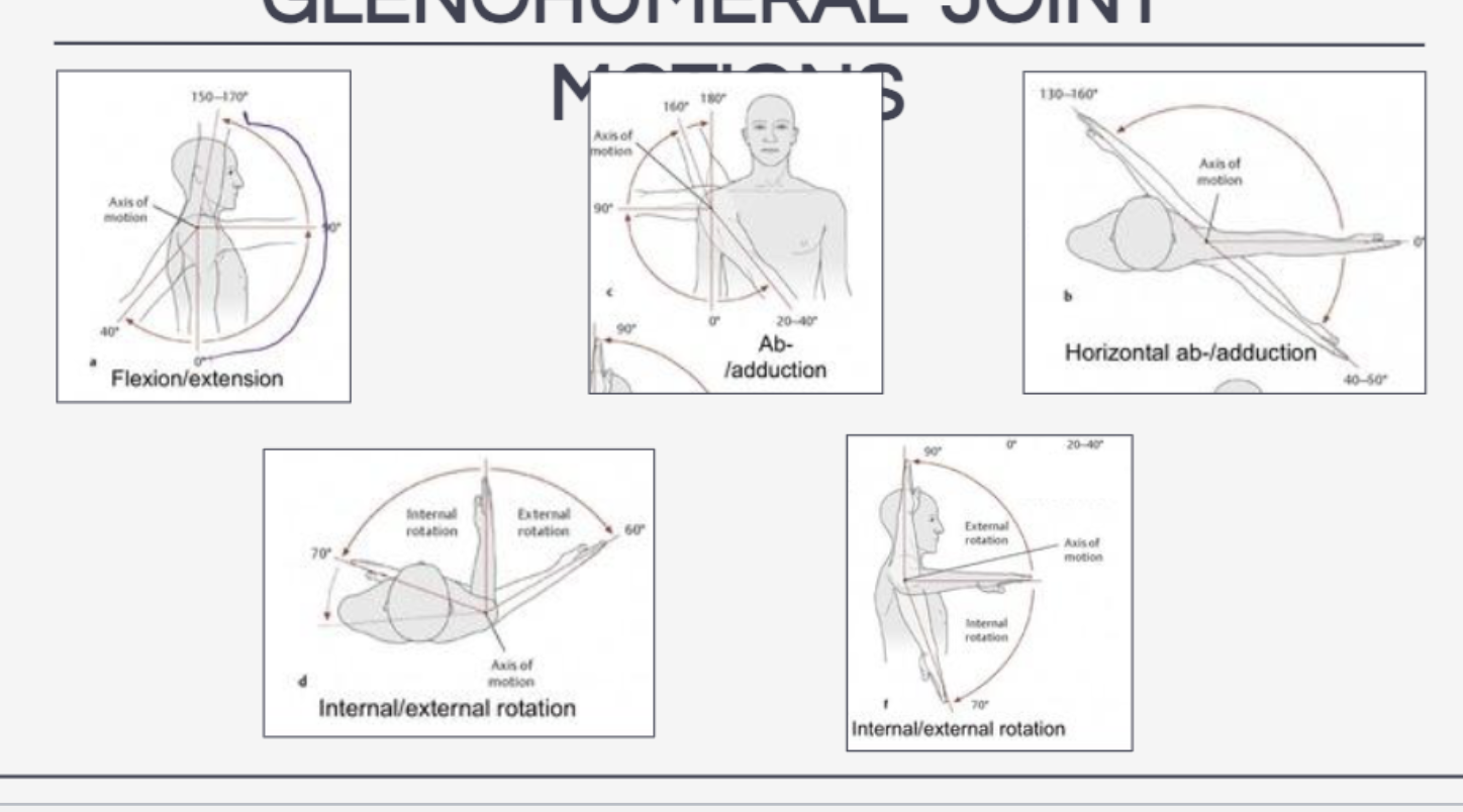
Glenohumeral Joint Motions
Flexion/extension
abduction/adduction
internal (medial) rotation/external (lateral) rotation
horizontal abduction/adduction
circumduction
Scapular Motions
elevation/depression
protraction/retraction
upward rotation/downward rotation

Pectoralis Major
Horizontal Adduction, Internal Rotation
Clavicular Head: Flexion, Abduction above 90 degrees
Sternal Head: Extension, Adduction

Pectoralis Minor
depression
protraction
downward rotation

Deltoid
Anterior: Flexion, Horizontal Adduction
Middle: Abduction, Horizontal Abduction
Posterior: Extension, Horizontal Abduction

Trapezius
Upper: BCS Extension, ICS Lateral Flexion, ICS Rotation, Elevation, Upward Rotation, Retraction
Middle: Scapular Retraction
Lower: Depression, Upward Rotation, Retraction

Latissimus Dorsi
extension
adduction
internal rotation

Levator Scapulae
Bilateral Cervical Flexion/Extension
Ipsilateral Cervical Lateral Flexion
Ipsilateral Cervical Rotation
Scapular Elevation
Scapular Downward Rotation

Rhomboid Major/Minor
retraction
elevation
downward rotation

Serratus Anterior
superior: protraction
inferior: protraction, upward rotation

Supraspinatus
stabilization
abduction

Infraspinatus
stabilization
external rotation
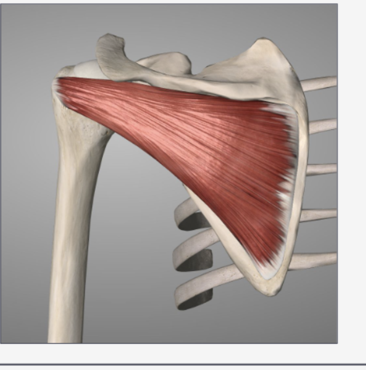
Teres Minor
stabilization
external rotation
adduction

Subscapularis
internal rotation

Scapulohumeral Rhythym
Scapula moves with humerus during shoulder elevation to maintain subacromial space and length tension relationship
Open vs. Closed Kinetic Chain in shoulder
Open
Partnering
Visual Aesthetic
Traditional Corrections
Technical Compensations
Closed
Ground Reaction Forces
Torsional/Rotational Forces
Active vs Passive Stability
Shoulder Injuries
impingement (bicep tendonitis, supraspinatus tendonitis)
instability (sublaxation/dislocation)
labral tears
muscle strains & ligament sprains
Cervical Spine
7
C1 (atlas)
C2 (axis)
Thoracic Spine
12
costal facets for spinal articulation with ribs
Lumbar Spine
5
sacrum & coccyx
sacroiliac joint
Sternum
manubrium sternal body & xiphoid process
Ribs
True (1-7)
False (8-10)
Floating (11,12)
General Vertebrae Landmarks
Vertebral Body
Pedicle
Lamina
Spinous Process
Transverse Process
Superior/Inferior Facet
Vertebral Foramen

Spinal Motions
occur at individual facet joint
flexion
extension
lateral flexion
rotation
Spinal Degrees of Motion
cervical spine: free in all
throacic spine: free in rotation, limited in remaining
lumbar spine: limited in rotation, free in remaining
Spine Ligaments
anterior longitudinal ligament
limits extension
posterior longitudinal ligament
limits flexion
Intervertebral Disc
annulus fibrosis (dough)
concentric Type l collagen
nucleus pulposus (jelly)
waster and type ll collagen
External Oblique
Bilateral Flexion
Ipsilateral Lateral Flexion
Contralateral Rotation

Internal Oblique
Bilateral Flexion
Ipsilateral Lateral Flexion
Ipsilateral Rotation

Rectus Abdominis
Bilateral Flexion
Ipsilateral Lateral Flexion
Posterior pelvic tilt

Transverse Abdominis
Ipsilateral Rotation
Support to Lumbar Spine via Thoracolumbar Fascia
Pulls Abdominal Wall Inward

Iliacus
Hip Flexion
Hip External Rotation
Anterior Pelvic Tilt

Psoas Major
Hip Flexion
Hip External Rotation
Anterior Pelvic Tilt

Erector Spinae
iliocostalis
longissimus
spinalis
Bilateral Extension
Ipsilateral Lateral Flexion
Ipsilateral Rotation

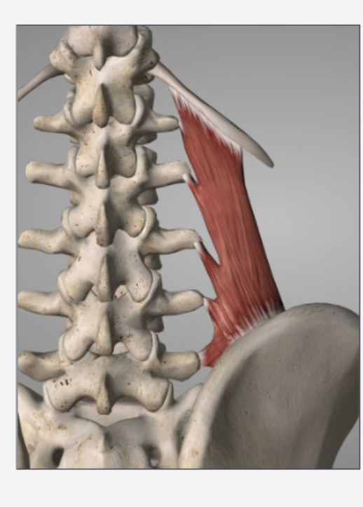
Quadratus Lumborum
Ipsilateral Lateral Flexion
Bilateral Extension
Fixes/Depresses 12th Rib
Stabilizes Spine & Pelvis
Multifidus
Bilateral Extension
Ipsilateral Lateral Flexion
Contralateral Rotation

Rotatores Longis/Brevis
contralteral rotation

Diaphragm
Increases intrathoracic volume, pushes abdominal viscera inferior
