BIOMI 2900 Midterm
 Call Kai
 Learn
 Practice Test
 Spaced Repetition
 Match
 Flashcards
 Knowt Play
1/419

There's no tags or description
Looks like no tags are added yet.
420 Terms
Robert Hooke
first to describe microbes
Antonie van Leeuwenhoek
first to describe bacteria
Pasteur
first to cultivate microbes, disproved spontaneous generation
inoculated sterile medium - no pathogens grew unless he added something
how did Pasteur disprove spontaneous generation?
Koch
developed pure culture techniques, proved that microbes cause disease
pure culture
a growing collection of cells derived from one type of microorganism
Koch's postulates
ID diseased animal, grow pathogen in pure culture, reinoculate pathogen into healthy animal (must cause disease!), reisolate pathogen (must be the same as the original)
Beijerinck & Winogradsky
developed enrichment culture technique, discovered microbial metabolic diversity
microbes!
which are more diverse, microbes or plants/animals?
Watson, Crick & Franklin
developed field of DNA structure
Sanger
developed DNA sequencing technique - helped us understand evolutionary history
Woese
developed the universal "tree of life"
rRNA, compared amongst many organisms
basis of the "tree of life"
Whitaker
developed an early model of the "tree of life" - showed most of evolution was microbial in nature
microbial
most of evolution was...
microbes!
which are older, microbes or plants/animals?
Haeckel
with Darwin, developed the earliest model of the "tree of life"
phylogeny
using molecular sequences to study the evolutionary history of life - reveals microbial diversity!
Mullis
developed PCR
Pace
developed cultivation-independent techniques with rRNA
light microscopy
limited by wavelengths of light... includes bright field, phase contrast and dark field
bright field light microscopy
light microscopy method w/ low contrast, need staining to see structures

phase contrast light microscopy
light microscopy method w/ increased contrast of live bacteria

dark field light microscopy
light microscopy method with increased contrast of refractile sample
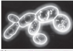
simple staining (used in bright field light microscopy)
stain cells all with one thing (e.g. crystal violet)

compound staining (used in bright field light microscopy)
stain different things differently in order to differentiate structures (e.g. Gram stain)

transmission electron microscopy (TEM)
pass e- (NOT light) through sample, visualize cell structures in sectioned samples
***100x more detail than light microscopy!
scanning electron microscopy (SEM)
coat specimen with metal and observed whether or not e' bound off (yields 3D black and white image)
fluorescence microscopy
can send light of any wavelength (not just white) - structures are visible if mcls are excited (emit light, photons) --> HIGH CONTRAST

centi
10^-2
milli
10^-3
micro
10^-6
nano
10^-9
pico
10^-12
femto
10^-15
100 micrometers
close to visible with the naked eye
0.02 micrometers
size of ribosomes
0.2 - 0.6 micrometers
size of bacteria
2 micrometers - 2 centimeters
size of eukaryotic microbial cells
10 micrometers
size of human blood cell
10-100 micrometers
size of most plant/animal cells
viscous sources >> inertial sources
why is it hard for microbes to move through fluids @
via diffusion (unlike humans)
how do microbes get necessary mcls?
(cell size)^2
variation of microbial metabolic rate
less, less
the greater the size of a microbe, the _______ its SA:V ratio and the ________ its rate
prokaryotic, eukaryotic, and viral
major types of microorganisms
prokaryotic cells
include bacteria and archaea, 0.2-700 micrometers, no organelles needed, no nucleus, and DNA forms "nucleoid"

eukaryotic cells
include just eukarya, 0.2 micrometers - 10 cm, has organelles, DNA in membrane-bound nucleus

endosymbiosis
hypothesis that chloroplasts and mitochondria are prokaryotic descendents which were engulfed by eukaryotic anscestors
***evidence = mitochondria and chloroplasts have BACTERIAL DNA, rRNA & lipids!
cytoplasmic membrane, cytoplasm, ribosomes, dsDNA genomes
things all types of cells share
viruses
NOT cells, 0.02 - 0.6 micrometers (SMALL!), have capsid shell &(potentially) envelope, obligate parasites
coccus

rod, bacilus

spirillum

spirochete
describes shape AND phylum, flagella on INSIDE of membrane, good at corkscrewing through things

budding & appendaged bacteria
help with attachement, PROECTIONS allow greated SA:V

rosettes
lots of budding/appendaged bacterial cells attached together
filamentous bacteria
long, skinny bead-shaped cells, each just a slice
ex) cyanobacteria

bacterial sheath
"house" in which to protect bacteria, effects relationship with nutrients and environment
cell envelope
the first things an antibiotic must encounter upon entering a cell
gram positive cell envelope
has teichoic and lipoteichoic acids (covalently bound to lipids), thick peptidoglycan layer (retains purple stain)

gram negative cell envelope
FAs present, has periplasm, LPS & outer membranes, turns pink with gram stin b/c of a thin peptidoglycan layer

periplasm
most enzymatic processes occur here, so many proteins are localized here
LPS / outer membrane components
porins, lipid A, core polysaccharide, O-polysaccharide
***the outermost surface of interaction, makes up MOST of cell wall!
lipid A
acts as an endotoxin for some pathogens in LPS layer of gram-negative cell envelope
O polysaccharide
provokes a strong immune response in people, found in LPS layer of gram-negative cell envelope
porins
transmit hydrophilic, low MW substances (differ in selectivity)
peptidoglycan
moveable, but gives protection and strength, unique to bacteria, chain of polysaccharides w/ peptide side chains, x-link = covalent peptide bond, can be L or D
direct crosslinks
transpeptidation in Gram negatives

peptide interbridges
transpeptidation in Gram positives

peptide bonds
vertical bonds in peptidoglycan
glycosidic bonds
horizontal bonds in peptidoglycan
autolysis, transpeptidase
how is a new peptidoglycan made?
transpeptidase
adds new peptide x-links to make new peptidoglycan
autolysins
cuts B-1,4 bonds of the polysaccharide backbone so new polysaccharide can be added
lysozyme
antimicrobial enzyme that cleaves the B-1,4 bonds of the peptide backbone
B-lactam antibiotics
antimicrobials that bind and inhibit transpeptidase
cytoplasmic membranes
act as permeability barrier, anchor proteins and conserve E via PROTON MOTIVE FORCE
proton motive force
H+ gradient drives E

nonpolar, small (e.g. O2, N2, H2O)***(+) nutrients the cell wants, via transporters!
what molecules can pass through the cell membrane w/o help?
polar, charged
what molecules can't pass through the cell membrane?
bacterial membranes
phospholipid bilyare w/ hydrophobic head/hydrophilic tail, glycerol-phosphate backbone, FA connected to head via ESTER BOND ***properties vary based on differing side chains & FAs
archaeal membranes
ETHER-linked phospholipids, lipids composed of ISOPRENE units (not FAs), have both GLYCEROL DIETHERS and TETRAETHERS ***may have lipid MONOlayers, BIlayers, or a MIX of both
integral membrane proteins
embedded in the membrane, can act as channels
peripheral membrane proteins
1 part anchored to the membrane, function associated with the membrane
active membrane transport
transport that uses energy (ATP or Proton Motive Force)
Lac permease (example of ACTIVE transport)
transports lactose to E.Coli via proton motive force

Phototransferase (example of ACTIVE transport, E.Coli)
type of group translocation --> substance = chemically modified during transport, used to transport GLUCOSE, FRUCTOSE and MANNOSE, requires 5 proteins ***E = from phosphate bond in phosphoenol pyruvate

ABC Transporter (ATP-binding cassette)
>200 diff systems, highly substrate-specific
*gram (-) have PERIPLASMIC binding pro
*gram (+) have SUBSTRATE binding pro

passive transport
transport via diffusion
across membrane, through integral membrane pro
diffusion can occur (2 ways)...
2 solutes go till = [ ] on each side of membrane
end result of diffusion
uniporters
1 mcl transported passively in 1 direction
antiporter
2 mcls transported passively in opposite directions
symporters
2 mcls transported passively in the same direction (1 of them is going down its concentration gradient)
archaeal cell envelope
***NO peptidoglycan! + usually no outer membrane, often have and S-layer, and some have pseudomeurein (regardless of presence of S-layer)
S-layer
tough, paracrystalline structure in archaeal cell envelopes, composed of pro or glycopro
pseudomurein
polysaccharide in archaeal cell envelopes, similar to peptidoglycan, but has NO D-AAs & has B-1,3 (not B-1,4) bonds between sugars
poly-B-hydroalkanoate (PHA)
internal cell structure -> polyester, C & E storage

glycogen
internal cell structure -> glucose polymer, C & E storage