Internal Environment: Circulation (2)
 Call Kai
 Learn
 Practice Test
 Spaced Repetition
 Match
 Flashcards
 Knowt Play
1/49

There's no tags or description
Looks like no tags are added yet.
50 Terms
Be able to differentiate between flow and velocity in terms of units and concept
Flow: CO is flow, volume of blood per minute
Velocity: d/t
Increasing which of the following factors in a tissue will increase blood flow to that tissue? (ICQ1)
Oxygen (dec. oxygen if tissue is hypoxic, arteriole will relax)
pH
Carbon dioxide
Sympathetic stimulation (didn’t do anything with it)
All of these are correct
ICQ Explanation
deals w/ arterioles
talking about regulation of blood flow to the tissues outside the autonomic nervous system → purely based on metabolism
doesn’t happen unless in repro
no sympathetic
tissue metabolic activity determines how much blood flow it gets
one of the signs of inc. metabolic activity is inc. CO2 production
oxidative phosphorylation
inc. hydrogen, make more acidity (pH dropping will inc. blood flow to tissue)
If a vessel has the conformation as shown below and blood is flowing from A to D, where is pressure the greatest? ICQ2
A
B
C (blood would flow both ways)
D
Pressure is the same at A, B, C & D

ICQ2
For C, blood would go from H → L pressure… resistance is higher at C
pressure is higher in capillaries
pressure in circulatory system is lowest at vena cava
pulmonary side → pulmonary vein
close to 0
If a vessel has the conformation as shown below and blood is flowing from A to D, where is flow the greatest? ICQ3
A
B
C
D
Flow is the same at A, B, C, & D

ICQ3
flow is liters/minute
CO on both sides of heart is same
shock: pressure becomes lower in the capillaries than it does in the veins
arterioles: controlling fraction of flow they get
lunch eaters sending higher fraction of CO to digestive system
Have lunch
metabolic activity of digestive system increases, it starts producing more CO2, using more oxygen
Blood flow to the arterioles to the GI tract dilate, open up to increase blood flow to that tissue
Did not trigger parasympathetic to dilate
Exercise
Increase temperature will increase blood flow to the skin
Increase temperature will increase muscle blood flow
Decrease pH will increase blood flow
Organ system itself gets to choose how much blood you need (intrinsic)
Input
Endocrine
Cortisol regulates a1 expression on smooth muscle of arterioles
CNS
Sympathetic
a1 on most arterioles and veins
B2 on arterioles to skeletal muscle and cardiac muscle
Parasympathetic
Only to reproductive organs
m2
Blood Flow
Dependent upon pressure gradients and vascular resistance
F = ∆P/R
Bernuolli’s Principle - deals with P (pressure foes from H → L)
Resistance (based on radius)
Viscosity
Surface area
Radius
Blood Flow (cont’d)
Radius - major determinant of resistance
Under smooth muscle control (i.e. sympathetic)

Poiseuille’s Law (conceptual formula, if this changes how does this impact flow?)
Flow rate = π∆Pr4/8nL
if values in the numerator go up, flow goes up
pressure, radius
if values in the denominator go up, flow goes down
viscosity

Arteries
Roles
Blood transportation away from the heart (anatomy)
Pressure reserve (physiology → arteries supply pressure bc they are plastic & recoil, keep the blood moving during diastole)
Functional anatomy
Large diameter
Low resistance
Elastic fibers
Collagen fibers
Arteries as a pressure reservoir

Arterial blood pressure

Regulation of Flow
Arterioles
role is to regulate CO to the diff. tissues
if everything is normal, organ systems get to choose
Pressure Drop
Starts at 93 mm Hg (MAP)
Ends at 37 mm Hg
had lunch, didn’t wait an hour & went swimming → putting your life at risk
blood going bc of lunch → digestive system
bc of swimming → muscles
could be a disaster
have enough blood to go around, tell CO to go up
give blood, eat lunch & go swimming → might not have enough
sympathetic nervous system will override in case of emergency

Regulation of Flow (cont’d) (in normal conditions w/out any emergency, based upon metabolism of target tissues)
Vasoconstriction
⬇︎ Tissue activity, CO2, H+, etc.
⬆︎ O2, sympathetic stimulation (tissue that is well oxygenated don’t need to send blood there, constricts & shunts blood to another part)
Cold, angiotensin II
ex: hypothermic
tissue ends up being hypoxic
heart is continuing to have a high oxygen demand & if they’re cold, heart doesn’t get its own blood supply as much… go into fibrillation)
Vasodilation
⬆︎ tissue activity, CO2, H+, K+, osmolarity, adenosine, prostaglandins
︎︎︎⬇︎ O2, sympathetic stimulation
Histamine, heat
body doesn’t use it physiologically to dilate
if we encounter something that triggers histamine, it will dilate
doesn’t inc. blood flow to a tissue
Regulation of Flow (cont’d 2)
Local control
Influences CO to organ (system)
Chemical mediators
Believed to act on vascular endothelium, not directly smooth muscle
NO- smooth muscle relaxation
Endothelin- smooth muscle contraction
Angiogenesis (creating new capillaries)
Histamine
90% of all cases of high BP have no discernible cause
endothelial cells are not doing their job properly, not sending right signal at right time
Regulation of Flow (cont’d 3) (stress relaxation if you stretch smooth muscle, it relaxes (stomach)
Local Physical Influences
Heat/Cold
Stretch
Arteriolar smooth muscle: stretch increases tone (not stress-relaxation response)
if we have inc. pressure & are stretching it, it’s going to reflexively constrict blowing out bc of too much pressure → myogenic effect
Decrease in stretch decrease in tone
Extrinsic Control
MAP regulation
MAP = CO x TPR (resistance)
conceptual
will calculate CO, HR, & SV
Regulation of Flow (cont’d 4)
Extrinsic control (external control, main site is autonomic nervous system)
Sympathetic tone (centrally mediated vasoconstriction)
Overridden in exercise
Norepinephrine
Cerebral vasculature is under local control
sympathetic tone is overridden by heat
body temp. is going up, dilating to skin to lose heat
sympathetic response should constrict to skin
BP during exercise doesn’t jump that much
rate & CO pick up, dilating (resistance drops)
in cooler temp., forcing skin to constrict
inc. BP higher than it should be during exercise
exercise & stress are similar, but not identical
BP does jump during stress
Cardiovascular Control Center (of autonomic nervous system)
Medulla
Regulation of MAP via baroreceptors
Normal Arteriolar Tone

Arteriolar vasoconstriction
low metabolic activity is going to constrict
don’t need blood going to that tissue bc it’s not metabolically active
What is Incorrect in the Image
The list incorrectly pairs metabolic factors with vasoconstriction. Those specific changes actually trigger vasodilation(opening the vessel, not narrowing it):
↓ Oxygen & ↑ Carbon Dioxide
Image claims: Causes vasoconstriction.
Correction: Low O2 and high CO2 indicate active tissue needing more blood flow, which triggers local vasodilation (arteriolar relaxation) to deliver oxygen and remove waste.
Vasoconstriction is caused by ↑O2 and ↓CO2 (low metabolic demand).
↓ Myogenic activity:
Image claims: Causes vasoconstriction.
Correction: Decreased myogenic activity (less stretch/pressure) leads to smooth muscle relaxation, causing vasodilation. Vasoconstriction is caused by ↑↑ Myogenic activity (increased stretch/pressure).
Notice how the handwritten pink note at the top contradicts the slide text by correctly stating: "low metabolic activity is going to constrict... don't need blood going to that tissue be it's not metabolically active."

Arteriolar vasodilation
all indicators of high metabolic activity is going to dilate bc you need more blood there

Autoregulation of tissue blood flow (Myogenic Effect)
capillaries are fragile, will tear apart if pressure is too high
one of ways we manage hypertension physiologically is inc. urine production
clinical sign: unable to sleep through night w/out having to get up to urinate
pressure goes up, arteriole will constrict a little, but not to bring it back to normal
if it doesn’t stay with flow, can’t get rid of extra volume, can’t bring pressure down

Factors affecting TPR

Exchanges across a continuous capillary wall
How do we get stuff from inside capillaries pushed into circulation so that it can go out & provide nutrients to the cell & pick up waste products

Diffusion
Interstitial fluid from capillaries
Passive diffusion (whether it’s water-soluble or lipid)
Down gradients
Solubility
Bulk flow
Ultrafiltration (pushing fluid out into interstitial space)
Reabsorption (pulling fluid back in & putting it on the venous side & sending it off to the heart)
Bulk flow across the capillary wall (Starling’s foces)
major force pushing out is BP
abbreviated Pc → called capillary BP
flow requires a change
no change in pressure (37 → 37)… no flow
has to drop
inward force is constant
what if this was a person living in the country with poor nutrition?
poor nutrition leads to liver failure
liver is responsible for producing most of plasma proteins
have low plasma proteins, low nutritional source for protein
value of 25 goes down
outward force stays the same, inward goes down
fluid production in interstitial space goes uo
edema
gathers in abdomen

Bulk Flow
Capillary blood pressure (hydrostatic pressure, Pc)
Outward force
Starts at 37 mm Hg, ends at 17 mm Hg
Determined by blood pressure
Plasma-colloid osmotic pressure (oncotic pressure, πp)
Inward force
proteins, ~25 mm Hg
Bulk Flow 2
Interstitial-fluid hydrostatic pressure (Pif)
Inward
Atmospheric
1 mm Hg
Interstitial-fluid colloid osmotic pressure (πif)
Outward
Protein in the interstitial spaces (negligible)
Bulk flow across the capillary wall (again)
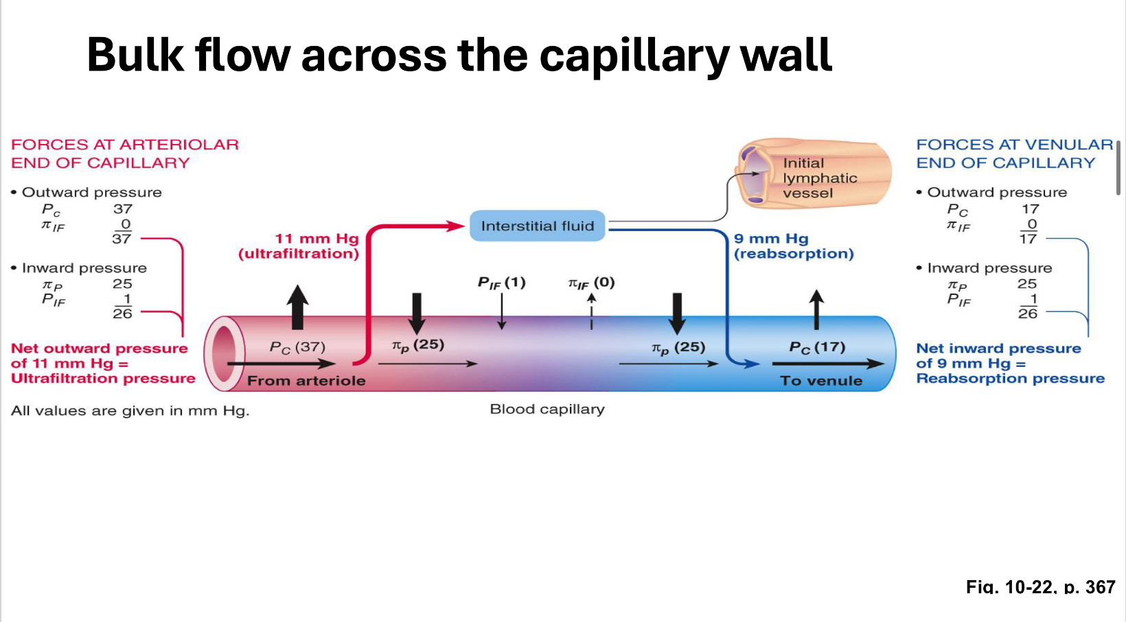
Bulk Flow 3
Net Exchange Pressure
(Pc + πif) - (πp + P if)
Positive number = ultrafiltration
Negative number = reabsorption
Problems lead to edema
Lymphatic System
Returns interstitial fluid to the heart
11 mm Hg on arterial-end of the capillary
9 mm Hg on venous-end of the capillary
2 mm Hg absorbed into Lymph Vessels
3 liters of lymph per day
7200 liters of blood in the capillaries per day
Anatomy
Slight overlap with endothelial cells
Allows fluid in, but not out
Can transport plasma proteins and bacteria
Travels to lymphnode
Edema
Reduction in plasma proteins
Decreases reabsorption
Increases permeability of capillary walls
Increased venous pressure
Blockage of lymph vessels (blocks lymphatic drainage)
interstitial fluid accumulation due to parasitic infection (microfilarin gets bigger & stuck)
any time tissues get pushed away from blood supply it dies
breast cancer
take lymph nodes out
axilla
lymphedema
Veins
Return blood to the heart
Serve as blood reservoir (64% of blood volume in veins)
Anatomy
Low level of smooth muscle
Low levels of elastic fibersHigh levels of collagen
64% of total blood volume
people can be hypertensive, but they don’t have swelling everywhere bc tissues are being protected

Factors that facilitate venous return

Venous Return
Venous Capacity (how much blood the veins hold)
Increase in capacity diminishes return
Blood volume
Sympathetic activity
Minimal constriction increases flow
Decreases venous capacity
if we have an arteriole & sympathetic squeezes it, flow, the volume of blood going through that arteriole drops
veins are like tubes of toothpaste with cap off
flow goes up
sympathetic stimulation to veins squeezes them & dec. volume they can hold
bc we have valves, only place blood can go back to is heart, inc. venous return
Skeletal muscle activity
Respiratory pump
Fig. 10-34, p. 376

Control
Short-term
Baroreceptor reflex (measured by carotid bodies, carotid sinus)
Long-term (long-term regulation of BP is through fluid control, sympathetic & parasympathetic are short-term)
Osmoreceptors & Volume receptors
Hypothalamus, kidney, atria
Alters salt and water balance to alter volume
Firing rate in the afferent neuron from the carotid sinus baroreceptor

In the capillaries, the greatest force that moves fluid from the interstitial space and returns it to circulation is:
Capillary hydrostatic pressure
Plasma colloid oncotic pressure
Interstitial fluid hydrostatic pressure
Interstitial fluid oncotic pressure
None of these are correct
Hypertension, Hypotension, & Shock
Hypertension
140/90
SECONDARY (10% of cases, can fix it if you treat, patient isn’t hypertensive anymore)
Due to definitive cause
Cardiovascular
Renal
Endocrine
Neurogenic
Hypertension
Primary (idiopathic, 90%, all we can do is manage symptoms, don’t know cause)
Salt management
Na+/K+ pump problems
Pressure on the medulla
Nitric Oxide
endothelial cells are not releasing nitric oxide, or smooth muscle is not responding to nitrous oxide
causes arterioles to open up, dilate (relaxer)
Digitalis-like compounds
Pathophysiology

Hypotension (drop in BP, so brain is not perfused adequately, give into gravity, loss of consciousness)
> 90/60 ±
Orthostatic (from standing too long)
Emotional stress
Shock (if hypotension goes bad enough, 4 types, perfusion is 0)
Hypovolemic
Cardiogenic
Vasogenic
Septic
Anaphylactic
Neurogenic
Circulatory Shock
MAP = CO x TPR
if resistance goes down, pressure goes down
if CO goes down, pressure goes down

The most common cause of hypertension is:
Poor diet
Lack of exercise
Sodium consumption
Na+ does not cause hypertension
if you have hypertension, Na+ is not a good idea
can contribute, will make heart failure worse… fail faster
Heart failure
Unknown
Pathophysiology 2
Positive feedback loops
Decreased tissue perfusion
Increased cellular death
Increased resistance in capillary beds
Decreased tissue perfusion
Decreased venous return
Decreased cardiac output
Metabolic acidosis
Increased interstitial fluid volume
Decreased venous return
Decreased blood pressure
Decreased coronary perfusion
Decreased blood pressure
Shock index = HR/SBP
SO2 = (HR x SV) x (1.39 x [Hb] x SaO2 + 0.03 x PaO2)

Three Positive Feedback Loops in Shock

Consequences and compensations of hemorrhage
