Therapeutic Approaches - Understanding and Management of Congestive HF in SA
 Call Kai
 Learn
 Practice Test
 Spaced Repetition
 Match
 Flashcards
 Knowt Play
1/14

There's no tags or description
Looks like no tags are added yet.
15 Terms
What is cardiac output?
CO = SV x HR
Cardiac output (CO) is the total volume of blood pumped by the heart's left ventricle (or right ventricle) into the circulatory system every minute.
Stroke Volume: Amount of volume the pump moves during cycle.
Stroke volume is the amount of blood ejected by a ventricle during each heartbeat (Cardiac Contraction)
Heart Rate: How many cycles your pump performs in a determined amount of time.
What affects stroke volume?
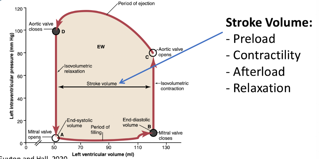
How are SV and HR related?
High HR decreases SV: When your heart beats too fast, the duration of diastole (the relaxation and filling phase between beats) is cut short. Because the ventricles don't have enough time to fill completely with blood, the volume of blood available to push out on the next beat drops.
Low HR increases SV: Conversely, a slower heart rate allows for a longer filling window. This increases the End-Diastolic Volume (the total blood pooled in the ventricle right before it squeezes).
**EXCEPT in exercise SV and HR both increase to accomodate
What impact does hemorrhage have on the body?
Neuro-endocrine response
Restore output: increase volume + HR
Maintain perfusion: vasoconstriction

What are the different ways to define the type of heart failure?
• There are different way to define the type of heart failure:
• Anatomical side affected → Site differences
• Development of clinical signs → Type of failure
• Dependent on cardiac architecture → Pathomechanismus difference
What is an example of forward failure?
Cardiogenic Shock:
• Very fast heart rate = less time to fill the heart → If not filled, cannot be pumped out.
• Cardiac remodelling → Tachycardia induced cardiomyopathy
• Clinical signs:
• Weakness
• Hypothermia
• Hypotension
Perfusion Issue → «FORWARD»
« The pump is not pumping»
Site: Left
Type: Forward
What does decompensation of the left side of the heart lead to?
A decompensation of the left side of the heart often leads to a clinical manifestation of left sided backwards failure.
The left side of the heart is a high pressure system. An increase in pressure affects the pulmonary veins.
How does cardiogenic pulmonary edema occur?

How does vasculature appear on x-ray?
Lateral x-ray
Veins appear - central and ventral, are distended and large compared to dorsal artery
Vein - black arrows
Arteries - white arrows
Pulmonary oedema - changes the opacity (More like soft tissue and fluid)

Cats can manifest a left sided heart failure with..?
Cats with left sided heart failure show lung oedema and pleural effusion
Dogs with left sided heart failure show lung oedema
Pleural effusion usually issue of right side of the heart in dogs
Describe an example of backward failure.
Cardiogenic Pulmoary Oedema
• Clinical signs:
• Tachypnoea/Dyspnoea
• Cough (Cats do not, dogs do - but near death cat will cough)
→ Fluid congestion inside of the lungs
«The pump is not sucking»
Myxomatous Mitral Valve Disease (MMVD)
• Mitral valve Insufficiency → Backflow → Increased Atrial Pressure → Cardiogenic Pulmonary Oedema
• Site = Left
• Type = Backwards
Summarize the difference between forward and backward failure.
Backward Failure
The Mechanism: The heart muscle is stiff or thick. It contracts normally, but it cannot relax properly to fill with blood.
Why it's isolated backward failure: Because it contracts normally, the heart manages to push enough blood forward to meet the body's baseline needs at rest. However, because it cannot relax, blood immediately backs up backward into the lungs (if left-sided) or the body (if right-sided).
The Symptoms: Fluid backing up into the lungs causing shortness of breath (dyspnea), coughing, or fluid backing up into the legs—all while organs like the kidneys and brain receive plenty of forward blood flow.
Forward Failure
The Mechanism: A massive, sudden insult severely cripples the heart’s ability to contract.
Why it's isolated forward failure: The drop in forward output happens so fast that the upstream venous system hasn't had the time to bottleneck and congest yet.
The Symptoms: Signs of profound shock due to lack of blood flow to vital organs. This includes a dangerous drop in blood pressure (hypotension), cold or clammy extremities, confusion (lack of blood to the brain), and a sharp drop in urine output (kidneys failing due to lack of perfusion).
If a patient has severe forward failure, the blood that isn't being pumped out will inevitably pool inside the heart, eventually causing backward congestion.
If a patient has severe backward failure, the high pressures will eventually stress the heart muscle so much that its ability to pump blood forward drops.

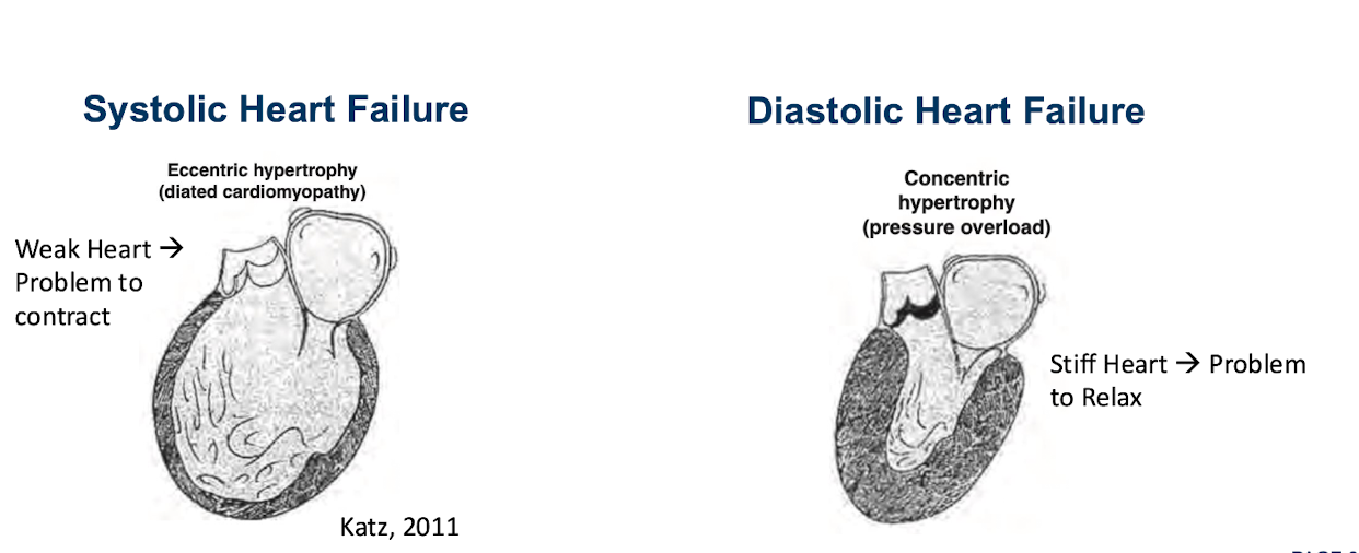
Describe systolic vs. diastolic hear failure.
**Hint at the phenotype causing the heart failure

What underscores the treatment of left-sided congestive heart failure?
• Oxygen supplementation
Ventilation?
• Loop diuretics - get rid of the excess fluid
Furosemide, Torasemide
• Systolic Dysfunction?
Pimobendan
Dobutamine?
• Arrhythmia treatment
• Evtl. ACE inhibitor/spironolactone (mostly DCM)
• Thoracocentesis if pleural effusion is present (cats).
Describe the difference between:
Forward vs. Backward Failure
Systolic vs. Diastolic Failure
Forward vs. Backward Heart Failure (Hemodynamic Classification):
Forward heart failure refers to the heart’s inability to pump out a sufficient volume of blood to meet the metabolic demands of the body (low cardiac output, leading to fatigue, confusion, or poor kidney perfusion).
Backward heart failure refers to the backup of blood behind the failing ventricle, causing elevated pressures in the pulmonary veins or systemic veins and resulting in fluid accumulation, such as pulmonary congestion/edema or peripheral swelling (edema/ascites).
Systolic vs. Diastolic Heart Failure (Mechanical/Contractile Classification):
Systolic heart failure (Heart Failure with Reduced Ejection Fraction, or HFrEF) occurs when the left ventricle loses its normal squeezing/contractile power, dropping the ejection fraction to 40% or lower.
Diastolic heart failure (Heart Failure with Preserved Ejection Fraction, or HFpEF) occurs when the heart muscle becomes stiff and cannot relax normally during the resting phase between beats, impairing proper filling even though the squeezing percentage (ejection fraction of 50% or higher) is preserved.