Developmental Bio Exam 2
 Call Kai
 Learn
 Practice Test
 Spaced Repetition
 Match
 Flashcards
 Knowt Play
1/66

There's no tags or description
Looks like no tags are added yet.
67 Terms
How do you maintain the tissues of your body in the face of damage? How do you continue to grow after embryogenesis?
Most multicellular organisms grow and change shape even after birth
Damage to your tissues and cells come in many different forms
External damage
Internal damage
Stem cells are our real-life regeneration
Stem cells allow the body to continue to generate new cells
Stem cells: a relatively undifferentiated cell from the embryo, fetus, or adult, that divides and when it does so, produces
One cell that retains its undifferentiated character and remains in the stem cell niche
A second cell that leaves the niche and can undergo one or more paths of differentiation
Single cell asymmetry
A single cell divides to produce another stem cell (self-renewal) and a daughter cell that will differentiate
These cells typically have potency, or power, to produce multiple types of differentiated cells
Population asymmetry
A group of cells divides to produce another stem cell (self-renewal) and a daughter cell that will differentiate
This method can be prone to sway in its proportions;
may not always have the same number of stem cells and daughter cells
Adult stem cell lineage
A stem cell divides and produces a daughter cell called a progenitor cell
relatively undifferentiated cells that are capable of a few divisions before differentiation
cannot self-renew indefinitely
sometimes move while dividing
Totipotent
Cell: Zygote
Source: Zygote
Most versatile cells, can differentiate into anything
Pluripotent
Cell: Embryonic stem cell
Source: Blastocyst (inner cell mass)
Tissues through gastrulation
Multipotent
Cell: Multipotent stem cell
Source: Embryo, adult brain
Embryo post gastrulation through adulthood, anything in a specific germ layer
Limited differentiation potential
Cell: Progenitor (example neuronal progenitor)
Source: Different tissue types (example brain or spinal cord)
Pluripotent stem cells come from the inner cell mass of the blastocyst
Trophectoderm
ICM (inner cell mass)
Primitive endoderm
Epiblast → embryo
Many different signaling mechanisms control whether a stem cell will divide
Paracrine signaling
Extracellular matrix (ECM) adhesion / mechanical force
Cell adhesion
Juxtacrine signaling
Endocrine signaling
Adult stem cells usually reside in specific locations in the organisim during development called niches
Stem cell niche - niche that houses stem cells and regulates stem cell
self-renewal
survival
differentiation of the progeny that leave the niche
One example of adult stem cell niche:
Small intestine crypts:
line the lumen of the small intestine
is a model stem cell niche
is adult stem cell lineage
use many of the signaling paradigms we have discussed to specify cell division and fate specification
Stem cells are asymmetrically dividing cells with the ability to differentiate into multiple lineages
Single-cell asymmetry
stem cell → committed cell
Population asymmetry (symmetrical differentiation)
stem cells
committed cells
Adult stem cell lineage
multipotent stem cell → committed cell → progenitor (transit-amplifying) cell → differentiated cells
Stem cell niche
Niche that houses stem cells and regulates stem cell:
self-renewal
survival
differentiation of the progeny that leave niche
Major example: intestinal stem cells
The Melton and Zhou labs
generated endocrine pancreatic cells through in vivo reprogramming of exocrine pancreatic cells
Etrastmus Darwin
Physician/naturalist
sexual reproduction via gamete recombination allows for an incredible amount of genome variation on which evolution can act
Sex is determined several different ways in animals
One common method is by the type of sex chromosome present in the organism
ex. Birds
female: ZW
male: ZZ
Sex is determined several different ways in animals (mammals)
Father: 44, XY → sperm: 22 X and 22 Y
Mother: 44, XX → eggs: 22 X and 22 X
Offspring genotypes: 44XX 44XY
Sex is determined several different ways in animals (insects)
Hymenopterans (bees, wasp, ants, ploidy level determines sex)
Flies
ratio of X chromosomes determines sex, not the presence of Y, XXY = would still be female due to ratio
How do we developmentally achieve different sexual phenotypes/morphs (mammals)
Mammals begin development with a bipotential gonad, start with vasculature if bipotential gonad that is a mosaic for different lineage (3 germ layers + germ cells) that can go XX (ovary → oocyte) or XY (testis → sperm)
Mammalian gonads begin development bipotentially
Mammalian gonads start as bipotential structures called genital ridges
Primordial germ cell migrate in mix with somatic cells
Mammalian gonads start as bipotential structures called genital ridges
primordial germ cells migrate in mix with somatic cells
Primary sex cords form:
either degraded during ovary development or progress development for mature testis
The bipotential gonad begins as a genital ridge, which is remodeled as sexual development progresses
initial sex chords → turn into primary sex cords → then either go to ovary development or testis development
For ovary development growth of cortical cords (secondary sex cords), ovarian follicles start, oogonium, ovarian medulla and ovarian cortex
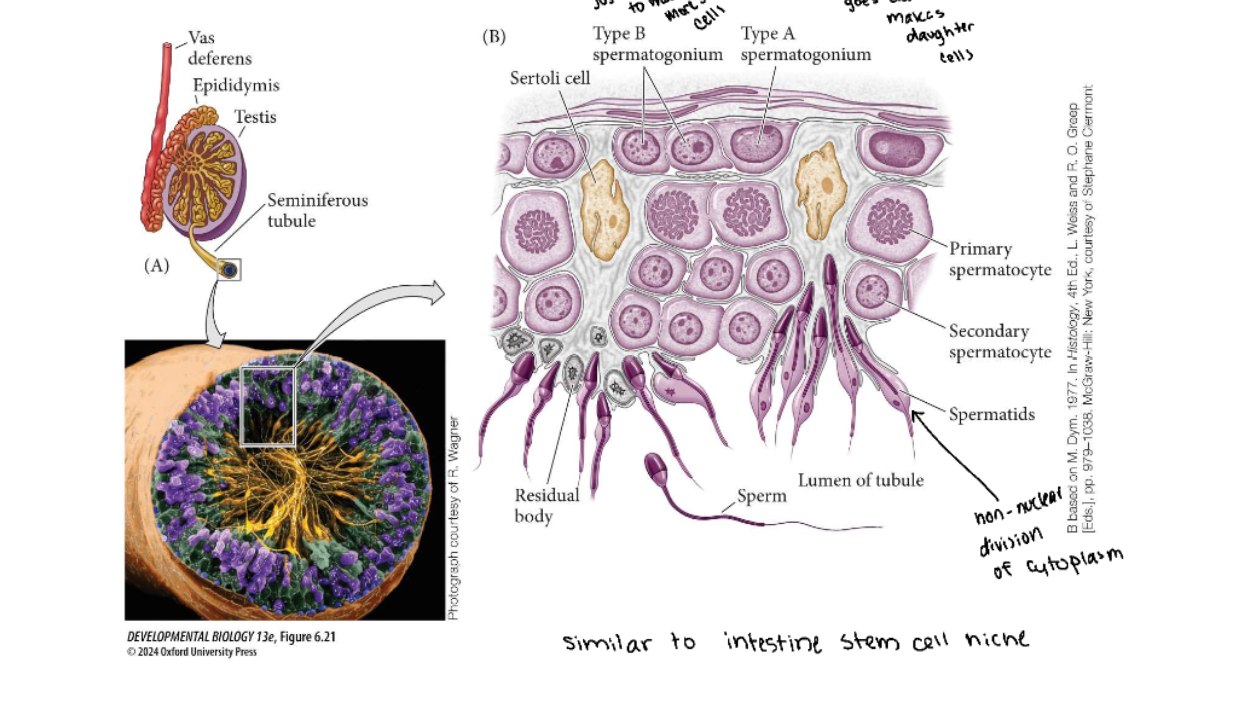
For testis continued primary sex cord development that then becomes seminiferous tubule housing spermatogonia
A differential gene expression cascade mediated by Sry (sex-determining region Y) and Wnt4 are thought to be at the heart of male/female sex determination

A more detailed genetic cascade for the generation of mammalian gonads

Mammalian gonads begin development bipotentially
Mammalian gonads start as bipotential structures called genital ridges
primordial germ cells migrate in mix with somatic cells
primary sex cords form
either degraded during ovary development or progress development for mature testis
The biopotential gonad makes a complex series of fate decisions based on autonomous and contextual differentiation
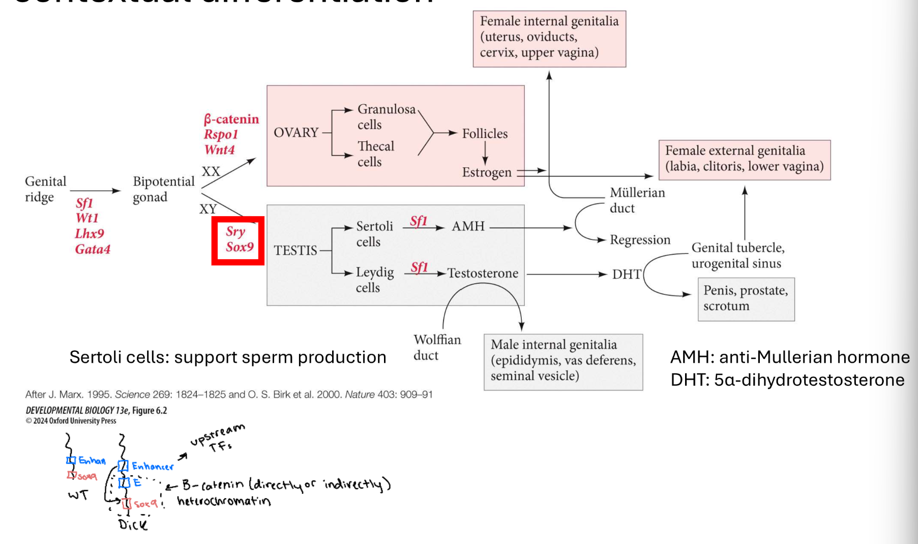
Sex-determining region Y (Sry) is one of the most important upstream regions factors in male sex determination

Sox9’s ability to promote testes development has been demonstrated experimentally

The development of gonads influences how the other sexual organs and structures develop from the bipotential genital ridge
Originally, a bipotential gonad develops, with both undifferentiated Mullerian ducts (female) and Wolffian ducts (male) present.

In addition to sex determination, X chromosomes carry other genes that can be transcriptionally affected by a process called x-inactivation
At fertilization
female with two X chromosomes
Early cleavage
both X chromosomes active in all cells
Implantation
random inactivation of one X chromosome in all cells of the inner cell mass, which will from the embryo
Retts syndrome
Barr bodies inactivate one X chromosomes
Sex phenotypes in mammals are also affected by the ability to generate and receive hormone signals
Mechanism of androgen receptors
testosterone is an androgen (masculinizing) steroid hormone that can travel to cells through the blood
In addition to genetic control of sexual determination, environmental factors play a major role in some species
Most turtles, and all crocodilians determine sex via environmental determinates
Specifically, they use temperature-dependent sex determination
Turtles and crocodiles only have even sex distributions at specific temperature ranges (hard to have even sex distribution with global warming)
Control of the sex-determination in these species is through Dmrt1
Dmrt1 controls the testes-determining cascade in many vertebrates
Amphibians, birds, fish
Mammals use Dmrt1 for maintenance of testes characteristics
A homolog for Dmrt1, doublesex, is important for Drosophila sex determination
Mammals use Dmrt1 for maintenance of testes characteristics

Methylation of Dmrt1 gene in Red-eared slider turtle eggs is correlated with temperature
MPT: Male producing temperature
male temp; low methylation of Dmrt1
FPT: Female producing temperature
female temp; high methylation of Dmrt1
If temperatures changed it causes methylation or unmethylation
High temperatures inhibit the transcription of KDm6B, a histone modifying protein
KDm6B (demethylase) can remove methyl groups from histones around genes like Dmrt1
allows for transcriptional activation of Dmrt1
Only transcribed heavily at male sex permissive temperatures
A temperature-sensitive Ca+ transport protein allows for a downstream phosphorylation of Stat3, which promotes the female sex-determination pathway
High temp turns on the breaks for females
When Stat3 is not phosphorylated it is not active

Once we have our gonads and secondary sexual phenotypes established, the gonads must control meiosis and gamete generation (Oogenesis)
Meiosis initiated once in a finite population of cells
One gamete produced per meiosis
Completion of meiosis delayed for months or years
Meiosis arrested at first meiotic prophase and reinitiated in a smaller population of cells
Differentiation of gamete occurs while diploid, in first meiotic prophase
All chromosomes exhibit equivalent transcription and recombination during meiotic prophase
Once we have our gonads and secondary sexual phenotypes established, the gonads must control meiosis and gamete generation (Spermatogenesis)
Meiosis initiated continuously in mitotically dividing stem cell population
Four gametes produced per meiosis
Meiosis completed in days or weeks
Meiosis and differentiation proceed continuously without cell cycle arrest
Differentiation of gametes occurs while haploid, after meiosis ends
Sex chromosomes excluded from recombination and transcription during first meiotic prophase
Retinoic Acid (RA)/(a major class of morphogens) is the upstream control for meiosis in germ cells
Female germ cells
normal:
RA → Stra8 → Meiosis
Male germ cells
Nanos2 added:
RA → Stra8 (doesn’t go to Meosis), Nanos2 transcription factor that represses Stra8 so meiosis does not continue
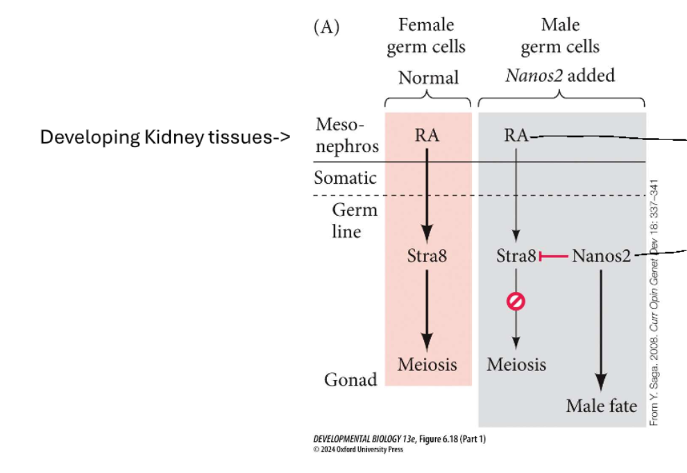
Male germ cells inhibit meiosis until puberty, where the Sertoli cells begin synthesizing RA

Sperm maturation occurs in stepwise fashion from outside the seminiferous tubule to the inner lumen
Mammalian oocytes divide rapidly before birth, but most die off; remainder halt as primary oocytes until systematic maturation at puberty

Oocytes are arrested at the first meiotic phase until puberty and the presence of luteinizing hormone allows the near completion of meiosis
Oocyte stage:
Primordial
arrested at diplotene
growth arrested
surrounded by single layer of granulosa cells
4c,2n
Germinal vesicle (GV)
arrested at diplotene
fully grown oocyte
surrounded by multiple layers of granulosa cells
4c,2n
Metaphase II (MII)
arrested at metaphase II
mature oocyte
developmentally competent
2c,n
Fertilization - the fusion of gametes to create a new organism
Combination of two different goals:
Sex - combining of genes derived from two parents
Reproduction - generation of new organism
Study of fertilization as we know it did not truly begin until mid-1800s
Sperm was discovered in the 1670s separately by Anton van Leeuwenhoek and Nicolas Hartsoeker
Leeuwenhoek initally thought the sperm were parasites, a line of thought that persisted in others for decades
Later, he and Hartsoeker both theorized each sperm contained a miniature pre-formed embryo or a “homunculus”
In 1840s Albert von Kolliker described the formation of sperm in the testes
known for anatomy and neuroscience
Karl Ernst von Baer and Alphonse Derbes show union of sperm and egg in sea urchins in 1847
The egg arrested at various stages of meiosis depending on species with completion occurring upon sperm entry
Primary oocyte:
roundworm
mesozoan
sponge
polychaete worm
clam worm
clam spisula
Urechis worm
Dogs and foxes
First metaphase
nemertean worm
polchaete worm
mollusk
core worm
many insects
seastars
Second metaphase
lancelet
amphibians
most mammals
fih
Meiosis complete
cnidarians (anemones)
Sea urchins
Our sea urchin eggs share much in common with mammalian eggs
Sea urchin:
female pronucleus
egg cell membrane
vitelline envelope
jelly coat
Mammal:
cumulus
polar body
ovum
zona pellucida (similar to vitelline envelope)
Cumulus
Mammalian eggs travel from the ovary inside a body of cells called a cumuls (cloud of supporting cells; similar to jelly coat)
Sea urchin fertilization involves the attraction of sperm to the egg, the acrosome reaction, and then membrane fusion
sperm contacts jelly layer
Acrosome reaction
Digestion of jelly layer
Binding to vitelline envelope
Fusion of acrosomal process membrane and egg membrane
Sperm are attracted to the egg in echinoderms via a molecule called resact found in egg jelly (sea urchins specifically)
Many species that undergo fertilization externally, particularly in aquatic environments, use chemotaxis to guide sperm to the egg
Resact experimentally deposited into a dish of sperm causes the sperm to swim toward the highest concentration of the molecule
Acrosome reaction
allows for the expulsion of enzymes for extracellular matrix digestion and exposure of membrane binding proteins
Bindin proteins found on sperm cell membrane are species specific
causes agglutination of eggs if same species
Hypothetical pathway of egg activation upon fertilization in sea urchins

Fast blockages to polyspermy are primarily achieved through change in membrane potential
the influx of sodium (Na+) to the egg cell upon initial sperm binding raises the membrane’s resting electric potential and prevents other sperm binding
Slow blockage of polyspermy occurs through raising of the fertilization membrane
Slow rising of the membrane creates a physical barrier to sperm entry; is considered slow in comparison to the membrane potential change
Calicum flash
Both slow raising of the membrane and fast membrane potential changes are thought to be tied to the calcium flash that activates upon the sperm binding

Mammalian fertilization processes are not as easy to observe
As opposed to sea urchin fertilization, all mammals and most terrestrial species have internal fertilization
difficult to directly observe
difficult to replicate precisely in vitro
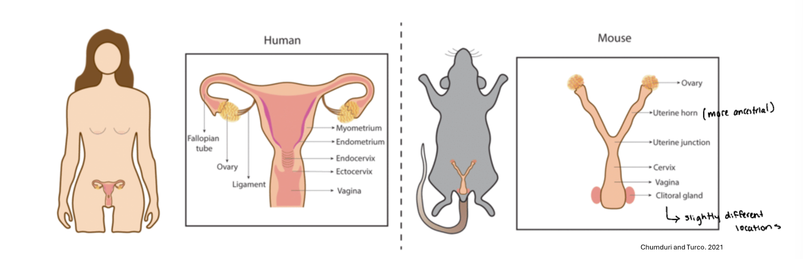
The cumulus-oocyte complex (COC) is translocated to the fallopian tubes
Fimbriae congested around site o COC expulsion on ovary
Sperm translocation is a number game with a few determining factors
200-300 million sperm ejaculated into vagina, but about a 1:1 million make it to oviducts
roughly 200 sperm expected to make it to the oviducts
Effected by:
Sperm motility - helps get through cervix
Uterine muscle contractions - 30 minutes to find oviducts
“too short to have been attained by even the most Olympian sperm relying on their own flagellar power” (Storey, 1995)
aided by cilial movement on cell surfaces
Sperm rheotaxis - detection of flow of fluid from oviducts to uterus
For mammalian sperm to participate in fertilization, they must undergo
Capacitation - gain of capacity for mammalian sperm to fertilize an egg
Gain capacity to:
recognize cues to guide them to the egg
undergo acrosome reaction
fuse with cell membrane
No capacitation, will not be able to move through cumulus
Capacitation involves reception of molecular cues that prepare sperm for fertilization
In reproductive tract, sperm are exposed to bicarbonate ions (HCO3-) and albumin proteins
Albumin - removes cholesterol, increases membrane fluidity (at high temps)
Bicarbonate - activates kinase pathways, removes acrosome reaction inhibitors
(if membrane is more fluid, it is easier to fuse; like bashing ice together vs putting water together, water combines better)

CatSper channels open to allow for hyperactivation, mediated by progesterone secreted by the cumulus
As sperm gets closer it moves faster
ABHD2: (abhydrolase domain-containing protein 2)
2AG: (2-arachidonoylglycerol)
Promotes loosey goosey
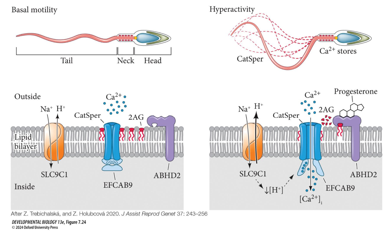
Hyperactivated sperm can detach from oviduct and detect temperature and chemical gradients
Capacitated sperm can orient to the ovum via two basic mechanisms:
Thermotaxis - detection of temperature gradients
Chemotaxis - detection of chemical gradients
progesterone secreted by cumulus cells
Sperm can bind to the zona pellucida through recognition of specific glycoproteins

Mammalian sperm bind to the cell membrane of the egg via
Izumo protein (on the sperm) and Juno protein (on the egg)
Sperm fuse to the egg membrane at a specific angle and the tail is then broken off and absorbed
