WSU BIOLOGY 315 Lab Exam 3
 Call Kai
 Learn
 Practice Test
 Spaced Repetition
 Match
 Flashcards
 Knowt Play
1/412

There's no tags or description
Looks like no tags are added yet.
413 Terms
Apex of Heart
On the bottom left side

Base of Heart
TA will choke heart

Right Atrium
What is this chamber(TA will tap it)

Right Auricle
What is this ft
Ear-like structure
Above atrium

Left Atrium
What is this chamber

Left Auricle
What is this ft
Above atrium

Right Ventricle
What is this chamber

Left Ventricle
What is this chamber

Anterior Surface of Heart
What is this region
Front of heart

Diaphragmatic Surface of heart
Where diaphragm meet apex
TA will wave below heart

Right Pulmonary Surface of Heart
Formed by right atrium
TA will use a chopping motion

Left Pulmonary Surface of heart
Formed by left ventricle
TA will use a chopping motion

Right Border of Heart
Opposite apex

Left Border of Heart
Apex side

Inferior Border of Heart
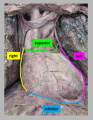
Superior Border of Heart

Coronary Sulcus
Upper depression

Anterior Interventricular Sulcus
Front side
Depression

Posterior Interventricular Sulcus
Back side
Depression

Aorta
Largest upper structure

Ascending Aorta
First part
Portion

Arch of Aorta
Follows ascending aorta
Portion

Pulmonary Trunk
collective structure

Left Pulmonary Artery
On the left side

Right Pulmonary Artery
On the back side

Superior Vena Cava
Upper, posterior

Inferior Vena Cava
Lower, posterior

Pulmonary Veins
Structure

Left Superior Pulmonary Veins
Upper tube, apex-side of probe

Left Inferior Pulmonary Veins
Lower tube, apex-side of probe

Right Superior Pulmonary Veins
Upper tube, probe side opposite the apex

Right Inferior Pulmonary Veins
Lower tube, probe side opposite the apex

Coronary Arteries
Upper boarder, a structure
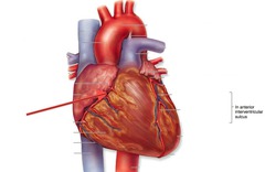
Right Coronary Artery
Under right auricle

Right Marginal Artery
Below right coronary artery

Posterior Interventricular Artery
Posterior, middle

Left Coronary Artery
Under left auricle

Anterior Interventricular Artery
Anterior, middle

Circumflex Artery
Squiggly, on left ventricle

Cardiac Veins
Posterior T

Coronary Sinus
Top of the T
Probe inside

Great Cardiac Vein
Left part of the T

Middle Cardiac Vein
Base of the T
Darker

Fibrous Pericardium
Outside of the flap

Serous Pericardium
Inside of the flap

Parietal Layer of Serous Pericardium
Shiny part of the flap

Visceral Layer of the Serous Pericardium
On the heart
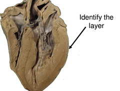
Endocardium
Inner layer

Myocardium
Thicker

Epicardium
Most outer layer
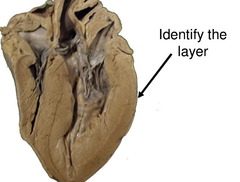
Interatrial Septum
Will be pinched

Fossa Ovalis
Less dark

Right Atrium
A chamber
Will be tapped

Right auricle
Ft
Ear shaped
Exterior shape
Pectinate Muscle
Dark, branching

Sinus Venarum
Lighter, smooth, internal

Opening of the Superior Vena Cava
Top of probe

Opening of the Inferior Vena Cava
Bottom of probe

Opening of the Coronary Sinus
Small space

Left Atrium
Two probes inside chamber

Left Auricle
Looks like an ear

Openings of the Pulmonary Veins
Where the two probes are
A collective space

Interventricular Septum
Whiter, below auricle

Muscular Part of the Interventricular Septum
Thicker, inferior

Membranous Part of the Interventricular Septum
Thinner, superior

Right Ventricle

Right Atrioventricular Orifice
Space with teeth

Tricuspid Valve
Probe through the right atrioventricular orifice

Papillary Muscles
The carrot itself, thicker

Tendinous Cords
Cords on papillary muscle
Greens of carrot

Trabeculae Carnae
Roots of the carrot

Conus Arteriosus
Cone shaped narrowing

Opening of the Pulmonary Trunk
Probe through large opening

Pulmonary Valve
The valve in the trunk

Semilunar Cusps of Pulmonary Valve
Components of the valve

Left Ventricle

Left Atrioventricular Orifice
Space with teeth on left side

Mitral Valve
Probe through left atrioventricular orifice

Aortic Vestibule
Narrowing approaching the aorta
Triangle shaped

Opening of the Ascending Aorta
Whole space
Includes aortic vestibule

Aortic Valve
Probe through

Semilunar Cusps of the Aortic Valve
Components of the aortic valve

Ascending Aorta (cadaver)
First inch toward head

Arch of Aorta (cadaver)
Has three splits

Brachiocephalic Artery
Right split off arch of aorta

Right Common Carotid Artery
Right medial split

Right Subclavian Artery
Right lateral split

Internal Thoracic Artery of right subclavian artery
Nub off subclavian

Left Common Carotid Artery
Left medial split
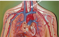
Left Subclavian Artery
Left lateral split

Internal Thoracic Artery of Left Subclavian Artery
Nub off left subclavian

Right Posterior Intercostal Arteries
By intercostal nerves
More round

Abdominal Aorta
Aorta after diaphragm

Left Renal Artery
White and visible

Right Renal Artery
White and under kidney

Celiac Trunk
First tube in abdominal aorta

Superior Mesenteric Artery
Second tube in abdominal aorta

Inferior Mesenteric Artery
Lower, cut in cadaver

Lumbar arteries
Under abdominal artery
Left gonadal Artery
Stringy
