Cardiac A&P/Fundamentals of EP
 Call Kai
 Learn
 Practice Test
 Spaced Repetition
 Match
 Flashcards
 Knowt Play
1/39

There's no tags or description
Looks like no tags are added yet.
40 Terms
Venous pathway from axillary vein to RA
Axillary vein → subclavian vein → brachiocephalic vein → superior vena cava → right atrium
*In the case of a persistent left SVC, a venous branch extends off the (brachiocephalic?) vein and feeds into the CS, in addition to the traditional SVC

Layers of the heart wall + function (deep to superficial)
Endocardium - smooth muscle to aid in blood flow, lines chambers and valves
Myocardium - thickest part of heart muscle, enables contraction and relaxation of cardiomyocytes
Epicardium - inner serous pericardium composed of fats, mesothelium, connective tissue to insulate the heart and protect it
Beyond the heart wall:
Pericardial space filled with fluid to lubricate and prevent friction; secreted from the serous layers
Outer serous pericardium
Fibrous pericardium - made of connective tissue; anchors the heart to the diaphragm and sternum, protects the heart from infection
Composition of the crista terminalis
Separates posterior/septal smooth tissue from anterior/lateral pectinate tissue
Triangle of Koch
Identify location of AVN in inferior RA subendocardium
Posterior boundary: CS os
Superior/anterior boundary: Tendon of Todaro
Inferior boundary: septal leaflet of TV
LV vs RV: how much thicker and general dimensions
LV: 2-3x thicker than RV
RV: ~6mm, LV: ~8-15mm
Most anterior structure of the heart? Most posterior?
RVOT/PA; CS
For each of the valves in our heart, what are the names of the cusps/leaflets?
TV: anterior leaflet + PM + mod band; septal leaflet + PM; posterior leaflet + PM
Pulmonic valve: anterior, left, and right cusps
MV: anterior leaflet + ALPM; posterior leaflet + PMPM
Aortic valve: RCC, LCC, NCC
What is an annulus?
A fibrous ring of tissue for all heart valves that is electrically inert and separates conduction between structures; part of the fibrous skeleton
For aortic and pulmonic annuli: sits at the base of the cusps and separates LVOT from aortic root (ventricular from aortic tissue) or RVOT from PA
For mitral and tricuspid annuli: the structure is saddle-like with leaflets hanging down from it
Coronary artery branches aorta through RCA and LCa
RCA
Sinoatrial nodal artery (or SVC branch): feeds SAN
Conal artery: feeds RVOT
Right marginal artery
Posterior descending artery - feeds inferobasal IVS
AV nodal artery - feeds AVN
LCA
LAD with branches - feeds anterior IVS, anterior LV, apex
Left Circumflex artery - feeds LA, lateral and posterior LV
Left marginal artery
Could have PDA if left or co-dominant
Atrial branches to feed RAA and LAA
Coronary vein architecture
From the CS:
Middle cardiac vein - runs along inferobasal IVS alongside PDA
Small cardiac vein - along RA/RV groove
CS splits into Great Cardiac Vein and Vein of Marshall
GCV → AIV
Left/right marginal veins
May have posterior branches
Describe each of the following congenital cardiac structural anomalies:
ASD/VSD
PFO
Persistent left SVC
Tetralogy of Fallot
Transposition of the Great Arteries
Notable holes in the interatrial or interventricular septa separating the chambers
Flaps of tissue fail to properly seal and close; leaves opening at FO; close in 75% of people
During development, left anterior cardinal vein fails to regress into vein and ligament of Marshall
Becomes “left” SVC instead that dilates and drains into CS
Still also have normal CS
Four defects: Hypertrophy of the RV; pulmonary stenosis at RVOT level; VSD; overriding aorta positioned over the VSD
Aorta and PA swap such that deoxygenated blood circulates to the body and oxygenated blood circulates to the lungs
General flow of conduction from SA node throughout heart
SA node → internodal tracts and Bachmann’s Bundle (with cell-to-cell to follow) → AV node → bundle of His → LBB and RBB → LBB includes LPF and LAF → Purkinje fibers
Describe structure/intrinsic rates/impulse delay of the following:
SA node
Internodal tracts
AV node
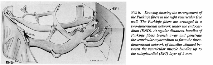
Purkinje fibers
Atrial, junctional, and ventricular foci
Positioned at high lateral SVC/RA junction; rate of 60-100 bpm at rest; contains ~10k pacemaker cells
Aggregated, parallel-oriented cardiomyocytes that enable preferential conduction; FP = anterior and middle tracts, SP = posterior tract
Delays impulse by 40 ms; cannot generate spontaneous impulses; gradual tissue transition from atrial to “midnodal” tissue
Run subendocardially up to 2mm below epicardium and send electrical impulses endo- to epicardium (see pic)
Atrial = 60-80 bpm, junctional = 40-60 bpm; ventricular = 20-40 bpm
His bundle (*start of ventricular structures) - describe structure and position
What coronary blood supply reaches the His bundle?
Starts at distal AV node → perforates central fibrous body → bifurcates at leftward side of IVS (interventricular crest) into LBB and RBB. Is about 10-20 mm long
Both the anterior and posterior coronary arteries

Name the four properties of cardiac cells
Automaticity: pacemaker cells have the ability to spontaneously depolarize without any input from the nervous system
Conductivity: facilitated by gap junctions to transmit molecules between cells that instigate APs
Selective permeability: cell membranes regulate what types of ions move in and out of cells at any given time
Excitability: the ability of the cell to respond to an electrical stimulus
Describe the 4 phases of an action potential for a cardiomyocyte
Resting State, Phase 4: Cell is at its resting membrane potential of -90 mV. Positive ions start trickling in via gap junctions to raise the membrane potential to -70 mV.
Depolarization, Phase 0: -70 mV is the threshold needed for voltage-gated sodium channels to open, rapid influx of sodium into the cell. When the action potential peaks, the sodium channels close. Slow leakage of calcium into the cell simultaneously.
Early repolarization, Phase 1: Potassium channels open, efflux of potassium out of the cell
Plateau, Phase 2: relative charge balance as potassium ions flow out and calcium flows (slowly) into the cell. Calcium influx actually prompts calcium release from the SR into the cell that facilitates contraction.
Final repolarization, Phase 3: calcium channels close, only potassium is exiting the cell to lower MP back to -90 mV. Reset sodium and potassium levels with Na/K pumps
Repeat Phase 4
Phases during which ARP and RRP occur for refractoriness
ARP: Phase 1 (immediately post-depolarization) - part of Phase 3 (final repol.)
RRP: Rest of Phase 3 until reaching Phase 4 (resting state)
Describe 3 phases of pacemaker cell action potential
Prepotential, Phase 4: Resting state at -60 mV, actually triggers voltage-gated sodium channels to open and allow SLOW influx of sodium into cell (i.e. funny currents) to increase MP to -40 mV
Depolarization, Phase 0: Reach threshold @-40 mV and calcium channels open, rapid influx of calcium into the cell
Repolarization, Phase 3: Calcium channels close, potassium efflux to restore membrane to -60 mV
Conduction velocities of:
AV node
Ventricular myocytes
Atrial myocytes
Purkinje fibers
AV node - 0.2 m/s
Ventricular myocytes - 0.4 m/s
Atrial myocytes - 1 m/s
Purkinje fibers - 4 m/s
Describe from left to right what each deflection of the P/QRS/T wave represents in terms of the action potential and the cardiac cycle
Start of P-wave: Atrial depolarization
During P-wave: Atrial contraction (systole)/plateau phase of AP
Baseline end of P-wave to onset of QRS: conduction delay in the AV node
QRS complex: Atrial repolarization/diastole, ventricular depolarization/systole and initial contraction
Baseline ST segment: ventricular plateau phase (continued contraction)
T-wave: ventricular repolarization and ventricular diastole (relaxation)
Describe what these ECG abnormalities may be indicating:
Enlarged P-waves
Enlarged Q portion of QRS complex
Flattened T-wave
Elevated T-wave
ST Elevation
ST Depression
Dilated atrium
Possible MI
Lack of oxygen (potential coronary artery disease)
Hyperkalemia (excessive K in blood)
MI
Insufficient oxygen for heart
Describe the structure of muscle in the heart, from lowest to highest structural complexity.
Myofilaments: actin, myosin, and titin. Arranged in end-to-end sarcomeres, the full length of which is referred to as a myofibril.
Multiple myofibrils, along with mitochondria, nuclei, the SR, T-tubules, sarcolemma, etc., compose a cardiac muscle fiber, or cardiomyocyte.
Cardiomyocytes are bundled together into fascicles.
Multiple fascicles compose the cardiac muscle.
What are transverse tubules in cardiac cells?
They stem off of the sarcolemma and transmit action potentials deep into the muscle fiber. There is one T-tubule per sarcomere, located at the Z-disk.
Structure and function of intercalated disks in cardiac muscle fibers?
Thickening of the sarcolemma (PM) that connects these fibers end to end.
Contain desmosomes (“glue”) and gap junctions (“doorways for ions”) intercellularly.

Describe the different bands of “color” found on a sarcomere
I band
A band
H zone
M line
Z-disc
I band - only actin
A band - width of myosin, includes both myosin and actin
H zone - only myosin (center)
M line - keeps myosin stationary
Z-disc - connects ends of actin

How are depolarization and contraction linked?
Depolarization of Na at the start of an AP triggers voltage-gated Ca channels to open
Influx of Ca triggers Ca release from SR
Calcium binds to troponin found on a strand of tropomyosin, unlocking troponin and exposing the binding sites for myosin heads
Myosin heads attach and pull, ratchet effect → CONTRACTION as z-discs shorten
Define coronary sulcus.
Which structure(s) rest within it?
A groove around most of the heart separating the atria from the ventricles that is deep to fat. (Think almost all the way around the heart, a transverse “ring”)
Contains coronary sinus, small cardiac vein, LCx, RCA
Define anterior IV sulcus.
Which structure(s) rest within it?
A groove on the anterior aspect of the heart, deep to fat, externally separates RV and LV
Contains LAD and AIV (or great cardiac vein)
Define posterior IV sulcus.
Which structure(s) rest within it?
A groove on the posterior aspect of the heart, deep to fat, externally separates RV and LV. Joins anterior sulcus at the apex of the heart.
Contains PDA and MCV
What is an auricle?
“Dog ear” flap on each atrial appendage that allows more blood to be stored in the atrium. The auricles of the RAA and LAA are on the more medial aspect.
High-level circulation of blood flow through vasculature.
Systemic veins → heart → PA to lungs → PV to heart → Aorta/systemic arteries → arterioles → capillaries → venules → veins
*Capillaries are assumed to be porous.
What is the functional (contractile) unit of a cardiac muscle cell?
Sarcomere
What is the resting MP and threshold for:
Cardiac myocytes
Pacemaker cells
-90 mV RMP; -70 mV threshold for AP
-60 mV RMP; -40 mV threshold for AP
What artery supplies the:
AV node
SA node
AV nodal branch from RCA (90%) OR the LCx (10%)
SA nodal branch from RCA (over 50%) OR the LCx (a bit under 50%)
Definitions and values/formulas for the following:
End-diastolic volume (EDV)
End-systolic volume (ESV)
Stroke volume (SV)
Cardiac output
Ejection fraction
Stroke index
Volume of blood in ventricles following atrial contraction, before ventricular systole. ~130 mL
Volume of blood in ventricles following ventricular ejection, after SL valves close but before AV valves open. ~60 mL
Volume of blood output (exiting ventricles) for one cardiac cycle: EDV - ESV
CO = Stroke volume x heart rate, amount of blood output per minute
EF = SV/EDV x 100%, the ratio of blood output per cardiac compared to the blood volume in the ventricles
SI = SV/body surface area
Describe when we hear the S1/S2/S3/S4 sounds.
Sounds
S1: near the start of ventricular systole (isovolumetric contraction) when turbulent blood in the ventricles forces the AV valves to close against increasing ventricular pressure
S2: when ventricular pressure drops below aortic/PA pressure and the semilunar valves close in the “ventricular ejection” phase, leads into isovolumetric relaxation
S3: during rapid V filling from atrium (passive, atrial pressure has exceeded ventricular pressure and opened AV valves)
S4: during atrial systole (“atrial kick”), final blood volume pushed into ventricles
When can isovolumetric contraction/relaxation occur?
When all four cardiac valves are closed, and the ventricles are neither gaining nor losing blood.
What pressure is required…
For the RA to open the TV
For the RV to open the Pulmonic Valve
For the LV to open the MV
For the LV to open the aortic valve and circulate blood to the rest of the body
2-8 mmHg (~10 mmHg)
15-28 mmHg (~20 mmHg)
5-12 mmHg (~10 mmHg)
~80 mmHg, LV pressure peaks between 90-140 mmHg
Define pulmonary wedge pressure
The pressure in the LA and LV during diastole, usually around 4-12 mmHg
What are trigones?
Connective tissue bridges between the annuli of the four cardiac valves