2- Principles of Differential Diagnosis and Biopsy of Pathologic Lesions (Dr. Oreadi)
 Call Kai
 Learn
 Practice Test
 Spaced Repetition
 Match
 Flashcards
 Knowt Play
1/81

There's no tags or description
Looks like no tags are added yet.
82 Terms
Define the following:
"The art or act of identifying a disease from its signs and symptoms"
Diagnosis
What are the fifteen steps for forming a treatment plan when identifying a lesion?
1. Recording the identifying data
2. History and physical examination
3. Chief complaint
4. HPI
5. Past medical history
6. Review of symptoms by system
7. Physical examination (radiographic)
8. Differential diagnosis
9. Working diagnosis
10. Medical laboratory studies
11. Dental laboratory studies
12. Biopsy
13. Consultation
14. Final diagnosis
15. Treatment plan
What type of imaging is the gold standard for aiding in diagnosis after clinical exam and med history?
Panoramic
When clinically identifying an oral pathology, you want to describe 3 things?
- Site
- Morphology
- Color

How would you describe this lesion?
- Lateral aspect of the right tongue
- Homogenous white plaque
- Non-wipeable
- No ulceration or bleeding
What lesion color should raise suspicion of malignancy?
Leukoerythroplakia
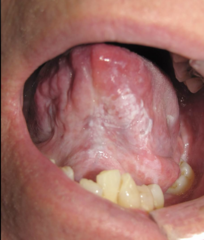
How would you describe this lesion?
Heterogenous white plaque on left ventral tongue and floor of mouth

How would you describe this lesion?
- Mixed leukoerythroplakia lesion
- Some ulceration
- On right anterior aspect of lateral tongue/posterior third to floor of mouth
All of the following are lesion characteristics that should raise suspicion of malignancy EXCEPT:
A) Leukoerythroplakia
B) Slow growth
C) Bleeding on gentle manipulation
D) Ulceration persisting for more than 2 weeks
E) Indurated/firm to touch
F) Fixation - immobile/attached to adjacent structures
B - RAPID growth
A lesion that has persisted over _______ should raise concern for malignancy
2 weeks
What is useful as diagnostic adjunct to the clinical examination and history of lesions within or adjacent to bone?
Radiographic examination
All of the following are what type of cyst of the jaw?
- Odontogenic keratocyst (KOT)
- Dentigerous cyst
- Eruption cysts
- Lateral periodontal cyst
- Gingival cyst
- Glandular odontogenic cyst
- KOT
- Ameloblastoma
Development cyst (Odontogenic)
All of the following are what type of cyst of the jaw?
- Nasopalatine duct cyst
- Nasolabial cyst
- Median cyst
Dentigerous cyst (Non-odontogenic)
All of the following are what type of cyst of the jaw?
- Radicular cyst
- Apical cyst
- Lateral cyst
- Residual cyst
Inflammatory cyst (Odontogenic)
T/F: If you see any lesion, it always requires a biopsy
False, not always necessary
What social habits of patients should raise suspicion for developing oral malignancy?
- Smoking
- Chronic alcohol intake
An ulcer in what location of the mouth should raise suspicion for oral malignancy?
- Floor of mouth
- Ventral lateral tongue
An ulcer for what duration should raise suspicion for oral malignancy?
4-6 weeks

What would you be suspicious of if you knew this 18 year old had a brief history of smoking?
Squamous cell carcinoma
Define the following:
Removal of tissue from a living individual for diagnostic examination
Biopsy
What are the 4 types of biopsies we can preform?
- Aspiration
- Cytology
- Incisional
- Excisional
What is the gold standard biopsy method?
incisional

A patient presents with a non-healing leukoerythroplakic ulcerated lesion on the ventral latereral border of the tongue. The lesion has been there for 3 weeks. What type of biopsy should you take?
A) Aspiration
B) Cytology
C) Incisional
D) Excisional
C) Incisional
All of the following are indications for what?
- Any lesion that persists for more than 2 weeks with no apparent etiology
- Any inflammatory lesion that does not respond to local treatment after 10 to 14 days (after removing local irritant)
- Persistent changes in surface tissue (lips or oral mucosa)
- Any persistent tumescence, either visible or palpable beneath relatively normal tissue
- Lesion that interfere with local function (fibroma)
- Bone lesions where inflammatory conditions can be ruled out
Biopsy

Define the following:
The use of a needle and syringe to penetrate a lesion for aspiration of cells or content
Aspiration biopsy
Your patient presents with a fluctuant mass in the area of the neck. You are pretty sure it contains fluid. What type of biopsy should you take?
A) Aspiration
B) Cytology
C) Incisional
D) Excisional
A) Aspiration
What biopsy is indicated?
- Lesions thought to contain fluid or any intra-osseous lesion before surgical exploration
- Location: Fluctuant mass in the soft tissues (deep or superficial) - consider surrounding anatomy
Aspiration biopsy
What type of biopsy?
- A 18-gauge needle is connected to a 5 or 10 ml syringe
- The area is prepped sterile
- The tip of needle may have to be repeatedly repositioned to obtain cellular sample
- The aspirated contents are smeared over glass slide and placed into container with fixative solution
Aspiration biopsy

What type of biopsy?
Aspiration biopsy
What type of biopsy?
- When a brush is placed in contact with oral epithelium and rotated with firm pressure 5 to 10 times
- The brush collects cells from all three layers of epithelium: superficial, intermediate and basal
- Brush is smeared onto glass slide and placed in container with fixative solution
Oral Brush Biopsy (cytology smear)

What type of biopsy?
Oral Brush Biopsy (cytology smear)
Cytology smears are evaluated by both pathologist and a computer. Which one determines if adequate specimen was obtained: pathologist or computer?
Computer
Cytology smears are evaluated by both pathologist and a computer. Which one determines if the cells are dysplastic or malignant: pathologist or computer?
Computer
Cytology smears are evaluated by both pathologist and a computer. Which one classifies the findings into three categories: negative, positive, or atypia: pathologist or computer?
Pathologist
T/F: Oral brush biopsies require either topical or local anesthesia to obtain from patients
False
T/F: You can do a brush biopsy as a substitute for a scalpel biopsy
False
What biopsy is indicated?
- Might be good for monitoring pts with chronic mucosal changes
- Does not require any topical or LA and results in minimal discomfort and bleeding
- Quick procedure
- Should serve as a trigger and not a substitute for scalpel biopsy
Oral Brush Biopsy (cytology smear)
What type of biopsy samples only a particular or representative area of the lesion?
Incisional biopsy
If the lesion is large or has different characteristics at different locations, what type of biopsy should you take?
A) Aspiration
B) Cytology
C) Incisional
D) Excisional
C) Incisional

What type of biopsy is indicated?
Incisional biopsy
T/F: If the lesion is large or has different characteristics at different locations, more than one area of the lesion may require biopsy
True
What biopsy is indicated?
- Lesion difficult to excise because of its extensive size
- Adjacent anatomic structures
- Suspected malignancy
Incisional biopsy
T/F: If a lesion is suspicious for malignancy, you should do an excisional biopsy to remove it from the patient immediately
False

Small lesions with high degree of suspicion should undergo what type of biopsy?
Incisional biopsy
The following are principles of what type of biopsy?
- Wedge shape
- Selected area that shows complete tissue changes
- Should include normal tissue (???)
- Necrotic tissue should be avoided
- Narrow better than broad
Incisional biopsy
Is normal tissue required with incisional biopsies?
No - only with excisional
What tool can be used to take a incisional biopsy?
Tissue punch
What type of biopsy?
- Removal of entire lesion
- Perimeter of normal tissue surrounding the lesion is also excised for many reasons:
- - Oncologic margin
- - Closure
Excisional biopsy
What are two reasons you would include a perimeter of normal tissue surrounding the lesion in an excisional biopsy?
- Oncologic margin
- Closure
What biopsy is indicated?
- Small lesions that appear to be benign
- Any lesion that can be removed completely without affecting function is best tx with excisional bx
- Pigmented and small vascular lesions
Excisional biopsy
Define the following:
The removal of the entire lesion, along with 2 to 3 mm of normal-appearing surrounding tissue, is excised
Excisional Biopsy
In an Excisional Biopsy, the entire lesion, along with ___-___ mm of normal-appearing surrounding tissue, is excised
2-3 mm

Your patient presents will a yellow 1cm x 1cm nodule on the lateral border of the tongue. It looks mostly like a lipoma.
What type of biopsy should you take?
A) Aspiration
B) Cytology
C) Incisional
D) Excisional
D) Excisional

What is placed here to help with healing?
Silver nitrate
T/F: LA should not be injected within the tissues to be removed
True
Which type of anesthesia do you use when block cannot be used?
Field block (1 cmm infiltration from lesion)
What are three tools to help with tissue stabilization during a biopsy?
- Pinch area adjacent to the lesion
- Instruments
- Heavy retraction sutures or towel clips to immobilize the tongue
Which of the following is not suggested when considering the principle of hemostasis during a biopsy?
A) Use of suction device
B) Gauze only
C) Gauze over suction device
A) Use of suction device = AVOID
T/F: Crushed specimen may be non-diagnostic and only delay definitive tx
True
The use of a ________ through the specimen is an excellent method for avoiding specimen trauma
Traction suture
Identification of surgical margins is indicated for which type of lesion?
Malignant lesions (confirmed)
What material is used to orient a surgical specimen?
Silk suture
A surgeon excises a confirmed malignant oral lesion and marks the specimen with a silk suture. What is the primary purpose?
A. To prevent tissue shrinkage
B. To identify the specimen's orientation
C. To reduce postoperative bleeding
D. To preserve the specimen
B. To identify the specimen's orientation
Where should the surgeon document the orientation of the surgical specimen?
On the pathology data sheet
What can the pathologist determine by evaluating the surgical margins of an oriented malignant specimen?
Whether the margins are involved and where
If you suspect an autoimmune disease is the cause of your patients oral pathologies, how should you take a biopsy?
Take two biopsies: (tissues must be immersed)
- Put one in 10% formalin solution
- And one in Michel's solution for immunofluorescence studies
While not a fixative, specimens can be held at room temperature for ____ days in transport medium before processing. This simple salt solution maintains pH, but does not kill most pathogens
5 days
For surgical closure, undermine the wound margins by at least the width of __________ in each direction which allows approximation of the tissue with little to no tension
The defect
For surgical closure, obtain primary closure, except in __________ surfaces which are allowed to heal by secondary intention
Attached mucosal
All of the following are biopsy techniques and surgical principles for what?
- Aspiration Biopsy of Radiolucent Lesions
- Mucoperiosteal Flaps
- Osseous Window
- Removal of Specimen
- Specimen Care
Intraosseous or hard tissue biopsies
Intraosseous/Hard Tissue Biopsy Technique: What is step 1?
Aspiration biopsy of radiolucent lesions
Intraosseous/Hard Tissue Biopsy Technique: What is step 2?
Mucoperiosteal flap
Intraosseous/Hard Tissue Biopsy Technique: What is step 3?
Osseous window
Intraosseous/Hard Tissue Biopsy Technique: What is step 4?
Removal of specimen and specimen care
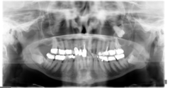
You patient presents with a pano exhibiting a well defined, corticated, unilocular radiolucency associated with the crown of an unerupted #16. What type of biopsy should you take first?
A) Aspiration
B) Cytology
C) Incisional
D) Excisional
A) Aspiration
What is used keeping the concave surface in contact with the osseous surfaces of the bony cavity?
Dental curette
For hard tissue biopsies, it may take _____ weeks before the path report is available due to the need to decalcify the tissue
2 weeks
T/F: For a benign process excised by the bx procedure, need to follow the pt with serial radiographs to monitor osseous healing. Definitive tx to be undertaken if incisional bx performed, or more aggressive lesion present
True
What all should you include on your biopsy sheet?
- All specimens need to be labeled
- History and clinical description
- Radiographs / pictures

After a biopsy, you can follow up with a patient within ___-___ days
7-10 days
If the pathology report does not corroborate the clinical impression of the lesion, what should you do?
The biopsy procedure should be repeated
What are 3 things to consider for referrals for biopsy?
- Health of patient
- Surgical difficulty
- Malignancy potential