Protein Post-Translational Modifications
 Call Kai
 Learn
 Practice Test
 Spaced Repetition
 Match
 Flashcards
 Knowt Play
1/50

There's no tags or description
Looks like no tags are added yet.
51 Terms
A Newly Synthesized Protein is not Necessarily a Finished Protein:
Translation produces a polypeptide with a defined primary sequence—but function depends on much more than sequence
Folding, proteolytic processing, covalent modification, trafficking, and degradation determine the protein's functional state
PTMs are one of the major mechanisms by which cells create distinct molecular states from the same gene product.
Think in terms of a protein lifecycle: synthesis → folding → localization → activation → regulation → turnover
Genome vs Proteome
The human proteome is vastly more complex than the human genome
Estimated that the human genome comprises between 20,000 and 25,000 genes
The total number of proteins in the human proteome is estimated at over 1 million
These estimations demonstrate that single genes encode multiple proteins

Over billions of years, organisms have acquired mechanisms:
Alternative splicing
Cotranslational modifications
Posttranslational modifications (PTMs)
These mechanisms allow genomes containing 20,000–25,000 genes to be converted into proteomes of more than one million proteins
enhancing the complexity and functional diversity of proteins in response to complex external stimuli and internal changes
Post Translational Modifications
More than 500 types of protein PTMs
These modifications play essential roles in various biological processes, including deoxyribonucleic acid (DNA) repair, signaling pathways, cell apoptosis, subcellular localization, protein–protein interactions, etc
Studies have shown that PTMs are closely associated with kinds of human diseases, such as neurological diseases (Alzheimer’s and Parkinson’s diseases, neuroblastomas and glioblastomas), breast diseases (breast cancer/tumors/carcinomas and mammary tumors/carcinomas) and blood diseases
Research on PTMs can facilitate the development of epigenetics, cellular functions and medical technology
Accurate detection is the primary task in PTM research
Modified Proteins are identified through experimentally, diverse techniques, such as radioactive label method, chromatin immunoprecipitation (ChIP), mass spectrometry (MS) and liquid chromatography
Verified PTM protein sequences were organized in known databases including UniProt, PLMD, dbPTM and PhosphoSitePlus
There are 908 917 experimental sites of more than 130 types of PTMs recorded in the dbPTM database
Phosphorylation contributes to the highest proportion of 62.83% (571 032), and acetylation and ubiquitination separately account for 15.12% and 13.04%.
Proteomic techniques are expensive, time-consuming and labor-intensive
The artificial intelligence industry is accelerating!
Computational proteomics use AI to predict PTMs
This new avenue greatly reduces the costs and presents reliable results, which has become a frontier topic in biology and bioinformatics
However, researchers should exercise significant caution. While AI-driven PTM site prediction is highly scalable, relying on these computational results without rigorous laboratory verification introduces heavy risks
What can a PTM Change?
Protein conformation
Protein activity
Protein charge and stability
Protein-Protein interactions (within and between cells)
Interactions with DNA and RNA
Binding affinity for proteins, DNA, RNA, lipids, or small molecules
Subcellular localization and membrane association
Protein half-life and degradation
Types of PTMs Based on the Nature of the Modification:
Complex molecules (irreversible/reversible)
Lipidation
Glycosylation
Proteins/peptides (reversible)
Ubiquitylation
Sumoylation
Chemical groups (reversible)
Methylation
Phosphorylation
Acetylation
Cleavage (irreversible)
Proteolysis
Timing of Post Translational Modifications
Immediately after (or during) translation, before folding is complete
Early PTMs affect protein folding efficiency and structural stability
E.g., proteolytic cleavage
Immediately after folding is complete
PTMs often direct proteins to their proper subcellular locations
E.g., glycosylation
After folding and subcellular localization
PTMs can activate or deactivate specific functions of the protein
E.g., phosphorylation
End of the protein life cycle
PTMs tag proteins for degradation, marking them for removal from the cell
E.g., ubiquitination
Cleavage of Polypeptides
Proteolytic cleavage is a PTM
Proteases hydrolyze specific peptide bonds in a precursor protein
Cleavage can activate an inactive precursor, remove a targeting sequence, expose a new terminus, or generate multiple products
Unlike many reversible PTMs, peptide-bond cleavage is usually irreversible in the cellular context
Specificity depends on protease recognition, substrate accessibility, and cellular compartment
Zymogen Activation
A zymogen, also called a proenzyme, is an inactive precursor of an enzyme
The pancreas initially synthesizes protease enzymes as zymogens to help prevent enzymes from inappropriately digesting proteins within the pancreatic cells
Enzymes like Trypsin are synthesized as proenzymes
For trypsin, trypsinogen is an inactive precursor translated by the rough endoplasmic reticulum and transported to the Golgi apparatus for sorting
Trypsinogen is always co-synthesized and packaged with a pancreatic secretory trypsin inhibitor (PSTI), which inhibits premature activation
Thus, there are two mechanisms in place to maintain the inactivity of the protease within the pancreas: (1) synthesis of the zymogen or proenzyme form, and (2) co-expression of a trypsin inhibitor protein that will bind and inhibit any prematurely cleaved trypsin until it has reached the small intestine
During packaging in the Golgi apparatus, trypsinogen and other digestive enzymes condense into core particles and are packed into zymogen granules
The condensed enzymes are stable, and minimal activation occurs within the zymogen granules
Once pancreatic cells receive secretory stimuli, zymogen granules are released into the pancreatic duct lumen, carrying digestive enzymes into the duodenum
Once in the duodenum, enteropeptidase activates trypsinogen by removing the trypsinogen activation peptide (TAP) from the N-terminal region

Proteolysis as Protein Maturation
Many hormones and neuropeptides are synthesized as inactive precursor polypeptides that require limited endoproteolysis to become biologically active
Example: Insulin
Insulin
A vital peptide hormone produced by the beta cells of the pancreas that is critical for glucose uptake for:
Metabolism, glycogen synthesis/storage, lipolysis regulation, and other processes related to cell growth
Synthesized as a precursor molecule and processed through a series of non-enzymatic and enzymatic events
How is Insulin made?
Expression of the INS gene leads to the translation of a 110-a.a. long precursor polypeptide ⇨ Preproinsulin
The first 24 a.a. signal peptide is cleaved and the rest of the peptide folds and forms disulfide bonds ⇨ Proinsulin
Removal of the 31 a.a. C-peptide (res. 57-87) creates the mature protein composed of two separate peptide chains ⇨ Insulin
Insulin is a secreted protein:
its processing is coordinated in the ER and Golgi apparatus
Glycosylation
A process in which sugar groups are transferred to proteins catalyzed by glycosyltransferases
Attached oligosaccharides are called glycans
Predominantly occurs in the ER and Golgi apparatus
One of the most commonly occurring PTMs: ~50% of all cellular proteins are glycosylated
Most glycans exist on the surface of cells and secreted proteins, with intricate and varied structures
The human genome contains approximately 700 genes related to glycosylation and deglycosylation, including enzymes, transporters, and chaperones
Changes in glycosylation patterns are linked to diseases
Occurs as proteins traverse through the secretory pathway
Step-wise processes add, remove, and trim sugar moieties
Functions of Glycosylation
Aids in protein folding and stability
Facilitates protein-protein/protein-ligand interactions
Helps mediate cell-cell adhesion and immune responses
Glycosylation requires the action of multiple enzymes located in different cellular compartments:
Glycosyltransferases → link sugars to proteins, forming glycan structures
Glycosidases → remove sugars, modifying and refining glycan structur
Glycans are primarily attached to either nitrogen or oxygen atoms on proteins:
N-linked glycosylation
O-linked glycosylation
N-Linked Glycosylation
The most common type of glycosylation in eukaryotes
~90% of glycoproteins contain N-linked glycans
Glycans are attached to the side chain nitrogen of an asparagine residue within a specific consensus sequence:
Asn-X-Ser/Thr
(X = any a.a. except Pro)
N-linked glycans tend to be large, bulky and diverse, requiring the action of many independent enzymes
Fully processed glycans will belong to one of the following three types:
High-mannose
Hybrid
Complex

O-Linked Glycosylation
Modifies the hydroxyl group of serine or threonine residues
Sugars are added one at a time as monosaccharides by specific enzymes
O-linked glycans are generally less branched and less diverse compared to N-linked glycans
O-linked glycosylation primarily occurs in the Golgi apparatus, but occasionally also in the cytoplasm and nucleus
Erythropoietin (EPO)
Protein hormone:
Stimulates red blood cell production
Produced in the kidney under hypoxic conditions
Binds to the EPO receptor on red blood cell progenitors in the bone marrow
Activates the JAK2 signaling cascade, which in turn initiates the STAT5, PI3K, and MAPK pathways
⇨ Differentiation, survival, and proliferation of red blood cells
Heavily glycosylated:
~40% of the total MW is from glycans
Three N-linked glycans and one O-linked glycan at Asn24, Asn38, Asn83, and Ser126
Glycosylation sites do not overlap with residues involved in receptor binding
The presence of N-glycosylations does not compromise the binding site to the erythropoietin receptor
Biological activity of incompletely glycosylated EPO is over 500-fold lower
Plasma half-life is significantly shorter due to increased rate of clearance from systemic circulation
Therapeutic Erythropoietin (EPO)
Can be used to treat anemia caused by Chronic Kidney Disease (CKD) and cancer
The recombinant EPO used for clinical treatment can be divided into four types according to their different glycosylation levels
It is also exploited as a performance-enhancing drug: To boost aerobic capacity (“blood doping”)
Lance Armstrong
Glycosylation is essential for the biological activity of EPO:
Cannot be produced in simpler organisms due to the lack of necessary glycosylation machinery (e.g., insulin in E. coli cells)
Produced in mammalian cells instead (e.g., CHO cells) to mimic the glycosylation pattern of endogenous EPO
Enables detection in anti-doping test
Newer variants have additional glycosylation sites engineered to increase the plasma half-life (and by extension the potency)
Protein lipidation can attach up to seven different types of lipids to proteins:
Fatty acids (FA)
Lipoic acids
Isoprenoids
Sterols
Phospholipids
Glycosylphosphatidylinositol anchors
Lipid-derived electrophiles
Protein lipidation can be divided into 2 categories:
Lipid modifications that occur in the cytoplasm or on the cytoplasmic side of membranes
S-palmitoylation, N-myristoylation and S-prenylation
Lipid modifications that occur in the lumen of secretory organelles, such as glycosylphosphatidylinositol (GPI) anchor and cholesterylation.
glycosylphosphatidylinositol (GPI) anchor and cholesterylation
Lipid attachment to proteins plays a crucial role in:
Regulating protein trafficking
Localization
Stability
Conformation
Interactions
Signal transduction by enhancing the hydrophobicity of proteins
An increasing number of studies have revealed that aberrant regulation of lipidation is implicated in the pathogenesis of various diseases:
Cancer
Neurological disorders
Cardiovascular diseases
Inflammatory diseases
Metabolic disorders
Infectious diseases
and others
This suggest the role of lipidation regulation in disease progression is promising for the development of potential therapeutic targets
Addition of Chemical Groups
Mostly reversible
Provides regulatory mechanisms
Many different types of modifications, e.g.,
Phosphorylation
Acetylation
Methylation
Glutathionylation
Sulfation
Formylation
Phosphorylation
Formed by adding a phosphate group from ATP to the side chains of amino acids by kinases
Usually turns hydrophobic nonpolar proteins into hydrophilic polar proteins
Phosphorylation is a reversible PTM, and the reverse process of phosphorylation is called dephosphorylation catalyzed by phosphatases
Phosphorylation modifications occur most commonly on the following residues:
Serine (86.4%)
Threonine (11.8%)
Tyrosine (1.8%)
It is important to note that kinases can also act on the side chains of other amino acids, such as cysteine, lysine, histidine, arginine, aspartic acid, and glutamic acid, although with reduced frequency
The protein phosphorylation system consists of:
Kinases
Phosphatases
Phosphorylation substrates
Phosphorylation‐binding proteins
Protein phosphorylation extensively affects:
Cellular stress response
Protein stability
Protein-protein interaction
Conformation
Protein activity
Localization
Transcriptional regulation
Signal transduction
Phosphorylation – Protein Kinases
Widely distributed in cells throughout the nucleus, cytosol, mitochondria, and microsomes
518 protein kinases have been identified and verified
Protein kinases are mainly divided into the following categories according to the type of amino acids on which protein phosphorylation occurs:
Serine/threonine protein kinases (STKs)
Protein tyrosine kinases (PTKs)
Dual‐specificity kinases (DSKs)
Histidine protein kinases (HPKs)
Phosphorylation in Aging
Aging is a process characterized by declines in both organism and organ functions
This process is characterized by cell cycle arrest, abnormal accumulation of senescent cells in tissues, and altered neurotransmission and response ability to external stimuli
Some protein phosphorylation significantly changes with age and has crucial physiological functions
Phosphorylation in Immune Regulation
Phosphorylation plays a crucial role in regulating innate and acquired immunity, a process that is coregulated by kinases and phosphatases
Phosphorylation and other PTMs work together to regulate the signaling networks of the immune system
Phosphorylation in Metabolism
Protein kinases transfer a phosphate group from ATP to specific amino acid residues (serine, threonine, or tyrosine) on metabolic enzymes, while protein phosphatases remove it
This structural change can turn a pathway on or off within seconds
For example, phosphorylation regulates key steps in:
Glycolysis
Citric Acid Cycle (Krebs)
Oxidative Phosphorylation (electron transport chain)
Glycogen synthesis
Lipid metabolism
Phosphorylation in Growth and Development
It is essential for the precise regulation of:
Cell proliferation
Cell cycle arrest
Differentiation into various cell types during embryonic development
Ex. Erk signaling dynamics
Extracellular signal-regulated kinase (ERK)
A member of the mitogen-activated protein kinase (MAPK) family, which is found in all eukaryotes and the ERK pathway plays a widespread role in the development
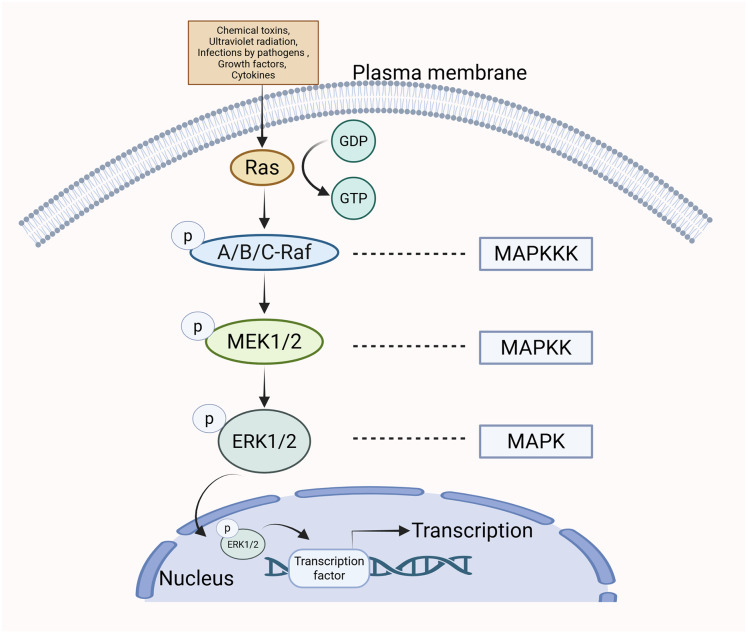
Diseases linked to Erk Signaling Cascade:
Cancers:
Lung Cancer: Driven by mutations in upstream components like RAS or RAF that lead to continuous ERK activation and uncontrolled cell proliferation
Breast, Ovarian, Pancreatic, and Colorectal Cancers: Frequently exhibit aberrant or hyperactivated ERK signaling, which promotes tumor growth, invasion, and metastasis
Thyroid and Brain Tumors: Associated with elevated phosphorylation and activation of the MAPK/ERK pathway
Neurodegenerative and Neurological Diseases:
Alzheimer’s Disease: Involves dysregulated ERK signaling linked to tau phosphorylation and beta-amyloid toxicity
Parkinson’s Disease: Linked to ERK involvement in glial neuroinflammation, L-DOPA-induced dyskinesia, and LRRK2 mutation-related apoptosis
Amyotrophic Lateral Sclerosis (ALS): Involves compromised signaling and motor-neuron viability issues tied to SOD1 mutations
Huntington’s Disease and Prion Diseases: Associated with pathway-driven neuroinflammation and cell stress responses
Multiple Sclerosis: Linked to microglial malfunction and neuroinflammation driven by inappropriate ERK overactivity
Cancer & Phosphorylation-Associated Targeted Therapies
Compared with traditional cytotoxic anticancer drugs, targeted anticancer drugs have the advantages of high efficiency, low toxicity, and strong specificity
Given the important roles of protein kinases in tumor growth and metastasis, if the kinase signaling pathway is effectively blocked, the malignant progression of tumors may be prevented
To date, 68 small molecule kinase inhibitors have been approved
EGFR: Erlotinib, Afatinib, Dacomitinib, Gefitinib, Osimertinib, Lapatinib, Veratinib
Lapatinib for Advanced or Metastatic Breast Cancer
Registered in Europe and in the U.S. in combination with capecitabine for patients with advanced breast cancer or metastatic breast cancer (MBC) whose tumors overexpress human epidermal growth factor receptor (HER)-2 (ErbB-2) and are progressive after prior therapy, which must have included anthracyclines and taxanes, and therapy with trastuzumab in the advanced disease setting
A potent reversible and selective inhibitor of the intracellular domains of the tyrosine kinases EGFR (HER-1) and HER-2 (HER: Human Epidermal Growth Factor Receptor)
Competes with ATP for the ATP-binding pocket
This leads to downstream blocking of the mitogen-activated protein kinase and phosphatidylinositol 3-kinase, Akt, and mammalian target of rapamycin dependent transduction pathways, resulting in growth arrest or apoptosis of tumor cells
Acetylation
Covalent attachment of an acetyl group
A process in which acetyl group donors, such as acetyl‐CoA and acetyl phosphate, covalently bind to the protein N‐terminus and lysine side chains, forming N‐terminal and internal acetylation
Most commonly occurs at the side chain nitrogen atom of Lys
It serves as a regulatory switch for protein conformation and activity changes
Protein acetylation is a dynamic equilibrium state regulated by two families of enzymes:
Lysine acetyltransferases (KATs)
Lysine deacetylases (KDACs)
Functional mechanisms of protein acetylation:
Altering protein conformation
Modulating protein-protein interactions
Regulating enzymatic activity
Altering protein-DNA interactions
Controlling subcellular localization
Influencing protein stability
Lysine Acetyltransferases and Histones
Histones are positively charged proteins in eukaryotic cell nuclei that package, organize, and regulate DNA
p300 are members of the KAT superfamily, primarily acetylating histones
By acetylating histones, p300 switches on DNA accessibility, enabling the binding of transcription factors to DNA and concurrent interaction with p300
Histone acetyltransferase activity of p300 leads to the opening of chromatin structure, allowing RNA polymerase II and transcription factors to bind, initiating gene transcription
Crystal structure of p300 Acetyltransferase Domain bound with Acetyl-CoA:
Active site contains a negatively charged cavity that binds unmodified Lys residues
Flexible activation loop has multiple Lys residues that can occupy this pocket, blocking access to substrate lysines and preventing enzyme activity
Once the lysines in the activation loop are acetylated, they lose their positive charge and can no longer bind to the negatively charged substrate pocket, relieving autoinhibition and enabling enzyme activity
Conformational and Functional States of p300: From an Autoinhibited Resting State to an Active Acetyltransferase:
Inactive: p300 adopts a compact, autoinhibited conformation in which the active site is occluded, resulting in poor engagement with DNA-bound transcription factors (TFs) and other co-regulators Nucleosomes remain compact, chromatin is in-accessible, histone acetylation is low, and gene transcription is repressed.
Active: p300 undergoes a conformational extension that relieves autoinhibition and exposes the active site. Binding of transcription factors and/or co-activators stabilizes the active conformation and promotes recruitment.
Active p300 acetylates nucleosomal histones, opening chromatin and increasing accessibility, thereby promoting gene transcription
Mechanisms of Gene Activation by Histone Acetylation
By weakening the protein-DNA interaction:
Positively charged N-term tails of histones bind tightly to negatively charged phosphate groups
Condensed, transcriptionally inactive nucleosome structure
Acetylation neutralizes the positive charge on histones, reducing their affinity for DNA
Relaxed, transcriptionally active nucleosome structure
Allows transcription factors to access the DNA
Ubiquitination
Covalent attachment of ubiquitin
Most commonly occurs at Lys
Side chain nitrogen is covalently linked to the C-term carboxyl group (Gly) of ubiquitin
Ubiquitination/SUMOylation
Can form polyubiquitin chains
C-term carboxyl group of each subsequent ubiquitin is linked to a Lys residue on the preceding molecule
Requires three types of enzymes:
E1 (activating enzymes)
E2 (conjugating enzymes)
E3 (ligases)
Note: Reversible by the action of deubiquitinating enzymes
The Guardian of the Genome – p53
Also known as:
Transformation-related protein 53
Tumor suppressor p53
Undergoes numerous reversible PTMs for precise control
Protects against cancer
Repairs DNA damage by regulating cell cycle and apoptosis:
Activates DNA repair proteins when damage is detected
Arrests cell growth upon DNA damage recognition until damage is fixed
Initiates apoptosis if DNA has sustained irreparable damage

P53: PTM Crosstalk
E.g., p53 ubiquitination and acetylation
Mechanism:
Unstressed Cells
p53 levels are kept low by ubiquitination of CTD Lys and continuous degradation
Stressed Cells (Exposed to Cellular Stress):
Upon cellular stress, phosphorylation of the TAD facilitates acetylation of the CTD Lys
Acetylation prevents ubiquitination of CTD Lys, thereby protecting p53 from proteasome-mediated degradation
⇨ One of many mechanisms of p53 regulation