The Child with Problems Related to the Production and Circulation of Blood - lecture 2 exam 3
 Call Kai
 Learn
 Practice Test
 Spaced Repetition
 Match
 Flashcards
 Knowt Play
1/43

There's no tags or description
Looks like no tags are added yet.
44 Terms
Functions of Blood
Pediatric blood production, function, and nursing care
Transport and regulation
- Red cells deliver oxygen and carry some carbon dioxide.
- Plasma transports nutrients, hormones, and waste.
- Blood helps regulate temperature and acid-base balance.
Protection and clinical meaning
- White cells support immune defense.
- Platelets and clotting proteins limit blood loss.
- Oxygen delivery depends on hemoglobin and tissue perfusion.
A normal oxygen saturation does not exclude anemia.
- just because the kid sats at 98% on room air doesnt mean they dont have anemia
speaker notes:
Explain that blood supports both transport and protection. Hemoglobin carries most oxygen, while plasma distributes dissolved substances. The circulation must move that blood to tissues. A child with anemia may have a normal pulse oximetry reading because the device estimates the percentage of available hemoglobin carrying oxygen, rather than the total hemoglobin concentration. Connect this distinction to pallor, fatigue, tachycardia, and activity intolerance. Ask students why normal saturation alone cannot establish adequate tissue oxygen delivery.
Components of Blood
Pediatric blood production, function, and nursing care
Cells and cell fragments
- Erythrocytes: hemoglobin carries oxygen.
- Leukocytes: immune defense against infection.
- Platelets: form the initial plug at an injury.
-- petechiae, deficient clotting factors
Plasma and clotting proteins
- Plasma is the liquid portion of blood.
- Albumin supports oncotic pressure.
- Clotting factors help form a stable fibrin clot.
- CBC results help identify affected cell lines.
-- infection
Match the abnormal component to the child's symptoms.
speaker notes:
Differentiate the platelet plug from the fibrin clot. A child with low platelets often develops petechiae or mucosal bleeding. Deficient clotting factors can produce deeper bleeding, including joint bleeding. White cell abnormalities require interpretation of the differential and clinical context. Review red cell count, hemoglobin, hematocrit, and platelet count as distinct measures. Use pediatric laboratory reference intervals because normal values vary with age.
Review of Red Blood Cell Count, Hemoglobin, Hematocrit, and Platelet Count
Review of Red Blood Cell Count, Hemoglobin, Hematocrit, and Platelet Count

Normal Pediatric RBC, Hemoglobin, and Hematocrit
Normal Pediatric RBC, Hemoglobin, and Hematocrit

Understanding Abnormal Findings
1. Red Blood Cell Count (RBC)
- Low RBC count may indicate anemia, blood loss, or decreased bone marrow production.
- High RBC count may occur with dehydration, chronic hypoxia, or polycythemia.
- Nursing consideration: Assess for fatigue, pallor, weakness, and decreased activity tolerance.
2. Hemoglobin (Hgb)
- Low hemoglobin suggests reduced oxygen-carrying capacity and may indicate anemia.
- High hemoglobin may be associated with dehydration or increased red blood cell production.
- Nursing consideration: Monitor for tachycardia, shortness of breath, pallor, fatigue, and signs of inadequate tissue oxygenation.
3. Hematocrit (Hct)
- Low hematocrit may indicate anemia, blood loss, or hemodilution.
- High hematocrit may indicate dehydration or increased red blood cell concentration.
- Nursing consideration: Assess hydration status, fluid balance, and signs of bleeding.
4. Platelet Count (PLT)
- Low platelet count (thrombocytopenia) increases bleeding risk, particularly when significantly decreased.
- High platelet count (thrombocytosis) may occur with inflammation, infection, iron deficiency, or bone marrow disorders.
- Nursing consideration: Monitor for petechiae, bruising, epistaxis, gum bleeding, and prolonged bleeding.
Pediatric Blood Production and Circulation
Pediatric blood production, function, and nursing care
Blood production changes with age
- Fetal blood formation shifts from early sites to liver and bone marrow.
- Bone marrow produces blood cells after birth.
- Erythropoietin stimulates red cell production.
- Hemoglobin composition changes during infancy.
Circulation supports oxygen delivery
- Fetal circulation relies on placental gas exchange.
- After birth, lungs oxygenate circulating blood.
- Right heart sends blood to the lungs.
- Left heart sends oxygenated blood to the body.
-- back up into lungs = right side heart issue
Interpret blood counts using age-specific reference intervals.
speaker notes:
Explain hematopoiesis as production of blood cells from marrow stem cells. The kidneys release erythropoietin in response to reduced oxygen delivery. Fetal hemoglobin declines as adult hemoglobin increases during infancy. A physiologic fall in hemoglobin occurs after birth and differs in premature infants. Avoid diagnosing anemia using adult ranges. Briefly connect the closure of fetal shunts and fall in pulmonary resistance to the cardiovascular slides that follow. Ask students to trace a red cell from the systemic veins through the lungs and back to systemic tissues.
Disorders of Blood Production and Function
Pediatric blood production, function, and nursing care
Red cell disorders
- Iron deficiency limits hemoglobin synthesis.
- Thalassemia reduces globin-chain production (inherited)
- Sickle cell disease alters hemoglobin and red cell behavior (inherited)
- Marrow failure can reduce several cell lines.
- GO TO RBC FUNCTION
Platelet, factor, and marrow disorders
- Immune thrombocytopenia reduces platelet counts.
- Hemophilia involves deficient clotting factor activity.
- Leukemia disrupts normal marrow function.
(anemia, thrombocytopenia)
- Symptoms may include infection, bleeding, or fatigue.
(bone marrow transplant)
GO TO PLATELET FUNCTION
Organize anemia by reduced production, blood loss, or destruction.
speaker notes:
Present these as overview examples rather than a complete diagnostic list. Iron deficiency can follow inadequate intake or blood loss. Thalassemia is inherited and should not automatically receive iron without confirmed deficiency. Sickle cell disease includes hemolysis and vascular occlusion. Leukemia can cause anemia, thrombocytopenia, and infection risk even when the total white count is elevated. Aplastic marrow disorders may cause pancytopenia. Emphasize that fatigue and pallor are nonspecific and require history, examination, and laboratory evaluation. Sources: NHLBI, Anemia diagnosis and causes, https://www.nhlbi.nih.gov/health/anemia/diagnosis and https://www.nhlbi.nih.gov/health/anemia/causes.
Disorders Affecting Blood Circulation
Pediatric blood production, function, and nursing care
Problems within vessels
- Sickle cell vaso-occlusion reduces tissue blood flow. (pain, organ death)
- Venous thrombosis can obstruct venous return.
- Central venous catheters can increase thrombosis risk. (threaded to heart so obstruction - pain, swollen limb: clot, urgent evaluation)
- Severe blood loss reduces circulating volume.
Connections to cardiac disorders
- Structural defects alter blood-flow pathways.
- Heart failure reduces effective circulation.
- Assess perfusion together with oxygenation. (cap refill and pulse ox)
- New neurologic or respiratory changes need urgent evaluation.*****
Blood disorders and heart disorders can both impair tissue oxygen delivery.
- dyspnea, weakness: urgent eval bc PE
speaker notes:
Distinguish hematologic obstruction from the congenital and acquired cardiac conditions covered later. Vaso-occlusion can cause severe pain and organ injury. A swollen painful limb, especially with a central line, warrants prompt evaluation for thrombosis. Sudden dyspnea, chest pain, focal weakness, or altered mental status requires urgent escalation. Do not assume all pain in a child with sickle cell disease is uncomplicated vaso-occlusive pain. Sources: CDC, Complications of sickle cell disease, https://www.cdc.gov/sickle-cell/complications/index.html.
Assessment of the Child With Hematologic Disorders
Pediatric blood production, function, and nursing care
History and family context
- Ask about fatigue, feeding, activity, and pain.
- Review diet, pica, medications, and recent illness.
- Ask about bruising, bleeding, and menstrual losses.
- Review family history and newborn screening.
(perfusion, hydration)
Focused examination
- Assess vital signs, perfusion, and hydration.
- Inspect pallor, jaundice, petechiae, and bruising.
- Assess respiratory effort and neurologic status.
- Check growth and abdominal enlargement.
(liver messed up: hepatomegaly)
Escalate active bleeding, poor perfusion, fever with SCD, or neurologic change.
speaker notes:
Use developmentally appropriate questions and a qualified interpreter when needed. Examine conjunctivae and oral mucosa as well as skin, recognizing that pallor varies with skin tone. Ask adolescents privately about heavy menstrual bleeding and medication use as appropriate. Trend vital signs and functional changes instead of relying on a single measurement. Splenomegaly, pallor, tachycardia, and weakness in sickle cell disease may indicate splenic sequestration. Fever may be the first sign of serious infection. Source: CDC, Fever, https://www.cdc.gov/sickle-cell/complications/fever.html.
Diagnostic Tests
Pediatric blood production, function, and nursing care
Initial blood evaluation
- CBC and differential: cell counts and hemoglobin.
- Reticulocytes: marrow response to anemia.
- Peripheral smear: cell size and morphology.
- Ferritin and iron studies: iron status
(increased iron and ferritin = inflammation)
Targeted testing
- PT/INR and aPTT: selected clotting pathways.
- Factor assays: suspected factor deficiency.
- Hemoglobin analysis: inherited hemoglobin disorders.
- Marrow examination: selected persistent abnormalities.
Interpret results with age, symptoms, trends, and the clinical question. (with peds bc it is different with children at different levels)
speaker notes:
A high reticulocyte count may reflect recovery, hemolysis, or blood loss, whereas an inappropriately low count suggests inadequate production. Ferritin can rise with inflammation, so interpret it alongside the clinical picture and other iron studies. Normal screening coagulation results do not exclude every bleeding disorder. Explain the reason for each test to the family, prepare the child for venipuncture, and minimize unnecessary blood loss. Marrow studies require procedural preparation, pain management, and consent processes according to policy. Sources: NHLBI, Blood tests and Anemia diagnosis.
Nursing Care of the Child With Blood Disorders
Pediatric blood production, function, and nursing care
Daily nursing priorities
- Trend perfusion, fatigue, pain, and bleeding.
- Balance activity with rest and nutrition.
- Use infection and bleeding precautions as indicated.
- Give prescribed therapy and monitor response.
(weight based)
Medication and transfusion safety
- Verify weight-based orders and medication checks.
- Teach safe iron use when prescribed. (stain teeth, constipation, stool softener)
- Follow blood-product identification protocols. (confirm and double check with a second nurse)
- Stop a suspected transfusion reaction and escalate.
(make sure kid isnt bleeding out, iron: GI upset, constipation, stool becomes dark)
Care priorities follow the child's condition and affected blood component.
(transfusion: reaction, blood tubing, right blood, right patient, type and cross match, second nurse check)
speaker notes:
Individualize precautions. For bleeding risk, reduce trauma, maintain pressure after venipuncture, and review medications that may worsen bleeding. For neutropenia, follow the prescribed infection prevention and fever plan. Oral iron can cause gastrointestinal upset and dark stools and must remain out of children's reach. During transfusion, monitor according to institutional policy. If a reaction is suspected, stop the transfusion, assess airway and circulation, maintain IV access with the appropriate solution and new tubing per policy, and notify the provider and blood bank. Source: NHLBI, Anemia treatment, https://www.nhlbi.nih.gov/health/anemia/treatment.
Sickle Cell Disease: Immediate Nursing Priorities
Pediatric blood production, function, and nursing care
Pain and hydration****
- Assess pain promptly using an age-appropriate scale.
- Give ordered analgesia and reassess effect.
- Maintain hydration without fluid overload.
- Give oxygen when hypoxemia is present.
(look at labs to make sure pt isnt drug seeking but be nonjudgemental, do not stall med administration for results)
Urgent complications
- Fever needs immediate clinical evaluation.
- Chest pain, cough, or dyspnea may indicate acute chest syndrome.
- Sudden weakness or speech change may indicate stroke.
- Rapid pallor and abdominal enlargement may signal sequestration.
Use the child's emergency plan and escalate new red flags promptly.
(fever is not normal for sickle cell, AAOx4, resp status)
speaker notes:
Avoid delaying analgesia while waiting for test results. Assess respiratory status, sedation, hydration, and response to medication. Routine oxygen is not a substitute for assessment when saturation is normal. Fever in sickle cell disease requires urgent evaluation because functional asplenia increases infection risk. Acute chest syndrome may involve fever, chest symptoms, hypoxemia, and a new pulmonary infiltrate. New focal neurologic deficits require emergency stroke evaluation. Sources: CDC, Fever and Acute chest syndrome, https://www.cdc.gov/sickle-cell/complications/fever.html and https://www.cdc.gov/sickle-cell/complications/acute-chest-syndrome.html.
Family-Centered Care
Pediatric blood production, function, and nursing care
Partnership and teaching
- Include caregivers in planning and daily decisions.
- Use clear language and teach-back.
- Adapt teaching to age, culture, and health literacy.
- Support the child's growing independence.
Home and school planning
- Review medicines and follow-up appointments.
- Provide a written symptom and emergency plan.
- Coordinate school accommodations and activity needs.
- Assess transportation, cost, and caregiver stress.
Ask the family to explain when and how they will seek help.
(#1 assessment tool)
use words such as nonadherence instead of noncompliance
speaker notes:
Caregivers know the child's baseline behavior and can identify subtle changes. Include children in decisions at their developmental level and support confidential adolescent discussions when appropriate. Ask families to demonstrate medication measurement and describe their response to fever, bleeding, or respiratory symptoms. Coordinate school attendance and activity plans without unnecessary restriction. Explore financial barriers and access to prescriptions. For inherited disorders, offer genetic counseling without assumptions about family choices. Source: CDC, Prevention and treatment of SCD complications, https://www.cdc.gov/sickle-cell/about/prevention-and-treatment.html.
Interprofessional Management
Pediatric blood production, function, and nursing care
Clinical treatment team
- Nursing coordinates assessment and education.
- Pediatrics and hematology guide diagnosis and therapy.
- Pharmacy supports medication and dosing safety.
- Laboratory and transfusion services support testing and blood products.
Support across settings
- Dietitians address iron and nutrition needs.
- Social workers help with resources and access.
- Child life supports coping during procedures.
- School nurses and genetic counselors support ongoing care.
Use one shared care plan with clear follow-up responsibilities.
speaker notes:
Clarify each discipline's role and identify who the family should contact between visits. The nurse communicates symptom trends, response to treatment, and barriers to adherence. Hematology directs disease-specific therapies and prophylaxis when appropriate. Pharmacists help assess interactions and dosing. Child life can reduce procedural distress. Dietitians help distinguish dietary needs from unneeded supplementation. Social work and school nursing can address missed classes, transportation, and caregiver burdens. Use structured handoff communication and confirm that referrals and follow-up appointments occur.
Overview References
Pediatric blood production, function, and nursing care
National Heart, Lung, and Blood Institute
- National Heart, Lung, and Blood Institute. (n.d.). Blood tests. https://www.nhlbi.nih.gov/health/blood-tests
- National Heart, Lung, and Blood Institute. (n.d.). Anemia: Diagnosis. https://www.nhlbi.nih.gov/health/anemia/diagnosis
Centers for Disease Control and Prevention
- Centers for Disease Control and Prevention. (2024). Complications of sickle cell disease. https://www.cdc.gov/sickle-cell/complications/index.html
- Centers for Disease Control and Prevention. (2024). Prevention and treatment of SCD complications. https://www.cdc.gov/sickle-cell/about/prevention-and-treatment.html
Additional topic-specific source links appear in speaker notes.
speaker notes:
These references support the new brief hematology overview. Use the linked NHLBI pages to review blood components and anemia evaluation. The CDC pages summarize sickle cell complications and preventive care. Clinical actions should follow the child's individualized plan, current orders, and institutional policy. The existing cardiovascular material continues after this section.
Cardiovascular System: Core Structure
Structure supports the heart's mechanical work
Four chambers
- Right atrium and ventricle receive and send blood through pulmonary circulation. Left atrium and ventricle receive and send blood through systemic circulation.
Valves
- Tricuspid, pulmonary, mitral, and aortic valves support one-way blood flow.
Heart muscle & coverings
- Myocardium contracts; endocardium lines chambers; pericardium surrounds the heart.
Electrical regulation
- SA node initiates the impulse; AV node and conduction pathways coordinate ventricular activation.
Vessels
- Arteries carry blood away from the heart; veins return blood; capillaries allow exchange.
speaker notes:
Teaching points (4 minutes):. Mechanical Pump — The Heart Muscle, (water pump in the house) heavy physical lifying to force water through the house.Electrical System — The Heart's Conduction System,( electrical wiring in the house)turn pump on and off in a synchronized rhythm. Vascular Circuit — The Blood Vessels( The plumbing pipes) contain the fluid being pushed by the pump. Ask learners to name which chambers serve pulmonary versus systemic circulation. Keep emphasis on foundations needed to understand pediatric differences and congenital flow patterns.
Trace the Blood Flow
BODY
Venae cavae
RIGHT HEART
RA → tricuspid → RV
LUNGS
Pulmonary valve → pulmonary arteries
LEFT HEART
Pulmonary veins → LA → mitral → LV
BODY
Aortic valve → aorta
Clinical connection: congenital defects alter the direction, amount, or route of blood flow
#1 indicator of issues in heart
tachypnea, fatigue during feeding, poor weight gain
L-->R: increase blood flow bc back up, will NOT BE BLUE bc it is recirculating
R-->L: decrease blood flow, BLUE, atrophy, decrease oxygen, decrease workload, decrease blood flow so blue!!!!
speaker notes
Ask one student to trace the full pathway, then ask the class what might happen if blood moves from the higher-pressure left side to the right side, if pulmonary outflow is blocked, or if oxygenated and deoxygenated blood mix. (3 minutes) Increased blood flow to the lungs (pulmonary overcirculation).
Increased workload on the heart.
Pulmonary congestion and possible heart failure.
Tachypnea, fatigue during feeding, and poor weight gain.
Risk of pulmonary hypertension if the defect remains significant and untreated.
Examples: Ventricular septal defect (VSD), atrial septal defect (ASD), and patent ductus arteriosus (PDA).
Teaching point: Left-to-right shunts usually do not initially cause cyanosis because oxygenated blood is recirculated through the lungs.
Expected student responses:
Increased pressure and workload in the right ventricle.
Right ventricular hypertrophy over time.
Reduced pulmonary blood flow when obstruction is severe.
Possible decreased oxygenation and cyanosis, especially when right-to-left shunting occurs.
Fatigue, exercise intolerance, and possible right-sided heart failure.
Examples: Pulmonary stenosis and pulmonary atresia.
Teaching point: Obstruction increases pressure behind the blockage. Severe obstruction may produce inadequate pulmonary circulation and hypoxemia.
Expected student responses:
Systemic oxygen saturation may decrease.
Cyanosis may occur.
Tissues may receive insufficient oxygen.
Chronic hypoxemia may lead to clubbing and poor growth.
The child may experience fatigue and exercise intolerance.
Examples: Tetralogy of Fallot, truncus arteriosus, and certain single-ventricle defects.
Teaching point: Mixing does not always produce cyanosis. The degree of hypoxemia depends on the anatomy, blood-flow direction, and amount of blood reaching the lungs.
pediatric cardiac differences
Regulatory
Heart rate and blood pressure vary with age. Autonomic and hormonal responses influence heart rate, vascular tone, and fluid balance.
Mechanical
The heart must maintain cardiac output as the child grows. Increased workload may show up as feeding fatigue, tachypnea, or poor growth in infants.
- systolic: contract
- diastolic: rest, fills
- stroke volume: blood ejected with each heart beat
Hemodynamics
Preload, afterload, contractility, heart rate, and rhythm influence cardiac output and tissue perfusion.
- blood flow, pressure, resistance
Nursing lens
Assess trends and age-specific behavior—not a single number in isolation.
- deteriorate when increase workload bc they cant handle it
- increase HR = SNS, exercise
- decrease HR = PNS
baby depends on heart rate heavily for cardiac output/function because they cant increase stroke volume
speaker notes
Explain regulatory, mechanical, and hemodynamic differences at the level in the syllabus outline. Cardiac output is affected by heart rate and stroke volume; preload, afterload, and contractility help explain why a child may deteriorate when workload rises. (4 minutes)
1. Regulatory Function — What Controls the Heart?
The regulatory system controls the heart's rate and response to the body's oxygen demands.
Sympathetic nervous system: Increases heart rate and contractility during stress, exercise, or decreased perfusion.
Parasympathetic nervous system: Slows heart rate through vagal stimulation.
Baroreceptors: Detect changes in blood pressure and help regulate heart rate and vascular tone.
Hormonal regulation: Epinephrine and norepinephrine increase cardiac activity.
Pediatric consideration: Infants depend heavily on heart rate to maintain cardiac output because their ability to increase stroke volume is relatively limited
Mechanical Function — How Does the Heart Pump?
The mechanical function involves contraction and relaxation of the heart muscle.
Systole: Ventricles contract, ejecting blood.
Diastole: Ventricles relax and fill with blood.
Stroke volume: The amount of blood ejected with each heartbeat.
Contractility: The strength of myocardial contraction.
When the heart muscle becomes weak or overworked, it may not pump enough blood to meet the child's metabolic needs.
Hemodynamics: What Changes Cardiac Output?
Preload
Ventricular filling/stretch before contraction. Excess volume can contribute to congestion.
Afterload
Resistance the ventricle must overcome to eject blood. Obstruction increases workload.
- fatigue, decrease perfusion, congested
Contractility
Strength of myocardial contraction. Reduced function can lower forward flow.
- decrease SV, CO, perfusion
Heart rate
Rate contributes to output; very fast or slow rates may reduce effective filling/output.
Cardiac output = heart rate × stroke volume
speaker notes
Ask students to predict how obstruction affects afterload and how impaired contraction affects forward flow. Reinforce that the clinical picture may include both poor perfusion and fluid congestion. (3 minutes)
Hemodynamic Function — What Affects Blood Flow?
Hemodynamics describes blood flow, pressure, and resistance within the cardiovascular system.
Factor
Simple Explanation
Pediatric Significance
Preload
Amount of ventricular filling and stretch before contraction
Excessive volume may contribute to pulmonary congestion and heart failure
Afterload
Resistance the ventricles must overcome to eject blood
Increased resistance makes the heart work harder
Contractility
Strength of ventricular contraction
Reduced strength decreases stroke volume
Heart rate
Number of beats per minute
Infants rely heavily on an adequate rate to maintain cardiac output
Focused Pediatric Cardiac Assessment
Gather history, observe first, then examine
History
Prenatal and birth history; known anomalies; feeding duration/fatigue; growth; respiratory symptoms; activity tolerance; family history.
Physical assessment
Heart rate/rhythm/sounds; murmur or thrill; pulses; extremity BP when indicated; color; capillary refill; respiratory effort; edema/hepatomegaly.
Red-flag cues
Cyanosis, tachypnea, sweating with feeds, poor weight gain, weak femoral pulses, syncope, altered behavior, reduced urine output.
Infants use energy to feed: fatigue, sweating, and tachypnea can be cardiac clues.
- observe work of breathing of infant
- get baseline to see changes
- compare pulses: perfusion/obstruction
speaker notes
Demonstrate an assessment sequence. Observe work of breathing and interaction before disturbing the child. Ask caregivers about baseline feeding and activity. Compare pulses and perfusion when obstruction is possible. (4 minutes)
Congenital Heart Disease: Classify by Flow
Start with the pattern, then connect it to the defect
Increased pulmonary flow
- Usually left-to-right shunting; pulmonary overcirculation and heart failure may occur.
- SOB
- L-->R
Obstructive flow
- Narrowing limits forward flow and creates pressure overload.
- fluid build up, increase BP
Decreased pulmonary flow
- Less blood reaches the lungs; cyanosis may occur.
- no perfusion
- R-->L
Mixed circulation
- Oxygenated and deoxygenated blood mix or circuits run in parallel; hypoxemia may occur.
speaker notes:
This classification mirrors the syllabus. Invite students to predict likely assessment cues for each pattern before naming individual defects. Explain that actual severity depends on anatomy and the degree of shunting or obstruction. (3 minutes)
Increased Pulmonary Blood Flow
Left-to-right shunting sends extra blood toward the lungs
Atrial septal defect (ASD)
Opening between atria; may be subtle or asymptomatic in childhood. Murmur or fixed split S2 may be noted.
Ventricular septal defect (VSD)
Opening between ventricles. Larger defects may cause tachypnea, sweating with feeds, poor growth, and heart failure.
Patent ductus arteriosus (PDA)
Persistent fetal connection between aorta and pulmonary artery. Continuous murmur, bounding pulses, or widened pulse pressure may occur.
Nursing focus: respiratory status • feeding tolerance • growth • signs of heart failure ***************
resp status, feeding, growth patterns, no cyanosis
speaker notes:
Use 5 minutes. Compare lesion location and expected manifestations. Small defects may be asymptomatic; larger shunts are more likely to cause pulmonary overcirculation and heart failure. Evaluation often includes echocardiography; management ranges from observation to catheter-based or surgical closure depending on significance.
Decreased Pulmonary Blood Flow
Tetralogy of Fallot (TOF)
Four features: VSD, pulmonary outflow obstruction, overriding aorta, and right ventricular hypertrophy. Clinical cues: cyanosis, murmur, hypercyanotic ("tet") spells (CARDINAL SIGN!!!!!!!!!!!!)
- TET SPELLS!!!!!!!!!!!
Tricuspid atresia
Tricuspid valve fails to form normally, preventing direct right atrium-to-right ventricle flow. Alternative pathways are required. Clinical cues: early cyanosis, tachypnea, feeding difficulty, poor growth.
Priority during a tet spell
Stay with the child • calm/minimize agitation • knee-to-chest • oxygen and emergency plan • escalate promptly (KNOW THISSSSSS)
Reduced pulmonary flow can produce cyanosis
R-->L
speaker notes:
Explain the pattern rather than memorizing isolated facts. For TOF, knee-to-chest increases systemic vascular resistance and can reduce right-to-left shunting. In ductal-dependent lesions, prostaglandin E1 may be ordered to maintain ductal patency while definitive care is arranged. (4 minutes)
Obstructive Congenital Defects
Obstruction creates pressure overload and may reduce downstream perfusion
Coarctation of the aorta
Narrowing of the aorta.
- Upper-extremity BP may exceed lower-extremity BP - Weak/delayed femoral pulses
- Cool lower extremities or poor perfusion
- Severe newborn cases may deteriorate as the ductus closes
Valvular stenosis
- Aortic stenosis: left ventricular pressure load; severe disease may cause chest pain, syncope, or heart failure (no perfusion to the brain)
- Pulmonary stenosis: right ventricular pressure load; severe obstruction may reduce pulmonary flow and cause cyanosis. (no perfusion to the lungs)
Assessment action: compare pulses and blood pressure in all four extremities when indicated.
(may need cath/surgery to decrease pressure)
speaker notes:
Ask what pulse and blood pressure findings would make students suspect coarctation. Discuss how the location of obstruction predicts consequences. Diagnostic evaluation may include echocardiography and other imaging as directed. Treatment depends on anatomy and severity and may include catheter intervention or surgery. (4 minutes)
Mixed Circulation Defects
Oxygenated and deoxygenated blood may mix—or circuits may run in parallel
Transposition of the great arteries
Aorta and pulmonary artery arise from the opposite ventricles. Survival before repair depends on mixing between circuits.
TAPVR / TAPVC
Pulmonary veins return oxygenated blood to the right side. An atrial-level communication is needed for systemic output.
Hypoplastic left heart syndrome
Left-sided structures are severely underdeveloped; systemic circulation is ductal dependent before staged palliation or transplant strategy.
Priority: recognize hypoxemia or poor perfusion and escalate for urgent specialist management.
speaker notes:
Use 3 minutes. Do not overload learners with surgical details. Emphasize recognition of cyanosis, poor perfusion, ductal dependence, and the need for rapid stabilization and pediatric cardiology involvement.
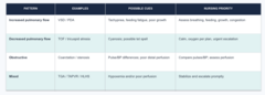
Quick Comparison: Pattern → Clue → Nursing Priority
KNOW THIS
speaker notes:
Have students use the table to explain why classification is useful. Ask: Which pattern suggests comparing femoral pulses? Which suggests knee-to-chest positioning? Which pattern may involve both hypoxemia and heart failure? (3 minutes)
Diagnostic Evaluation: What Each Tool Contributes
Select tests based on the child's presentation and suspected condition
History & physical
Growth and feeding trends, vital signs, oxygen saturation, pulses, murmurs, perfusion, and respiratory effort.
Echocardiography
Defines cardiac anatomy, shunts, valve function, chamber size, and ventricular function.
- picture
ECG & chest imaging
ECG assesses electrical patterns and possible chamber enlargement; chest imaging may show cardiomegaly or pulmonary congestion.
Laboratory studies
Individualized to assess perfusion, renal function, electrolytes, inflammation, or infection.
Cardiac catheterization
Can measure pressures/oxygen saturation, define anatomy, and provide selected interventions.
- need consent, NPO, baseline
speaker notes:
Emphasize that no test replaces bedside assessment. Explain that echocardiography is central for structural/function evaluation, ECG contributes rhythm/chamber information, and cardiac catheterization can be diagnostic or interventional. Before catheterization, verify consent, allergies, NPO status, baseline pulses, and family understanding. (4 minutes)
Pediatric Considerations: Cardiac Catheterization
Before and after the procedure
Before
• Explain in developmentally appropriate language • Verify consent, allergies, and NPO status • Document baseline vital signs and distal pulses • Assess family understanding and anxiety
After
• Monitor cardiorespiratory status and vital signs • Check puncture site for bleeding/hematoma • Reassess distal pulses, color, temperature, refill, sensation • Position access extremity as ordered; monitor hydration and urine output
Urgent cue: bleeding, loss of distal pulse, cool/pale limb, or worsening perfusion.
- NOTIFY THE DOCTOR!!!!!
speaker notes:
Use 3 minutes. Reinforce that a cool, pale extremity or loss of pulse is not a routine finding. Follow facility protocols for observation, extremity positioning, and discharge instructions.
Heart Failure: Recognize the Pediatric Pattern
Forward-flow problems and congestion can occur together
Poor cardiac output
Tachycardia, fatigue, cool/pale extremities, restlessness, decreased urine output, delayed capillary refill; hypotension may be late/severe.
Pulmonary congestion
Tachypnea, dyspnea, retractions, crackles, feeding/activity intolerance, respiratory distress.
- fluid in lungs
Systemic congestion
Hepatomegaly, edema, ascites, rapid weight gain; infant clues include sweating with feeds and poor growth.
- organ enlargement, weight, respiratory, feeding, growth
Trend daily weight, intake/output, respiratory status, perfusion, and feeding tolerance.
(meds, rest, nutrition, underlying cause)
speaker notes:
Connect heart failure manifestations to pediatric assessment. Ask learners to identify signs of poor output versus congestion. Management is cause-specific and may include medications, rest/energy conservation, nutrition support, and treatment of the underlying defect. (4 minutes)
Nursing Care Plan: From Cues to Actions
Individualize to the child's condition, age, development, and family
Recognize cues
Respiratory effort, oxygenation, pulses, perfusion, feeding, growth, urine output, edema, behavior.
Analyze & prioritize
Is the child unstable? Is oxygenation, systemic perfusion, or fluid balance threatened?
Plan interventions
Monitoring, prescribed therapies, nutrition/feeding support, rest, family education, and referrals.
Evaluate response
Compare trends with baseline: work of breathing, saturation, perfusion, weight, output, and tolerance.
VSD: resp, feeding safety, heart failure
speaker notes:
This is the care-planning framework for the third syllabus outcome. Ask students to create a brief care plan for an infant with a large VSD who is tachypneic and sweating during feeds. Expected priorities include respiratory assessment, feeding safety, monitoring for heart failure, prescribed treatment, and caregiver teaching. (4 minutes)
Management & Family-Centered Care
Care is collaborative and developmentally appropriate
Support physiology
Administer prescribed therapy safely; monitor response and adverse effects; support oxygenation, fluid balance, nutrition, and rest.
Prepare child & family
Use plain language, play and age-appropriate explanations; invite questions; explain procedures honestly.
Coordinate resources
Collaborate with pediatric cardiology, nursing, pharmacy, nutrition, social work, and community resources as needed.
Use teach-back and ask about language, cultural practices, transportation, cost, and support needs.
speaker notes:
Discuss culturally competent and family-centered care without assumptions. Ask what the family understands, preferred language, barriers to follow-up, and how the plan can fit routines and resources. Explain the importance of teach-back and shared planning. (3 minutes)
Acquired Heart Disease: Kawasaki Disease
Acute inflammation with risk to the coronary arteries
Recognition
Persistent fever with a compatible combination of: - Bilateral nonpurulent conjunctival injection
- Red/cracked lips or strawberry tongue
- Rash and extremity changes
- Cervical lymphadenopathy
- Most often affects children younger than 5 years.
- FEVER, STRAWBERRY TONGUE,
Acquired Heart Disease: Infective Endocarditis
Microorganisms adhere to susceptible endocardial surfaces and form vegetations
Risk context
Certain congenital defects or prosthetic material, prior endocarditis, central venous catheters, and other cardiac risk conditions.
Clinical cues
Persistent fever, malaise, fatigue, poor appetite, weight loss, new/changed murmur, petechiae, or embolic/immune findings.
Evaluation & treatment
Blood cultures and echocardiography support diagnosis. Prolonged IV antimicrobial therapy is common; monitor for valve dysfunction, heart failure, emboli, and renal effects.
- culture before antibiotics to know what type to give pt
Nursing priority: obtain ordered cultures aseptically before antimicrobials when possible.
speaker notes:
Use 4 minutes. Explain the sequence: vulnerable endocardial surface, bacteremia, adherence, vegetation formation, potential valve injury/embolization. The nurse does not independently diagnose; follow orders for cultures and therapy, monitor complications, and teach line care and medication completion when applicable.
Cardiomegaly & Cardiomyopathy
Distinguish an enlarged heart from disease of the heart muscle
Cardiomegaly
An enlarged cardiac silhouette or heart size finding. It is a clue that requires interpretation alongside symptoms, examination, and imaging—not a complete diagnosis by itself.
Cardiomyopathy types
Dilated: enlarged ventricle with impaired systolic function. Hypertrophic: thickened myocardium; filling/outflow may be impaired. Restrictive: stiff ventricles limit filling.
Clinical cues & care
May include fatigue, dyspnea, poor feeding/growth, heart failure, dysrhythmia, or exertional syncope. Management is individualized; monitor function, perfusion, fluid status, and family history.
Ask about family cardiomyopathy, transplant, or unexplained sudden death.
speaker notes:
Use 4 minutes. Explain that cardiomegaly is a finding, whereas cardiomyopathy refers to myocardial disease. Diagnosis may include echocardiography, ECG, imaging, and selected laboratory or genetic evaluation. Treatment depends on cause and type; advanced cases may need specialist therapies.
Case Application: Choose the Priority
speaker notes:
Allow 4 minutes total: 2 minutes for pairs and 2 minutes for debrief. The case is an application of the outline, not a diagnostic test. Students should recognize the cluster and describe focused assessment and timely provider communication.

Case Application: Acquired Disease
speaker notes:
Allow 4 minutes. Scenario A: Kawasaki disease; coronary artery evaluation and prompt specialist-directed treatment. Scenario B: infective endocarditis; ordered blood cultures with aseptic technique before antimicrobials when possible, cardiac evaluation, and monitoring for heart failure/embolization.

key points
TAKEAWAY 1
Classify congenital defects by pulmonary flow, obstruction, and mixing.
TAKEAWAY 2
Interpret pediatric cues in context: feeding, growth, respiratory effort, perfusion, pulses, and oxygenation.
TAKEAWAY 3
Use diagnostic modalities to define anatomy/function and guide treatment; bedside assessment remains essential.
TAKEAWAY 4
Build a family-centered, developmentally appropriate care plan and collaborate across the care team.
speaker notes:
Close (3 minutes). Ask students to respond to the exit check. Revisit each learning outcome and clarify that students should be able to describe classification, findings, diagnostics/management, care planning, and collaboration. Total planned timing is approximately 90 minutes including discussion and case activities.
Correct answer: B. The infant is showing heart-failure symptoms and increased energy expenditure with feeding. Nursing care should conserve energy and support nutrition without worsening respiratory distress. Forcing feeds can increase oxygen demand and aspiration risk. Daily weight trends are clinically useful when ordered.
A 3-month-old with a large ventricular septal defect becomes tachypneic and diaphoretic during feeds and is gaining weight poorly. Which nursing action is the priority?
a. Encourage the infant to finish the full prescribed feeding volume at each feeding.
b. Allow rest periods, monitor respiratory effort, and use the prescribed high-calorie feeding plan.
c. Place the infant flat after feeding to decrease energy expenditure.
d. Limit daily weights because they may increase family anxiety.
THINK FIRST
Think: feeding is work for an infant with heart failure.
Correct answer: A. Knee-to-chest increases systemic vascular resistance and can reduce right-to-left shunting during a hypercyanotic spell. Calm the child, provide oxygen and prescribed therapy, and escalate rapidly if the episode does not resolve.
A child with Tetralogy of Fallot suddenly becomes very cyanotic and irritable while crying. Which action should the nurse take first?
a. Place the child in a knee-to-chest position and begin calming measures.
b. Encourage the child to walk to improve venous return.
c. Offer a large oral feeding to increase circulating volume.
d. Lay the child flat with the legs extended.
THINK FIRST
Do not increase agitation or oxygen demand during a tet spell.
Correct answer: C. High-dose IVIG can interfere with the immune response to measles- and varicella-containing vaccines. CDC guidance lists an 11-month interval after the 2 g/kg IVIG dose used for Kawasaki disease unless the clinician determines another plan. Reinforce individualized vaccine reconciliation after IVIG.
Which discharge statement by the parent of a child treated with IVIG for Kawasaki disease indicates a need for further teaching?
a. "I will keep the cardiology and echocardiogram follow-up appointments."
b. "I will give aspirin only as prescribed and call before adding over-the-counter medicines."
c. "My child can receive the scheduled MMR vaccine next week because the fever is gone."
d. "I will call for new chest pain, trouble breathing, or a return of significant fever."
THINK FIRST
IVIG can change the timing of some live vaccines.
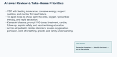
Answer Review & Take-Home Priorities
speaker notes
name one "must-not-miss" cue from each major pattern: pulmonary overcirculation, cyanosis, obstruction, infection/inflammation, and dysrhythmia. Remember that good pediatric cardiac nursing combines physiologic assessment with developmentally appropriate, family-centered care.
Overcirculation = Tachypnea
Cyanosis = Hypoxemia
Obstruction = Weak pulses
Infection/Inflammation = Fever
Dysrhythmia = Abnormal heart rate or rhythm