Genes/Genomics + Fetal lifespan
 Call Kai
 Learn
Learn Practice Test
 Spaced Repetition
 Match
Match Flashcards
 Knowt Play
1/93

There's no tags or description
Looks like no tags are added yet.
Name | Mastery | Learn | Test | Matching | Spaced | Call with Kai | Chat |
|---|
No analytics yet
Send a link to your students to track their progress
94 Terms
What are Chromosomal Anomalies?
Structural/Numerical changes in autosome/s3x chromosomes
During mitosis/meiosis
Occurs prenatally, before implantation, or postnatally
Why do chromosomal anomalies matter? (and important components of care)
They can lead to:
Spontaneous Abortion
Fetal demise, neonatal death/hospitalization
Physical deformities or intellectual issues
Accurate identification + genetic counseling + prevention + treatment
Numerical chromosomal disorders and r/x factors:
Aneuploidy: abnormal #
Monosomy/Trisomy
R/x:
Mom >35, higher BMI
Previous child w/trisomy
Unexpected recombination
Smoking, Radiation
Folic acid deficiency
Major Autosomal Trisomies:
Trisomy 13 → Patau syndrome
Trisomy 18 → Edwards syndrome
Trisomy 21 → Down Syndrome
Trisomy 21 etiology, r/x, and s/s
1 in 700/800 babies
Increased r/x w/Maternal age
However, due to younger clients having more prengnancies, more newborns w/Trisomy 21 are from younger clients
Median lifespan is 47 years
S/s:
Face/Head:
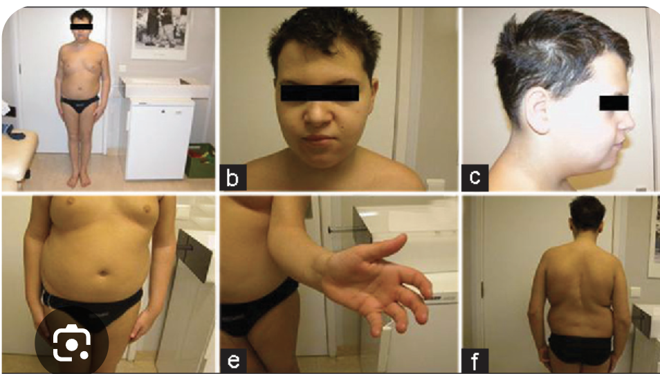
Flat facial appearance, flat nose
Wide-set eyes, upward slant
Small ears, Short neck
Protruding Tongue
Extremities:
Small hands, feet
Single palmar crease
Muscle tone:
Hypotonia
Trisomy 21 Associated Health Problems:
May lead to:
VIsion, Hearing issues
Sleep Apnea
Congenital Heart Defects: Affects 50% of neonates
ToF, AVSD, VSD, PDA
Why are children with Trisomy 21 at an increase feeding/aspiration concern?
Hypotonia → poor muscle control → dysphagia
Congenital heart defects → ^ respiratory rate & effort → fatigue and poor endurance → issues coordinating breathing AND swallowing → increased aspiration r/x when eating
Other factors:
Protruding tongue
Poor lip seal
Fatigue
Absent Feeding cues
Describe the Normal Structure of the Chromosome:
P: short arm
^
Centromere: holds both arms together
Also determines shape and reference point for gene location
v
Q: long arm
Telomeres: DNA sections at end of chromosome
Protects chromosome during DNA replicaiton

Structural Chromosomal Anomalies and Types
Results from improper repair when chromosome breakes OR imbalanced exchange of chromosome material
Types:
D:.
Deletion: DNA segment loses one or more nucleotides
Duplication: DNA segment produces 1 or more copies
Translocation: chromosome splits and the 2 pieces reattach to other chromosomes
Inversion: segments splits off and rejoins same chromosome in opposite direction
Ring: fused together ends of chromosomes → forms ring
S3x Chromosome Anomalies and Types:
Affects 1:400 births
Turner Syndrome: 45 X
Klinefelter syndrome: 47 XXY
Triple X syndrome: 47 XXX
48 XXY
XYY syndrome
Turner Syndrome and s/s:
Females (45 X)
Short Stature, Wide chest, Webbed neck Low hairline
Early ovarian loss
Lymphedema, Amenorrhea
Structural kidney and heart defects

Klinefelter Syndrome and s/s
Occurs in Males (47 XXY)
Tall stature, Long extremities
Female-pattern body hair distributions
Delayed puberty → Small testes → infertile
Developmental Delays
Increased Breast cancer r/x

Triple X Syndrome and s/s
Occurs in females (47, XXX)
Increased height
Learning issues, delayed speech/language, behavioral issues
Hypotonia, Seizures, Kidney abnormalities
XYY Syndrome and S/s:
Occurs in Males (XYY)
Taller than average
Delayed learning, speech/language, and motor development & Behavioral issues
Hypotonia, Hand tremors
Asthma, Seizures, Scoliosis
Larger teeth
What is Multifactorial inheritance? (and examples)
Complex disorders resulting from multiple genes + environment + lifestyle
“polygenetic inheritance”
T2DM, Heart disease, Higher BMI
Makes it difficult to predicts due to many factors and families often sharing same environment and lifestyle
Unifactorial / Single-Gene Disorders, types, and rules:
Caused by a DNA mutation in ONE gene
More predictable inheritance patterns
Autosomal Dominant:
1 mutated copy may cause disorder OR a new/”de novo” mutation can occur
Possible no family h/x due to de novo mutation
E.g. Huntington and Marfans
Autosomal Recessive:
Both gene copies must contain mutation
May skip generations due to possible carriers
E.g. CF, Sickle cell or PKU
X-linked dominant
1 copy causes this
can occur with females (XX) and males too (XY)
“Fragile X syndrome”
X-linked recessive:
One mutated X gene in male can cause this due to them only having 1 X (XY)
Female needs both Xs mutated (XX)
'“Hemophilia”
X-Linked Rule:
Due to father only giving Y chromosomes to son, no male-to male transmission occurs
Father may pass his X chromosome to daugher though
Phenylketonuria (PKU) and screenings & t/x:
Autosomal recessive that involves the PAH deficiency/absence → Phenylanine not broken down → accumulates → high levels dmg brain → severe intellectual disability
Screening:
First day of life via routine newborn a/x
T/x:
Phenylanine-restricted diet
What are carcinogens?
something capable of contributing to cancer development by causing or promoting harmful genetic changes.
Cancer may occur randomly or due to carcinogen exposure over many years
Is cancer inherited?
no
What Is Preconception Carrier Screening? How is it performed? What are the types?
Genetic testing done before pregnancy to check whether someone has a mutated gene
Checks if mother/father may be carriers
Done via blood, saliva, or cheek tissue testing
Types:
Targeted Carrier Screening: screens based on ethnicity, race, or family h/x
Tay sachs is more common amongst Ashkenazi/eastern Europe ancestry
Expanded Carrier Screening: screens for many conditions w/out race/ethnicity in play
What Can Clients Do With a Positive Carrier Result?
Option 1 → Proceed with pregnancy
Should choose more frequent prenatal testing
Option 2 → IVF
Allows embryo to be tested for disorders before transfer back to uterus
Option 3 → Donor egg/sperm
Choose reproductive material of a donor who is not a carrier
Option 4 → Don’t proceed
Who should be offered prenatal testing and who is considered high risk?
Everyone should be offered prenatal testing despite risk levels
High Risk:
^ Mom age
Personal genetic disorder
Family h/x
Aneuploidy
Previous spontaneous abortion or fetal demise
Exposure to dangerous meds/substances
First trimester testings:
Done around 10-13 weeks of gestation
Cell Free Fetal DNA
no fetal r/x from blood collection
Done as early as 10 weeks
Screens for:
Trisomy 21, 18, & 13
Nuchal Translucency (NT): US screening
Checks for amount of fluid in back of neck
Done around 11-13 weeks
Screens for:
Trisomy 21 + 18 and cardiac anomalies
Helpful when using HcG and PAPP-A testing too
CVS: invasive and early DIAGNOSTIC procedure that collects placental tissue sample
Done around 10-13 weeks
Can also determine fetus gender
CVS Routes, Risks and Precautions:
Routes:
Transcervical: through cervix → chorionic villi
Transabdominal: through abdomen → chorionic villi
Uses US for guidance
R/x:
Bleeding, Infection
Spontaneous abortion, membrane rupture
Inconclusive results
If Rh negative mom + Rh positive fetus
^ r/x for fetal hemolytic disease with CVS
If patient taking Anticoags → ^ r/x for bleeding
caution
What four screenings does second trimester have
Quad screen
Anatomy ultrasound/scan
AFP
Amniocentesis
What is Quad screen?
Quad screen a blood test that measures:AFP + HCG + Estriol + Inhibin-A
A-H-E-I
Elevated INHIBIN-A increases ability to predict trisomy 21 risk and avoid false positive results
Done 15-22 weeks
Estimates risk for:
Trisomy 18 + 21 & neural tube defects
If positive = US + Amniocentesis
Anatomy Scan/US
Done 18-22 weeks; a/x spine + face + heart + abdomen & limbs
Checks for fetal growth, sex, and any possible malformations
Alpha-Fetoprotein:
Can be done as part of the Quad test OR by itself
Done around 14-22 weeks
Optimal around 16-18 weeks
Recommended for:
Family h/x
>35 Mom age
Exposure to dangeous meds/substances
DM
Checks for:
Spina bifida
Trisomy 18 + 21
Amniocentesis:
DIAGNOSTIC prenatal test; confirms/rule-out genetic concerns
Done 2nd trimester or if needed, 3rd trimester
Steps:
US guides it (prevents injury)
Needle inserted via abdmoen
Enter amniotic sac
Fluid removed
Analyzed
Third Trimester Genetic Testing:
No specific genetic testing done apart from previous ones:
Cell-Free DNA
Can be done up till birth
US
Harder to see due to fetus changing positions
Amniocenetesis
Reasons for testing:
Intrauterine infection
Fetal anemia or lung maturity
Collecting DNA for paternity testing
Newborn Screening: What Happens?
Done 1-2 days after birth
Blood Tests: heel is pricked and drops placed in special paper
Results avaliable after 5-7 days
For metabolic/genetic screenings
Hearing Screening: tiny earphones placed and connected to equipment → evaluates newborn’s response to sound
Cardiac screening: O2 saturation measured to check for Critical congenital heart defects
What Conditions Are Screened for Newborns?
Endocrine Disorders
Organic Acid Disorders: impairs body’s ability to break down protein
Fatty Acid Oxidation Disorders: impairs ability to convert lipids into energy
May lead to coma, seizures, or death if nutrition is not met
Amino acid metabolism disorders
PKU
Hemoglobin Disorders
Critical Congenital Heart Defect Screening; how is it performed? result types too?
Checked before newborn discharge
I/x congenital heart disease while bebe is in a nonemergent sate
How is it performed?
Right hand (preductal)
Lower extremity (postductal)
Results:
Infant passess if:
O2 sat > 95%
and difference between readings is <3%
Not normal if result is outside expected range and inconclusive
Repeat screening in 1 hour → if abnormal again → another hour later
What is the difference between a genetic counselor/provider role and RN role in regards to genetics & genomics?
Primary provider is responsible for discussing:
Why test is done
Risks, Benefits
Alternatives
May also assist with family planning before getting conception
Provides resources:
Emotional support, community, treatment resources
RN:
Helps pt understand, prepare, pariticipate and receive appropriate support/referrals
Key pregnancy terms:
fertilization → zygote → blastocyst → implantation → embryo
Zygote: single cell formed when sperm fertilizes the ovum
Gestational age: age of pregnancy from first day of LMP
Fetus: developing human; after embryonic period
Embryogenesis: building embryo at start of fertilization
Blastocyst: early developing structure that implants to uterus
5 days after fertilization
Implantation: attachment of blastocyst to uterine lining
week 4 of gestation
Menstrual Cycle to Fertilization:
Weeks 1-2 of cycle
FSH → matures ovarian follicle → produces estradiol → high amounts = ^ LH → ovulation → ovum released → fertilization → zygote
Periods of Prenatal Development
Period | Approximate gestational timing | Major event |
|---|---|---|
Pre-embryonic period | Through the end of week 4 | Fertilization, cell division, blastocyst formation, implantation Day 56: most prominent in embryo injury |
Embryonic period | Week 5 through week 10 | Major organs and body structures develop Most vulnerable to teratogens as major organs are still developing |
Fetal period | Beginning week 11 through birth | Growth and maturation of existing structures |
The Four Blastocyst Membranes
Membrane | Main function | Memory |
|---|---|---|
Amnion | Produces/contains amniotic fluid | Amnion → Amniotic |
Chorion | Contributes to placenta formation | Chorion → placenta |
Allantois | Helps remove nitrogenous waste | Allantois → waste |
Yolk sac | Provides early nutrition and gas exchange before placenta develops | Yolk sac → early support |

What is zona pellucida and how does it relate to implantation?
Zona pellucida surrounds developing blastocyst
prevents from prematurely attaching to fallopian tube
After implantation occurs
Implantation:
Around 4 weeks of gestation
Endometrium is alr thick + vascular + prepared for blastocyst arrival
END OF PRE-EMBRYONIC PERIOD
Embryonic Period and Major developments:
Rapid organ and body system development
Weeks 5-10
Cell multiplication increases; differentiation occurs
One group of cells → nerve cells
Another group → muscle cells
Another → blood cells
MOST vulnerable to teratogens
Major Developments:
Blood cells
Kidneys
Nervous System
Muscles
Limbs
Heart
GI
Respiratory structure
Reproductive structures
Sensory Structures
Teratogenic types:
Certain meds
Chemicals
Radiation
Cannabis
Illicit drugs
Alcohol
Rubella infections
Poor immunization
Week 5 of Embryonic development:
MK
Limb buds appear
Muscles start developing
CV
Heart begins developing
Blood cells develop
Resp
Trachea and bronchi form
GI
GI begins developing
GU
Urinary system begins developing
Bladder and Kidneys start forming
Ovaries begin developing
Neuro
Nerve cells develop
Brain and Spinal cord begin developing
Sensory
Inner and middle ear begin developing
Week 6-7 of Embryonic development:
MK
Arms + hands & legs + feet begin forming
Movement begins
Tissues that will turn to bones start developing
Bone formation begins
Continued muscle formation
CV
Embryonic cardiac activity starts
HR: 110-120/min
Blood starts pumping via major vessels
Resp
Lung dev. begins
GI
Mouth starts forming
Bowel temporarily moves outside abdomen during development
Neuro
Continued brain dev.
PNS nerve ganglia seen
Tissues develops INTO spinal cord
Sensory
Retina and nostrils start forming
Week 8 of Embryonic development:
MK
Limbs become longer
Hands & feet form
Fingers & toes start forming
Neuro
Brain continues dev.
Week 9 of Embryonic development:
MK
Elbows form
Finger and toes become visible
Resp
Breathing movements seen
Sensory
Nipples develop
Hair follicles develop
GU
Male: testosterone begins developing
Male genitalia start forming
Week 10 of Embryonic development:
MK
Cartilage Replaced with bone cells
GI
Bowel begins turning back into cavity
Sensory
Facial features become more distinct
Eyelid becomes more developed
Outer ear begins developing
Fingerprints form
GU
Kidneys function
Urine production starts
Uterus and fallopian tube develop
Amniotic Fluid role between pregnancy stages:
Early pregnancy: fluid comes from
Maternal serum
AMniotic cavity
Coelomic fluid
~Week 10:
Urine from baby accumulates in placenta and increases amniotic fluid amount
Later pregnancy:
Mostly from:
Fetal urine & fetal lung secretions
GI secretions, Placenta, Umbilical cord
Why Does the Fetus Have Amniotic Fluid?
Protection: cushions fetus against maternal trauma
Protects umbilical cord against compression
Antibacterial properties → infection protection
Allows growth: provides room to grow
Nutrient dense
Vitamins
Proteins
Electrolyes
Immunoglobulins
How does Amniotic fluid mainatin balance/homeostasis:
Fluid entering system (urine or lung secretions)
Fluid leaving system (fetal swallowing + intramembraneous absorption)
Both maintain balance in total amniotic fluid
What is AFI?
Amniotic fluid index: US that assesses (a/x) # of fluid
Polyhydramnios
Oligohydramnios
Both types lead to poor fetal outcomes
Functions of the Placenta
Respiratory function: transfer o2 to fetus
Nutrition function: transfers nutrients to fetus
Waste-removal: moves waste to maternal circulation
Endocrine function: produces pregnancy-supporting hormones
Immunological function: transfers mom’s antibodies → fetus
Where is the placenta? What can cross its barrier?
Upper portion of uterus
IF placenta implants in lower uterine segment → pregnancy issues
Checked via US
Certain meds, Alcohol, Nicotine, Infections/viruses (rubella), Anti-Rh antibodies
How is the umbilical cord composed? What are its veins and arteries? Possible abnormalities?
Composition:
2 arteries + 1 vein
Umbilical vein: placental → fetus; carries oxygenated blood
Umbilical artery: fetus → placenta; carries deoxygenated blood
Surrounded by wharton’s jelly
Embryonic connective tissue protecting the vessels
Cord normal inserted in center of placenta
Abnormalities:
Marginal insertion: inserted at edge of placenta
Abnormal vessels: Sometimes, vessels may spread out before reaching site → X Wharton’s Jelly protection
Increases risk of
Fetal/mom hemorrhage
Use US to evaluate everything related

How does fetal circulation work and what are the shunts?
Lungs not working as primary gas exchange site so, bebe receives o2 from umbilical vein
Ductus venosus: bypasses liver
Foramen ovale: bypasses lungs
Ductus arteriosus: bypasses lungs
Follow blood in fetal circulation
Placenta → Umbilical vein → Ductus venosus → Inferior vena cava → RA → foramen ovale → LA → LV → Aorta → Body
When blood returns from upper body and enters Right atrium (RA), this is the path:
RA → RV → pulmonary artery → Ductus arteriosus → Aorta

Ductus Venosus, Foramen Ovale, Ductus Arteriosus
Ductus Venosus: Bypasses liver and alllows o2 blood to enter inferior vena cavsa
This is done because liver does not function before birth
Foramen Ovale:
Between Right atrium and left atrium
Allows bypass of lungs
Due to lungs not working before birth
Closes after birth due to pressure changes occuring when bebe begins breathing
Ductus Arteriosus:
Pulmonary artery ←> aorta
After birth:
Closes because blood flow changes and o2 levels increase
Why does fetal hemoglobin and fetal erythropoetin matter?
HbF (Fetal Hgb) is used to compensate for hypoxic environment fetus is in
This Hgb type has a higher affinity for oxygen → oxygen binds easier
Low o2 environment stimulates fetal kindeys to produce erythropoeitn → RBC stimulation
What are the two major phases of fetal development?
Embryonic period:
Major body systems and organs develop
Done by week 13
Really rapid growth
Fetal Period:
Starts week 11
organs & body system begin to mature and function
After this, bebe can survive outside womb
Survival rates and growth throughout weeks
22–24 weeks: survival is possible in some settings but rates are low
27 weeks: still preterm; source reports approximately 94% survival
31–34 weeks: rapid growth continues
35–37 weeks: continued weight gain and development of sleep patterns
Cardiovascular System development:
Week 5
Cardiac loop forms
Septa begin developing
Weeks 5–7
Septa are present
Heart may be seen via transvaginal US
Heart continues dividing into its major sections
End of week 5
Heart is developed enough to pump blood
End of week 6
Atrioventricular canal develops
Right and left sides of the heart become separated by the atrioventricular septum
Week 6
Beating heart may be visible through transvaginal ultrasound
Weeks 6–7
Heart rate becomes regular
Approximately 110–120 beats/min
Week 37
Heart and blood vessel development is considered complete
Respiratory System development
Week | Development |
|---|---|
5 | Trachea and esophagus begin developing |
7 | Right and left bronchi begin forming |
8 | Structures that will eventually become mature lungs are present |
9 | Breathing movements can be seen |
16 | Respiratory tree formed; epithelium lines airways; cilia begin developing |
17 | Intrapulmonary arteries present |
20 | Lamellar bodies develop; gas-exchange portion of lungs develops
*Start of growth surge* Dexamethasone (or other steroids):
|
24 | Lungs begin producing surfactant
|
24–birth | Lung surface area and terminal sacs continue developing |
32 | Surfactant becomes more effective at reducing risk of atelectasis |
34 | Rhythmic breathing becomes evident
|
36 | Final division of respiratory tree |
After birth | Lung development continues |
GU development:
Week 5
Urinary tract begins developing
Kidney structure is present by the end of the week
Week 6
Renal pelvis develops
Kidney lobes form
Collecting tubules and major calyces develop
Week 7
Bladder and urethra continue developing
Week 10
Kidneys begin producing urine
Week 32
Kidneys are considered fully formed
Reproductive Development
Male development
Week 9
Testosterone production begins
Male reproductive structures begin developing:
Epididymis
Vas deferens
Ejaculatory duct
Seminal vesicles
Week 12
Scrotum develops
Week 28
Testes descend into the inguinal canal
Week 33
Testes descend into the scrotum
Female Development
Week 5
Ovaries develop
At birth, ovaries contain approximately:
1–2 million follicles
Week 10
Uterus forms
Fallopian tubes form
By week 12
Genital development is complete but continues to grow in 3rd trimester
Fetal sex can be identified by ultrasound
Neurological System early development
Neural tube
Around week 4:
Neural tube closes
Incentivize to take folic acid before it closes
Motor nerve fibers appear
The neural tube becomes the precursor of the:
Brain + spinal cord
Weeks 5–8
Major neurological development occurs:
Brain vesicles develop
These contribute to the:
Forebrain
Midbrain
Hindbrain
Parasympathetic ganglia develop
All 12 cranial nerves can be detected
Embryo responds to touch and light
Neurological later development
Weeks 10–12
Sympathetic trunk appears
Facial reflexes develop
Upper-extremity reflexes begin
Major brain structures can be seen
Week 15
The fetus can demonstrate:
Startle/Moro reflex
Twitching
Swallowing
General limb movements
Weeks 17–20
Hormonal response to pain develops
Rapid eye movement appears
Quiet and active sleep periods occur
Weeks 26–29
Major maturation occurs:
Parasympathetic nervous system develops rapidly
Gross muscle movements are visible
Hormone production occurs
Functional brain patterns are present
Fetus can control body temperature
Myelination is completed
Hearing Development
Around week 4
Internal ear begins developing.
Week 5
Cochlear duct develops
Primordial cochlea develops
Week 16–28
Eustachian tube develops significantly
Week 18
Auditory canal becomes formed and patent
Week 23
Internal ear reaches approximately adult size
Week 26
Fetus can hear
Eye Development
Starts around week 4
Neuro tube closes → optic grooves develop → optic vesicles form → optic cup develops → retina begins forming around week 7 → iris + cilliary body develop → lens forms → week 26: eyes developed
Gastrointestinal System development
Week 5
Digestive structures are distinguishable
Week 7
Mouth begins forming
Week 11
Swallowing begins
Muscle layers are present throughout intestines
Week 12
Intestinal glands begin developing
Pancreas begins producing insulin
Week 14
Continuous mucous membrane forms throughout intestinal tract
Liver begins producing red blood cells
Week 18
Liver and pancreas begin producing secretions
Week 22
Meconium is visible in intestinal tract
Week 24
GI absorption begins functioning
Week 32
GI absorption reaches maturation
Musculoskeletal System Development:
Weeks 6–7
Bone formation begins
Muscle formation occurs
Hands and feet begin forming
Bone → Muscle → Limb
Week 9
Toes have formed
Week 12
Upper extremities and associated muscles are present
Week 14
Fetus can make a fist
18–20 weeks
A client pregnant with their first child may feel fetal movement
Weeks 23–25
Bone marrow begins producing blood cells
Week 37
Bones and muscles are fully developed
When is fetal movement heard? What should mom do?
During 2nd trimester
Early as 16 weeks; around 18-20 weeks (mostly this range is for first pregnancies)
Sadovsky method:
count kicks for 30 mins or longer
Integumentary Development
Weeks 11-14:
Toenails & fingernails present
Weeks 15-18:
Lanugo appears
Weeks 19-22:
Lanugo present everywhere
Eyebrows present
Weeks 23-26:
Fat storage begins
FInger/footprints form
Eyelids open & close
Weeks 27-30:
Covered with vernix caseosa (thick white subtance covering body)
Eyelash and eyebrows well-formed
weeks 35-37:
Less skin wrinkles
Subcutaneous tissue develops
Weeks 38-40:
Lanugo only in under arms and shoulders
Fingernails elongate
Breast buds appear
Hair is thicker & coarser
what is Spontaneous Abortion (Miscarriage)? What may mom experience?
Pregnancy loss occuring naturally:
Usually around 1st trimester; w/out warning
Mostly due to fetal chromosomal abnormalities and increased mom age
Mom may experience bleeding, cramping, passage of tissue
Possibly no pain/bleeding at all
The 5 Types of Spontaneous Abortion
Threatened abortion: pregnancy is still viable
50% of times it may lead to spontaneous abortion
Findings:
Mild-mod abd cramping
vaginal bleeding/spotting
Cervix remains closed
US shows viable pregnancy
Priorities:
A/x dilation, US, B-hCG, UTI possibility
Anxiety/depression
Inevitable abortion:
Findings:
Vaginal bleeding
Cervical dilation
Cramping, no pregnancy tissue passed yet
Incomplete Abortion:
Cervix dilated
Cramping + bleeding
Some tisse remains and some is loss
Complete Abortion:
Uterine cramping and bleeding
All products of conception have passes
US shows empty uterus
Missed Abortion: Fetus has died but body hasn’t expelledit
No signs of life, no uterine contraction
No vaginal bleeding
S/s of pregnancy may stil occur or regress
D/x:
B-hCG and pelvic US
If B-hcG > 3000 → pregnancy should be visible
falling numbers = nonviable pregnancy
Not every miscarriage will have pain or bleeding
Risk factors (r/x) for Complete spontaneous abortion:
Most common:
Fetal chromosomal abnormalities
Mom:
Diabetes
Celiac disease
Autoimmune disorders
Maternal age ≥35
Previous pregnancy loss
Uterine structural abnormalities
Hypertension
Malnutrition
Lifestyle:
Illicit drug use
Cigarette smoking
Exposure to Arsenic or Lead
Large amounts of caffeine
Alcohol consumption
Infection
Parvovirus B19
Zika virus
Cytomegalovirus
Syphilis
Specific risk factors for Threatened or MIssed abortion:
Threatened:
Maternal age ≥35
First pregnancy
Hyperemesis gravidarum
Placenta previa
Gestational diabetes
Low socioeconomic status
Missed:
Maternal age ≥30
BMI >24
Embryo/fetus measuring larger or smaller than expected
“size discrepancy”
Important tests done to check for abortion:
Test | Purpose |
|---|---|
Ultrasound | Examines uterine contents and pregnancy viability |
Quantitative β-hCG | Evaluates pregnancy progression/viability |
Vaginal cervical examination | Determines cervical dilation |
Urinalysis | Especially important with threatened abortion because UTI can be associated |
Blood type/Rh status | Determines need for Rho(D)-immune globulin |
Rh Status, and how it should be given?
If mom Rh-Negtive → RhoGAM
If not given → alloimmunized after exposed to Rh + fetal blood → in future pregnancy → erythroblastosis fetalis
Timing:
W/in 72 hours of spontaneous abortion, termination or vaginal bleeding
What things should a RN educate patient what may be considered signs of abortion? What follow ups would be done?
Heavy vaginal bleeding + severe cramping + fever
Saturating 1-2 pads per hour for 2 hours
Follow ups:
Transvaginal US, B-hCG, continuous monitoring
Acetominophen for pain meds
ASSESS for:
Depression, Anxiety, Grief, PTSD → counseling, support groups, support
What are the 3 treatment options of spontaneous abortions?
Conservative Management:
Monitoring for spontaneous passage of products
80% of abortions pass by themselves
Medical Management:
Misoprostol (^ uterine contractions) or used with Mifepristone (blocks PG which stops pregnancy):
Takes 3 days for meds to aid complete expelling of abortion tissues
Report soaking 2 pads in 1 hour to provider if taking Mifepristone
DIlation and Curetage: removes tissue from uterus
Done when there is:
Infection, hemodynamic unstable, severe anemia
Bleeding disorder, CV disorders
Can be conjucted with meds
Complications of Spontaneous Abortion Treatment
Infection and Hemorrhage
Other:
Hypovolemic shock
DIC
Retained products of conception
Excessive vaginal bleeding
Surgical complications can include:
Cervical laceration
Uterine perforation
Reasons for Induced Abortion:
Undesired pregnancy
Maternal health concerns
Fetal congenital anomalies
Medical conditions that make pregnancy dangerous
Therapeutic abortion
Maternal medical reasons
Fetal nonviability
Conditions That Can Make Pregnancy Dangerous
Cardiovascular
Severe cardiovascular disease
Left ventricular heart failure
Renal
Severe kidney disease
Acute renal failure
Pulmonary
Pulmonary arterial hypertension
What is Selective Reduction?
Done to lower number of fetuses in mutlfetal pregnancy due to higher risk of having preterm birth and pregnancy complication
Before an induced abortion, what should be done?
Confirm pregnancy via:
Vaginal US or abdominal US
Medication Abortion (what meds, common effects, contraindications, and complications)
Mifepristone → Misoprostol
Mifepristone: stops PG → X pregnancy
Misoprostol:
Cervical dilation, contractions, vaginal bleeding, passage of clots, & expulsion of contents
Taken 24-48 hrs AFTER mifepristone
Common effects:
Cramps, bleeding, clots, N/V, Diarrhea, Fever/Chills, Headaches
Most intense pain occurs 2.5-4 hours after misoprostol → lasts 1 hr
Bleeding may occur for several weeks after
Contraindications:
IUD, ectopic pregnancy
Anticoagulation therapy
Chronic adrenal failure
Porphyria, Hemodynamically unstable
Complications:
Pain, Infection
Heavy bleeding
>2 pads in 2 hrs
If products not passed within 24 hrs → provider
Procedural Abortion
Procedure | Typical gestational range in source |
|---|---|
Manual vacuum evacuation | 4–10 weeks |
Suction curettage | 6–14 weeks |
Dilation and extraction | 14–24 weeks |
Procedural Abortion Complications
Minor
Vasovagal response
Heavy bleeding
Unsuccessful procedure requiring repeat suction
Serious
Uterine perforation
Infection
Sepsis
Hemorrhage
Post-Abortion Teaching
Avoid putting anything in the vagina for the period specified by the provider/facility instructions
Use contraception if pregnancy prevention is desired
Keep follow-up appointments
Contact provider if:
Soaking 2 pads in 2 hours
Clots larger than a lemon for ≥2 hours
Temperature ≥38°C (100.4°F) for 24 hours
Pregnancy s/s
Moderate–severe abdominal/back pain
Vomiting/diarrhea lasting >24 hours
Foul-smelling vaginal discharge
What is the classical triad of post-abortion complication:
PAIN + BLEEDING + LOW-GRADE FEVER
Most likely due to retained products of conception
Infection may develop → septic shock
v BP, ^ HR, ^ /vTP
Interventions to prevent deterioration:
Large-bore IV access, o2 therapy
Monitor blood loss, VS, psychosocial status
Continued bleeding and poor contractions
When to Call Rapid Response with abortion complications? what members included?
Develops acute vaginal bleeding
Has increasing abdominal tenderness
Develops a high fever
Becomes unresponsive
Stops producing urine
Team:
Obsterician, Radiologist, General surgeon, Urologist, Infection disease, RN
Sepsis vs Localized Infection relating to pregnancy issues
Localized:
Foul-smelling discharge
Abdominal pain
Fever
Sepsis:
Hypotension
Altered mental status/disorientation
Hemodynamic instability
Organ dysfunction
Discharge Teaching After Pregnancy Complications
Use contraception to prevent pregnancy if desired
Complete prescribed antibiotics
Increase fluids to approximately 64 oz/day
Rest and avoid overexertion
Avoid strenuous exercise
Avoid alcohol for 48 hr or while taking narcotics
Avoid putting anything in the vagina for the specified period
Intrauterine Fetal Demise (IUFD) / Stillbirth, causes, r/x, and s/s
Fetal death occuring at or beyond viability
Causes:
Fetal growth restriction (FGR)
Placental abnormalities
Trisomy 21,18,13 and Turner
R/x:
Placental abruption, previa, single artery, abnormal cord insertion
Mom:
DM, Preeclampsia, HTN, Obesity
Antiphospholipid syndrome
>35 years, first pregnancy with older age
Multiple gestation
Fetal:
FGR, abnormalities
>40 weeks
Late start to prenatal care
Lifestyle:
Tobacco, Alcohol, Illicit drugs
S/s:
Asymptomatic or
Decreased fetal movement or non
Client may “feel” smthing is wrong
IUFD d/x & care
D/x:
Fetal doppler or external electronic fetal monitoring or US
Also ask when they last felt a fetal movement
CBC, Blood type + rH, HBsAG Coag
Syphillis, HIV, Rubella, Urine toxicology
Check umbilical cord
Culture of mom and fetus placenta
Care
EMPATHY + PRIVACY + RESPECT + CHOICE
Emotional Support
Respect family wishes
If they want to hold, bathe, name, or photograph baby
Memory box, clothing, foot prints, religious ceremonies
A/x for possible PPD, Anxiety, depression, or dysfunctional grieving
Vaginal birth is prefered
T/x:
If <24 weeks → D&E
>24 weeks → misoprostol + oxytocin
Or balloon cathether: ripens cervix for labor
12 hours to work
Breast Care
Stop milk production
Possible breast milk donation
Reduce exposure to other newborns crying
Follow up with support systems and birth control or emotional recovery