Introduction to Immune Response
 Call Kai
 Learn
Learn Practice Test
 Spaced Repetition
 Match
Match Flashcards
 Knowt Play
1/81

There's no tags or description
Looks like no tags are added yet.
Name | Mastery | Learn | Test | Matching | Spaced | Call with Kai | Chat |
|---|
No analytics yet
Send a link to your students to track their progress
82 Terms
Cytokines functions
Mature dendritic cells
↑ Macrophage function
↑ selectin
↑ vascular permeability
↑ function of other immune cells (survival, proliferation, cytotoxicity)
Role of selectins
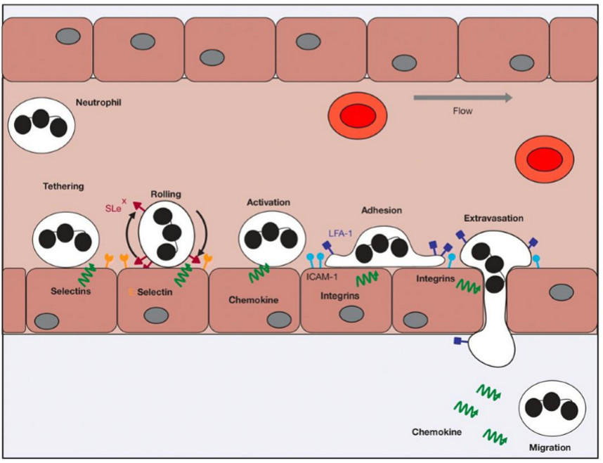
Receptors on endothelial cells that bind to ligands on neutrophils causing them to decelerate on the blood vessel wall
Give 2 examples of cytokines
interleukins (IL-1)
TNF
Chemokines functions
Attract other immune cells
Chemokine example
IL8
How do chemokines attract inflammatory cells into tissues
By increasing their motility
Pluripotent haematopoietic stem cells can go down what 2 lineages to become what types of cells
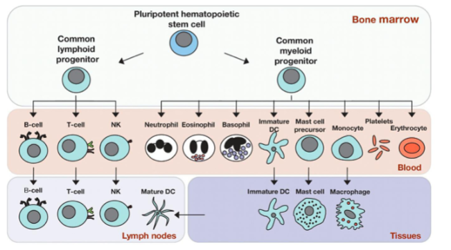
What’s the most abundant white blood cell
Neutrophils - comprising well over 50% of WBC’s
Name the 3 main cells of the myeloid lineage
Neutrophils
Macrophages
Dendritic cells
Neutrophils role
phagocytosing and killing microbes
secrete cytokines & chemokines
(make pus)
Process of neutrophil extravasation
Neutrophils develop from where
Bone marrow
Role of integrins
Strong adhesion molecules on neutrophils that facilitate their active migration into tissues
Macrophages are activated by what
PRRs (pathogen recognition receptors) (toll-like receptors) on cell surface
Endosomes (in other cell compartments) that recognise PAMPs (Pathogen Associated Molecular Patterns) & DAMPs (Danger Associated Molecular Patterns)
Give 3 examples of PAMPs/DAMPs
Examples include certain sugars not seen in humans e.g. lipopolysaccharide (LPS/endotoxin), nucleic acids (DNA/RNA), factors released factors by dying cells
Macrophages develop from where
Bone marrow
Macrophage general response to activation
The danger signal activates the macrophage promoting phagocytosis of microorganisms & abnormal cells
The macrophage begins to secrete cytokines and chemokines.
Presents antigens to T cells
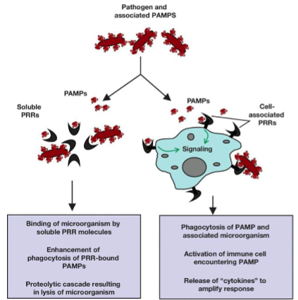
4 specific things released by macrophages
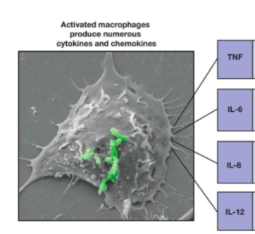
Role of TNF
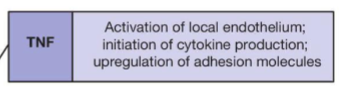
Role of IL-6
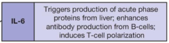
Role of IL-8
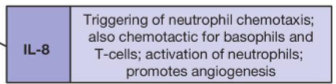
Role of IL-12
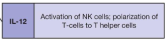
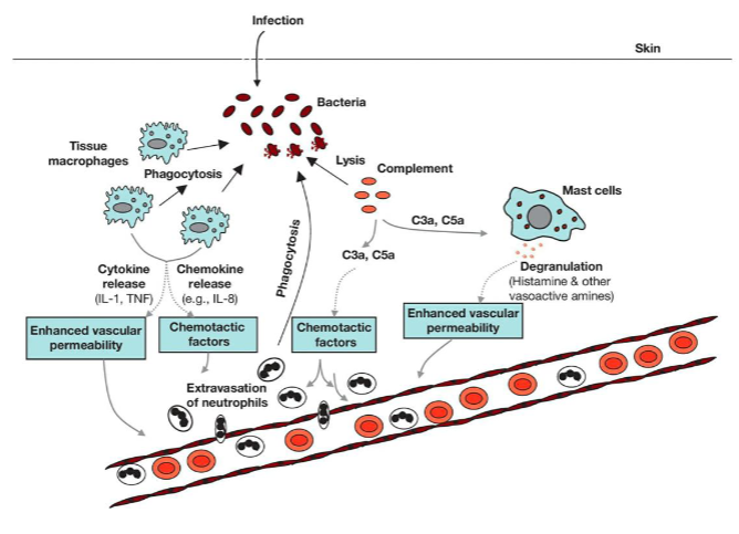
In the complement system associated with macrophages, what is the role of C3a & C5a
Induce inflammation & chemotaxis
What happens when C3a & C5a activate mast cells
Degranulation → mast cells release histamines & other vasoactive amines → enhances vascular permeability
Role of dendritic cells
Present antigens to T cells
Dendritic cells develop from where
Bone marrow
Name the 3 main cells of the lymphoid lineage.
Which are involved in innate / adaptive immunity?
NK cells (innate)
T cells (adaptive)
B cells (adaptive)
How do NK responses generally differ from T & B cell responses
T & B cells make specific receptors to recognise antigens because they are adaptive - NK cells don’t
How do T & B cells make specific receptors to recognise antigens
Genetic recombination of limited precursors & germline DNA
What type of receptors do NK cells use (where do they come from)
They are germline encoded - not rearranged
So they don’t recognise specific receptors
What activates NK cells
Missing self: MHC doesn’t reach KIR (cells that lack MHC class 1 molecules)
Stress induced: Have high stress induced molecules - abnormal proteins
ADCC: Antibody-dependent → antibodies recognised by Fc receptors
What is KIR
Strong inhibitory receptors on NK cells that inhibit NK when they recognise MHC
Source of antibodies on cells recognised by Fc receptors
B cells
NK response to activation
Kill with cytotoxic granules containing lytic proteases - perforin & granzyme
Kill with death ligands - TRAIL & Fas
Release cytokines
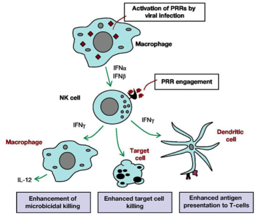
How do NK cells, macrophages and dendritic cells influence each other
Macrophages produce cytokines → activate NK cells → release cytokines → mature dendritic cells → enhance macrophage function
Where do T cells come from
Bone marrow precursors → Thymus → T Cells
What activates T cells (generally)
TCRs (T cell receptors) recognise a combination of antigen‐derived peptides and MHC
What specific antigens do CD4 and CD8 T cells recognise
(give 3 examples of each)
CD4 T cells → MHC class II antigens (HLA-DR, DD, DQ)
CD8 T cells → MHC class I antigens (HLA-A, B, C)
Function of CD4 and CD8 T cells
CD4 → Help other immune cells & limit unwanted immune response
CD8 → Kill infected cells
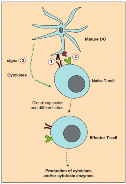
Explain the 1st of the 3 signals necessary to fully activate T cells
Signal 1: TCRs bind to antigens → CD4/8 activates ITAMs on CD3z → CD3 activates T cells
Explain the 2nd of the 3 signals necessary to fully activate T cells
Signal adhesion molecules (LFA-1 [on T cell] & ICAM-1 [on APC/tumour cell]) → cause tight binding of APC & T cell → Co-stimulatory molecules (on the APC) bind to CD28 (on T cell) → Prevents apoptosis of T cell & provides 2nd signal → T cell activated
Explain the 3rd of the 3 signals necessary to fully activate T cells
Cytokines released → Clonal expansion & proliferation → T cell activated
What allows T cells to stay active for a long time
Adhesion molecules (LFA1 & ICAM1) form an immune synapse
Why must T cell activation be controlled
Can lead to autoimmune disease
What 3 different T cells have what 3 different responses to activation
Helper T → Activates B cells & helps other immune cells
Regulatory T → Limits excessive/unwanted immune response
Cytotoxic T → Kill infected cells
How are T cells deactivated
“Off switch” molecules: PD1 on T cells binds to PDL1 on APCs → Phosphates dephosphorylate signalling molecules → inhibit kinase activating signals → turn off T cells
Competitive inhibition: CTLA4 competes with CD28 (both on T cells) to bind to CD80/CD86 on APCs → Limits prevention of apoptosis
Tregs: Tregs express ligands for immune checkpoints (PDL1/CTLA4) → release suppressive cytokines & eliminate CD4 & CD8
What problem can occur with Tregs
They can be overactive in cancer
Where do B cells develop
Bone marrow
How are B cells activated
Helper T cells
or
Follicular dendritic cells present antigens
What 2 cells can B cells differentiate into when activated - what role does each one carry out
Memory B cells - wait to mount rapid response if antigen presents again
Proliferation into plasma cell that makes antibodies → Binds to antigen → Neutralise APC / Mark for destruction by NK cells / macrophages
What decides what role B cells go into
CD4 T cells
CD40L → Memory B
CD40 → Plasma cell
Name the 2 regions of an antibody & their function
Fab region → Antigen binding (variable)
Fc region → links complement & effector cells (NK cells → ADCC / Macrophages → ADPC)
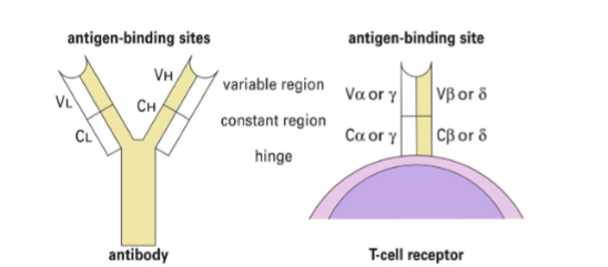
How are TCRs similar to Fabs
Both have 2 different peptide chains
Both have variable, constant & hinge regions
How do TCRs differ from Fabs
TCR = membrane bound with 1 antigen binding site
Fab = not membrane bound. Has 2 binding sites
Do both B and T cells exhibit immunological memory
Yes
What immune cells are involved in humoral immunity
B cells
(Humoral immunity = the part of the adaptive immune system that uses B cells and antibodies to fight pathogens in body fluids)
What type of molecules can antibodies be
Antigens can be proteins, carbohydrates, lipids, nucleic acids, small molecules released in a soluble (secreted) version
Via what mechanism do B cells have the ability to make millions of antibody molecules capable of recognizing numerous antigens
Via somatic recombination B cells have the ability to make millions of antibody molecules capable of recognizing numerous antigens
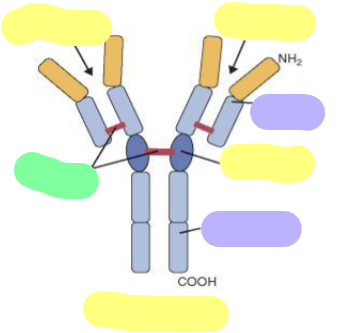
Yellow = Region
Purple = Heavy/Light Chain
Green = Other
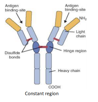
The BCR (B Cell Receptor) is a complex containing an antibody on the surface of the B cells, which is linked to signalling molecules, e.g. ….
tyrosine kinases
Strength of B cell activation depends on what
how good a fit the BCR is for the antigen
How is the BCR used in B cell cancer treatment
Some B cell cancers are treated by using kinase inhibitors that block signalling through the BCR
Compare Primary vs Secondary Immune Response
Primary response: slower, lower antibody levels
Secondary response: rapid, strong, long-lasting
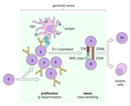
Where do Follicular Dendritic Cells present antigens to B cells
In the germinal centre of lymph nodes
How does B cell selection for antibody production work
Only B cells that receive the strongest survival signal (best antigen:antibody fit) are selected and survive.
Others undergo apoptosis (programmed cell death)
When the B cells are selected what other cell selects whether they become antibody producing Plasma cells or Memory B cells
Helper CD4 T cells provide additional survival signals helping the selected B cells to mature into either antibody producing Plasma cells or Memory B cells
Following antigen recognition what do B cells do to increase their efficiency & to make the tightest fit possible to antigen
B cells rapidly proliferate and mutate their Ig genes to make the tightest fit possible to antigen
MHC stands for what
major histocompatibility complex
What is MHC & what does it do
MHC is a genetic locus that controls histocompatibility
In early transplantation experiments, the donor and recipient were found to need the same MHC locus (tissue type) in order to avoid graft rejection. Why?
MHC molecules are recognized by T cells, via their T-cell receptors (TCRs) and unless the donor and recipient are matched for these molecules the host T cells see the donor cells as foreign and induce rejection
Alternatively, donor T cells see host cells/tissues as foreign and cause graft versus host disease.
True / False: MHC genes are highly polymorphic
True
MHC antigens in humans are aka what
HLA antigens (human leukocyte antigens)
The T‐cell receptor (TCR) complex has how many chains, called what
2 - alpha and Beta
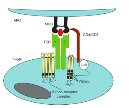
Co‐clustering of CD4 or CD8 with the TCR complex facilitates signal propagation though what mechanism through what molecule
through phosphorylation of immunoreceptor tyrosine‐based activation motifs (ITAMs) within the CD3 ζ chain
Clinical significance of CD3 ζ (zeta)
an important component of artificial TCR’s e.g. CAR-T
In what stage of T cell activation (which signal involving which molecule) can T cell receive too much activating signal? What happens then?
In T cell activation by signal 2 (using CD28)
A strong activating signal, if not balanced by a strong anti-apoptotic signal from an anti-apoptotic protein (Bcl-xL) can induce apoptosis - activation‐induced cell death)
Name 2 Immune Checkpoint Receptors and their Ligands
Immune Checkpoint Receptors (PD-1 and CTLA4)
Ligands (e.g. PD-L1 and CD80/86)
How do immune checkpoints inhibit activating signals
Activation of immune checkpoints → phosphatases dephosphorylate signalling molecules → inhibit activating signals
Do phosphatases & kinases work together or oppose each other? Explain
Phosphatases oppose the action of kinases.
Kinases phosphorylate amino acid substrates, typically inducing cellular activation e.g. tyrosine kinases
Phosphatases inhibit activating signals
When are CTLA-4 and PD-1 upregulated on activated T cells
usually after prolonged activation
What type of T cell is often overactive in cancer
Tregs → impaired immune response