Chapter 5: T-Cell Mediated Immunity
 Call Kai
 Learn
 Practice Test
 Spaced Repetition
 Match
 Flashcards
 Knowt Play
1/25

There's no tags or description
Looks like no tags are added yet.
26 Terms
T-Cell Activation
Multiple TCRs need to be triggered for T cell activation to occur (high avidity)
CD3 and zeta produce the same signals in all clones
LFA-1
A type of intergrin proteins in T cell that bind to ICAM-1 on APC for adhesion
CD28
Receptor on T-cells that binds to B7 costimulation for sufficient T cell activation
CTLA-4 & PD-1
Inhibitory receptors of T cells, prevent immune response against self antigens
Compete with CD28 for binding of B7
Lck
Protein tyrosin kinase
Non-covalently bind to CD4/CD8 coreceptor
When MHC binds to coreceptor, it phosphorylates ITAM of CD3 and zeta chain
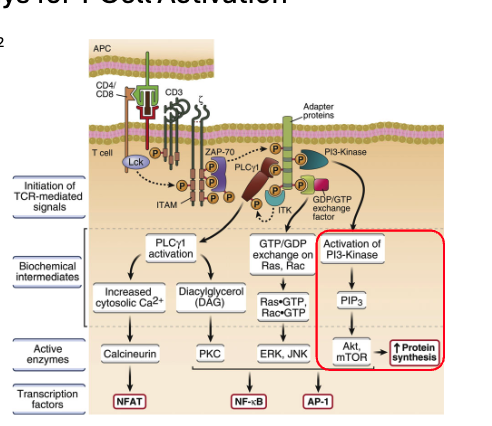
ZAP-70
Phosphorylated ITAM on zeta, become active by Lck phosphorylation
Phosphorylates PLC-gamma

Phospholipase-C Gamma (PLCγ)
Phosphorylated by ZAP-70
Enzyme that facilitates: PIP2 → IP3 + DAG
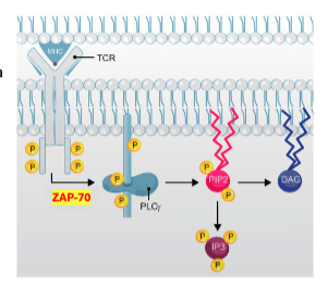
Inositol 1,4,5-triphosphate (IP3)
Releases calcium from ER to cytosol to activate enzyme calcineurin

Calcineurin
Dephosphorylates transcription factor NFAT

NFAT
Activated by calcineurin
Migrates from cytosol to nucleus and supports transcription of IL-2

Cyclosporine
Immunosuppressive drug that prevent graft rejection
Inhibits calcineurin phosphatase activity → suppress NFAT
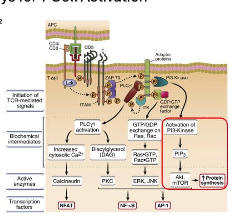
Diacylglycerol (DAG)
Activates protein kinase C (PKC)

Protein Kinase C (PKC)
Phosphorylates inhibitor κB
nuclear factor-κB is released and move into nucleus
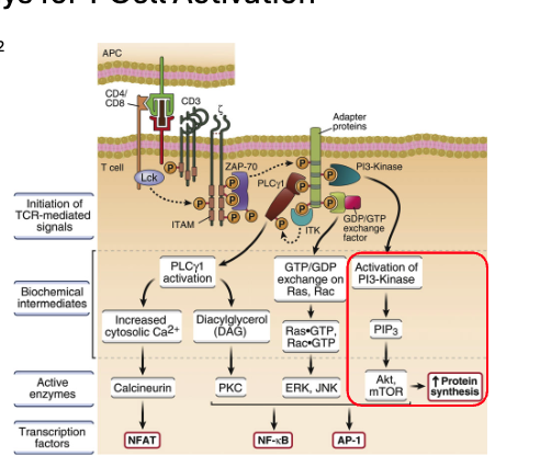
GTP-Binding Ras/Rac Protein
Exchanges GDP for GTP on Ras & Rac
These enzymes activate kinases ERK & JNK

ERK & JNK
Kinases that activated by Ras/Rac GTP
Formation of transcription factor AP-1, which enters nucleus and enhances transcription of T cell genes

Phosphatidylinositol-3 Kinase (PI-3)
Phosphorylates PIP2 → PIP3

Phosphatidylinositol-3,4,5-triphosphate (PIP3)
Converted by PI-3 kinase
Activates serine-threonine kinases Akt, and mTOR that promotes cell survival

ATP Metabolism in T Cell
Naive T cell is resting so it has low glucose uptake. Generate ATP via oxidative phosphorylation in mitochondria
Activated T cell requires high glucose uptake, ATP via anaerobic glycolysis
Interleukin-2 (IL2)
Autocrine secreted by activated T cell
Naïve T cells: only β and γc chains → low-affinity IL-2R
Activated T cells: produce α chain → high-affinity IL-2R
Stimulates survival and proliferation of T-cells
Clonal Expansion of T Cells
Cytotoxic T cells expand 1000 times more than HTL
CD8 functions by direct contact whereas CD4 secrete cytokines
CD40 Ligand
Expressed when T cells activated via CD28-B7 complex
Most important surface protein of effector HTL cells that makes APC better at stimulating by binding to CD40 on B cells, macrophages, dendritic cells
Migration of T Cell
Naive T cells migrate between blood and PLO (lymph, spleen, MALT) until they encounter DC that displays Ag
Effector cells: migrate to sites of infection
Naive T Cell Migration
Express receptor L-selectin and chemokine receptor CCR7
Enter LN through high endothelial venules (HEV), once entered lymph node retains the cells in paracortex (T-zone) due to expression of CCR7 & L-selectin
High Endothelial Venules (HEV)
Lined with endothelial cells that express lectins (carbohydrate ligands) that bind to L-selectin on naive T cells
Also display chemokines that get recognized by CCR7 (transduces signals to activate integrin)
Sphingosine-1-Phosphate (S1P)
Higher levels in blood than inside lymph node
When naive T cells in the LN, the low-S1P environment induces receptor S1PR1 to exit LN toward higher concentration via efferent vessel
If naive cells encounter Ag, the S1PR1 is suppressed temporarily so the cells stays longer to undergo expansion and differentiation.
Naive vs Activated T Cell Receptors
Naive has L-selectin receptor, but activated cell has E and P ligands
Naive has CCR7 receptor, but activated cell has CXCR3 receptor