Ansc 230 Exam 6
 Call Kai
 Learn
 Practice Test
 Spaced Repetition
 Match
 Flashcards
 Knowt Play
1/71

There's no tags or description
Looks like no tags are added yet.
72 Terms
plasma
liquid fraction of blood
- contains: water, triglycerides, fats, carbs, ions, proteins, dissolved gasses, hormones, wastes
Blood composition
55% plasma and 45% formed elements (RBC, WBC, and platelets)

Hematocrit (packed cell volume)
percentage of whole blood volume composed of RBCs

Bilirubin
produced as a breakdown product of hemoglobin
- gives plasma its yellow coloring
jaundice
yellowing of the skin and the whites of eyes that happens when the body does not process bilirubin properly
the red blood cells of mammals are typically shaped as _ _ and _ when mature
biconcave disks
anucleate
(live in blood circulation for ~ 110-120 days)

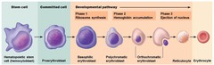
Erythropoiesis
proliferation and differentiation of small population of hematopoietic stem cells resident in the bone marrow (liver, spleen and bone marrow in fetus) into mature red blood cells
Erythropoietin (EPO)
glycoprotein secreted by the kidney and stimulates erythropoiesis in the bone marrow
Erythropoietin mechanism
- under hypoxic conditions, the kidney will produce + secrete EPO to increase the production of RBCs
- EPO binds to the erythropoietin receptor (Epo-R) on the surface of progenitor cells -> this results in differentiation and proliferation of the erythroid cell line

interstitial fluid transport
lymphatic capillaries in body tissues reabsorb interstitial fluid (becomes lymph) which ultimately returns to the blood plasma
blood plasma in capillaries --> interstitial fluid --> lymph in lymphatic pathway --> lymph returns to blood plasma
lymphatic capillaries characteristics
1. run parallel to blood capillaries in all body tissues
2. incomplete basement membrane, endothelial cells overlap one another
3. low pressure allows interstitial fluid to enter lymphatic capillaries
lymph movement
moves from lymphatic capillaries, to afferent lymph vessels, to lymph nodes, to efferent lymph vessels, to venous circulation

Hemostatic regulation
damaged blood vessel
- vasoconstriction
- endothelial cells activated: dissociate + increase in #
- thrombocyte activation: form projections on surface, cluster together, clot formation, RBC WBC trapped in clot, remodeling of clot
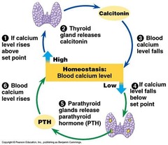
lymphoid organs/tissues
- lymph nodes - dendritic cells, lymphocytes (B-cells & T-cells), fixed macrophages
- thymus
- tonsils
- MALT (Mucosa Associated Lymphoid Tissue)
- spleen
spleen
blood flows through the spleen - removes damaged cells
- storage site for red blood cells
- activating lymphocytes
spleen anatomy
- capsule: outer covering
- trabeculae: elastic fibers and smooth muscle (houses arteries, veins and nerves)
- parenchyma
a) red pulp: fixed macrophages & collagen fibers (reticular mesh network) + packed red blood cells (venous sinusoids)
b) white pulp: lymphocytes
3 classes of lymphocytes
T cells, B cells, NK cells
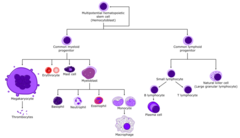
Hematopoiesis
blood cell formation - starts with Hemocytoblast in bone marrow
1. Lymphopoiesis: common lymphoid progenitor made from Hemocytoblast
a) if leaves bone marrow before differentiation: forms NK cells (reside in spleen, tonsil, thymus, LNs)
b): if stays in bone marrow before differentiation: forms B cells (reside in circulation) + forms T cells (to thymus for maturation)
2. common myeloid progenitor cell from Hemocytoblast
a) Thrombopoiesis: megakaryocyte creates Thrombocytes (platelets)
b) erythropoiesis: proerythroblast forms reticulocyte (RBCs)
c) Mast cell - histamine production
d) myeloblast: basophils, eosinophils, neutrophils, macrophages
How immune cells travel
1. carried in blood
2. diapedesis: small fenestrations form in capillary wall, allows cells to emerge
3. ameboid movement: cells physically interacting with surrounding tissues
4. chemotactic signaling via chemical messages
iron deficient anemia
insufficient iron in the diet, interferes with hemoglobin production
megablastic anemia
- deficiency in Vitamin B12 (critical for Erythropoiesis)
- Macrocytes (abnormal RBCs), last 30-50 days
- atrophy of part of gastic mucose (lose parietal cells); these cells produce intrinsic factor which is critical for B12 absorption
aplastic anemia
non-functional bone marrow
leukopenia
lower than expect proportion of white blood cells in circulation
lymphoblastic leukemia
cancerous cells derived from common lymphoid progenitor cell - overproduction of malformed B / T cells
myeloid leukemia
cancerous cell derived from common myeloid progenitor cell - overproduction of malformed myeloid cells
immunity
1. physical barrier: mucus
2. acquired immunity: T cells + B cells; antibodies activate T cells - target pathogens for destruction
3. MHCII: antigen presentation, found on antigen presenting cells, B-cells present antigens using MHCII- recognized by T-cells - cytokines released - activate B-cells release antibodies
5 classes of antibodies
IgG, IgM, IgA, IgD, IgE
Functions of antibodies
- agglutination
- recruitment of NK cells and macrophages
- neutralization
- activation of compliment system (innate immune system, compliment cascade)
digestion
breakdown of foods by chemical and physical processes to yield components that can be absorbed by the body
Absorption
movement of nutrients from the lumen of the GI tract into epithelial cells of the GI tract, and ultimately into the blood stream
prehension
grabbing and grasping of foods with the tongue, teeth and lips
mastication
chewing food into a bolus that can be swallowed
deglutition
swallowing of bolus of food
teeth
1. incisors: cutting
2. canines: tearing
3. premolars: grinding
4. molars: grinding
tongue
position bolus of food for swallowing
salivary glands function and location
produce saliva
- sublingual (under tongue), mandibular (lower jaw), parotid (near ear)

saliva contains the enzyme
amylase
- hydrolyzes a1-4 linkages in starch

mucus
viscous; lubricates and protects epithelial cells that line the GI tract
urea
nitrogen source for rumen bacteria, anti-foaming properties
3 phases of swallowing
1. voluntary: bolus pushed to pharynx, touch receptors activated motor neurons release acetylcholine
2. pharyngeal phase: pharynx contracts, push bolus to upper esophageal sphincter (UES); nitric oxide and vasoactive intestinal peptide (VIP) cause the UES to relax
3. esophageal phase: bolus passes by UES; acetylcholine release causes smooth muscle of esophagus to contract; bolus arrives at lower esophageal sphincter (LES); nitric oxide and VIP allow LES to relax -> bolus to stomach

achalasia
failure to relax the lower esophageal sphincter
regions of the stomach
- cardia: entrance to stomach (cardiac sphincter)
- fundus: domed portion
- body: w/ greater & lesser curvatures
- pyloric: pyloric sphincter (valve)

parietal cells
gastric gland cells that produce HCL (proton pumps H+/K+ ATPases) in response to several stimuli, including acetylcholine release
G cells
gastric gland cells that produce gastrin in response to acetylcholine (acts on parietal cells, chief cells, ECL cells)
- gastrin acts on parietal cells -> stimulate HCL release
chief cells
gastric gland cells that produce pepsinogen (upon stimulation by gastrin)
- when exposed to HCL, converts pepsinogen into pepsin; an enzyme that breaks down protein
Enterochromaffin-like (ECL) cells
gastric gland cells that produce histamine (upon stimulation by gastrin)
- histamine acts on the H2 receptor on parietal cells -> causes HCL release
Tagamet
H2 receptor antagonist
Prilosec
inhibits H+/K+ ATPases
4 components of ruminant stomach
1. rumen, reticulum, omasum, = forestomach, fermentation
2. abomasum = true stomach, digesta moves here after fermentation
small intestine regions
duodenum (digestion), jejunum (absorption), ileum (absorption)

enteroendocrine cells
small intestine cells (duodenum) that produce cholecystokinin (CCK)
- amino acid metabolism, fatty acid breakdown, promote release of pancreatic enzymes, inhibit stomach emptying
S cells
cells of the duodenum and stomach that produce secretin
- causes pancreatic enzyme release and bicarbonate release to GI tract
what stimulates secretin release from S cells
low pH
secretin blocks
H2 receptors in parietal cells (prevent HCL release)
peristaltic contractions (gut motility)
- interstitial cells of Cajal - pacemaker of cells of the GI tract that create slow waves of contractions
- Acetylcholine: increase strength and frequency of contractions
- NO and VIP: decrease strength and frequency of contractions

Meissner's plexus
enteric nervous system
- under mucosa
- control capillary beds in GI tract, secretions by mucosa, lymphatics
Auerbach's plexus
enteric nervous system
- within the muscularis of GI tract
- controls contractility of GI tract
chyme
contents from stomach that enters the intestine
absorption in the small intestine
- large amount of surface area = efficient absorption
- villus found throughout
- villi covered in epithelial cells called enterocytes (which are covered by microvilli)
- villi covered by cells with microvilli increase surface area of intestine ~600X
endocrine portion of pancreas
alpha cells = glucagon release
beta cells = insulin release
exocrine portion of pancreas
- acinar cells = produce digestive enzymes
- ductal cells = role in neutralizing pH in GI tract
Trypsinogen is converted to trypsin by
Enterokinase, a brush border enzyme on duodenal and jejunal mucosa
liver absorption of fat
- liver produces bile (ions, bicarbonate, water)
- common bile duct transports bile from liver/gall bladder to GI tract; joins sphincter
- bile emulsifies fats + forms micelles that can be absorbed by brush boarder
- enterocytes absorb micelles, produce chylomicrons
- chylomicrons excreted to center of villus
- capillary network + lymphatic vessel move absorbed chylomicrons to liver by hepatic portal vein
- hepatic portal vein brings absorbed nutrients from GI tract to liver
- liver absorbs nutrients, recycles bile salts, detoxifies blood
liver sinusoids are lined by
hepatocytes (liver cells that absorb nutrients and bile salts) and Kupffer cells (fixed macrophages)
blood detoxification by liver
blood flows through liver sinusoids where hepatocytes and Kupffer cells detoxify/clear the blood
- blood from hepatic portal vein moves through the liver sinusoids to the central vein of the liver, then the blood is routed to the vena cava, and back to the heart
bile canaliculi
ducts between hepatocytes that collect bile
alkaline tide
In gastric parietal cell, HCO3- leaves the cell in exchange for Cl- going in the cell (blood pH rises after eating)
* carbonic anhydrase *

Lymphopoiesis: common lymphoid progenitor made from Hemocytoblast
a) if leaves bone marrow before differentiation: forms NK cells reside in spleen, tonsil, thymus, LNs
b): if stays in bone marrow before differentiation: forms B cells reside in circulation + forms T cells to thymus for maturation
common myeloid progenitor cell from Hemocytoblast
a) Thrombopoiesis: megakaryocyte creates Thrombocytes (platelets)
b) erythropoiesis: proerythroblast forms reticulocyte (RBCs)
c) Mast cell - histamine production
d) myeloblast: basophils, eosinophils, neutrophils, macrophages
voluntary phase of swallowing
bolus pushed to pharynx, touch receptors activated motor neurons release acetylcholine
pharyngeal phase of swallowing
pharynx contracts, push bolus to upper esophageal sphincter (UES); nitric oxide and vasoactive intestinal peptide (VIP) cause the UES to relax
esophageal phase of swallowing
bolus passes by UES; acetylcholine release causes smooth muscle of esophagus to contract; bolus arrives at lower esophageal sphincter (LES); nitric oxide and VIP allow LES to relax -> bolus to stomach