exercise rx: exam 2
 Call Kai
 Learn
 Practice Test
 Spaced Repetition
 Match
 Flashcards
 Knowt Play
1/35

There's no tags or description
Looks like no tags are added yet.
36 Terms
special considerations for pregnancy
absolute contraindications for pregnancy
hemodynamically significant heart disease
incompetent cervix or cerclage
intrauterine growth restriction
multiple gestational at risk of premature labor
persistent 2nd or 3rd trimester bleeding
placenta previa after 26 or 28 wks of gestation
preeclampsia or pregnancy induced hypertension
premature labor during current pregnancy
restrictive lung disease
ruptured membranes
severe anemia
uncontrolled or poorly controlled HTX
uncontrolled thyroid disease
uncontrolled T1D
unexplained persistent vaginal bleeding
other serious CVD, respiratory, or systemic disorder
warning signs to stop exercising during pregnancy
vaginal bleeding
loss of amniotic fluid from vagina indicating rupture of membranes
dyspnea/shortness of breath prior to exertion and/or that does not resolve with rest
regular + painful uterine contractions
dizziness, feeling faint, or headache
chest pain
calf pain or swelling
decreased fetal movement
preterm labor
exercise rx guidelines & recommendations for pregnancy

what exercises to prescribe for pregnancy

HR zones for pregnancy

time + voltage interpretation on rhythm strip
one small square (vertical) = 0.1 mV
one large square (vertical) = 0.5 mV
one small square (horizontal) = 0.04 secs
one large square (vertical) = 0.2 secs
normal time of each interval + segment
PR interval = 120-200 ms (3-5 small boxes)
QRS complex = 80-120 ms (2-2.5 small boxes)
QT interval = 350-450 ms
genesis, path sequence, & termination of wave of depolarization
p wave = atrial depolarization
qrs complex = ventricular depolarization
t wave = ventricular repolarization
pr interval = the time from start of atrial depolarization to start of ventricular depolarization
pr segment = the time from end of atrial depolarization to start of ventricular depolarization
qt interval = the time from start of ventricular depolarization to end of ventricular repolarization
define P waves, QRS, T waves, PR interval, PR segment, QT interval, ST segment, TP segment, J joint

calculate HR
large box method
small box method
6 sec method
which method to default to for arrhythmias
mall box method
1500/# of small boxes between R waves
Large box method
300/# of large boxes between R waves
6-second method
count cardiac cycles in a 6-second interval and multiply by 10
define normal sinus rhythm
SA node depolarizes
depolarization spread through atria
delay at AV node
bundle of his depolarizaes
L & R branches depolarizes
purkinje fibers depolarize
60-100 bpm
one pa wave for each qrs complex
p wave should be positive in lead 2
define tachycardia
100+ bpm at rest
define bradycardia
<60 bpm at rest
define regular rhythm
consistent b/w r waves
define regularly irregular
clear + distinct pattern but every contraction is not spaced equally
define irregularly irregular
erratic pattern + erratic spacing
define arrhythmias (why do they happen)
any disturbance in the rate, regularity, site of origin, or conduction of the cardiac electrical impulse
can be a single, aberrant (or even prolonged pause b/w beats) or a sustained rhythm disturbance that can persist for the lifetime of the patient
not every arrhythmia is abnormal (i.e. athletes with abnormal heart rates/rhythms)
causes
hypoxia & ischemia (damage)
sympathetic stimulation
drugs
electrolyte imbalances
enlargement
what are the 5 types of arrhythmias
arrhythmias of sinus origin - electrical activity follows usual conduction pathways but is too fast, too slow, or irregular
ectopic rhythms - electrical activity originates from a focus other than SA
reentrant arrhythmias - electrical activity is trapped within an electrical racetrack whose shape & boundaries are determined by anatomic or electrical MI configurations
conduction block - electrical activity originates from in SA node & follows the usual pathway but is blocked or delayed
preexcitation syndromes - electrical activity follows anomalous conduction pathways that bypass normal ones providing electrical shortcut
3 basic rules for EKG interpretation
Depolarization towards the positive pole will get a positive deflection
Depolarization towards the negative pole will get a negative deflection
Depolarization directly thru a positive and negative pole will get a biphasic complex
distinguish b/w limb leads + chest leads
limb: I, II, III, aVR, aVL, aVF
inferior: II, III, aVF
high lateral: I, aVL
forgotten lead: aVR
chest: V1-V6
septal: V1, V2
anterior: V3, V4
low lateral: V5,V
einthoven’s triangle
Each side of the triangle represents a distinct bipolar lead measuring the voltage difference (potential difference) between two extremities:
Lead I: Connects the Right Arm (negative) to the Left Arm (positive) at 0°.
Lead II: Connects the Right Arm (negative) to the Left Leg (positive) at +60°. (This lead most closely aligns with the heart's normal electrical conduction axis, making it the primary rhythm-monitoring lead).
Lead III: Connects the Left Arm (negative) to the Left Leg (positive) at +120°
Electrical Axis: By combining Leads I, II, and III (along with augmented unipolar leads like aVR, aVL, and aVF), clinicians calculate the average direction of the heart's electrical dipole in the frontal plane (normally between -30° and +90°).
Deflection Direction: Electrical impulses traveling toward a positive electrode produce an upward (positive) deflection on the ECG tracing, while impulses moving away produce a downward (negative) deflection
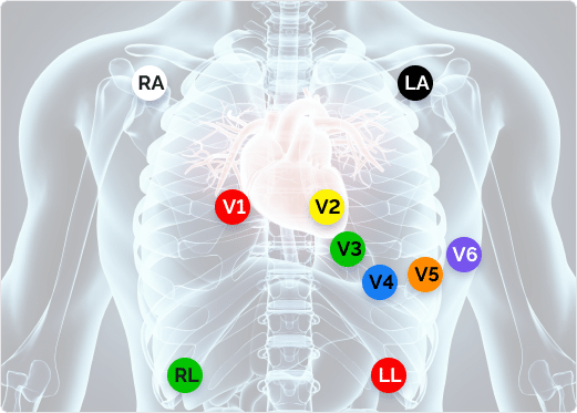
12 lead ECG placement
hexaxial system - quadrant method
Normal axis can usually be determined very readily by examining leads I and aVF
If the QRS complexes in these leads are positive, then the axis is normal.
The QRS complexes of leads I and aVF are very positive, consisting of mainly large R waves
This implies that the net vector of ventricular depolarization is traveling toward the positive poles of these leads
The problem is that Lead I positive and aVF negative doesn't automatically mean abnormal left axis deviation.
causes of axis deviation
ventricle or atrial hypertrophy
LBB
pulmonary conditions (COPD, embolism)
usually RAD
MI
fascicular block
obesity + stature
what is angina
the classic symptom of cardiac ischemia
described as diffuse chest pain or pressure that may radiate to the neck, arms, or back, or back + may be accompanied by shortness of breath, nausea, vomiting, dizziness, or diaphoresis
underlying pathophysiology: narrowing of coronary arteries by atherosclerosis
impedes blood flow to the heart muscle
less common causes: aortic stenosis & hypertrophic cardiomyopathy
what is unstable angina
angina that is present with much less or even no physical exertion & typically lasts much longer & is more severe
what is MI
blood flow is stopped or limited
stemi: ST segment elevation MI
non stemi: ST segment does not elevate but most often will be depressed
present in MI & unstable angina
how to diagnose MI
history & physical exam
cardiac enzyme determination
ekg
stemi vs non stemi and J-point elevation (what are the norms)
stemi:
during acute stemi the ekg often evolves thru 3 stages
T wave peaking followed by T wave inversion
ST segment elevation
appearance of new Q waves
non stemi:
most often caused by either nonocculsive thrombosis of a major coronary artery or complete occlusion of a small offshoot of one of the major coronary arteries
unlike stemis they can involve less than the entire thickness of the heart muscle & can be localized to a particular region by a single coronary vessel like stemis
only ekg changes: t wave inversion + st segment depression (no elevation)
j point
common in young healthy people
leads V2/V3: st elevation
men < 40: >2.5mm
women (all ages): >1.5mm
CABGs - on/off pump
if pump is on:
O2-poor blood is directed to machine to become oxygenated and machine pumps blood
O2-rich blood is returned to the body skipping the heart & lungs
The surgeon can work on a still heart to attach a healthy blood vessel that routes blood around the blockage.
Once the grafts are complete, the heart resumes pumping, and circulation is transferred back to the heart.
Why use it: The heart is still while the surgeon works, providing a stable surgical field and good visibility
if pump is off:
A mechanical stabilizer reduces movement in the targeted area while the rest of the heart continues beating.
The surgeon connects a healthy blood vessel to create a new route around the blocked coronary artery while the heart continues pumping
The surgeon checks the graft and completes the operation without needing to restart the heart or disconnect from a heart-lung machine
Why use it: It avoids the need for cardiopulmonary bypass and temporarily stopping the heart. However, operating on a moving heart is technically more challenging
CABG: CBPI
The least invasive method for revascularization is a catheter-based,
percutaneous intervention
A doctor inserts a thin, flexible tube called a catheter into a blood vessel, usually in the wrist or groin.
The catheter is guided toward the coronary arteries using X-ray imaging. Contrast dye helps the doctor see where the artery is narrowed.
A small balloon at the catheter's tip is inflated at the narrowed area, pressing plaque against the artery wall and widening the opening.
Often, a mesh stent is expanded inside the artery to help keep it open after the balloon is deflated and removed
CABPG: stent
Plaque made of cholesterol, fats and other substances builds up inside the coronary artery, reducing blood flow to the heart muscle
A catheter carries a balloon-mounted stent to the narrowed area. The balloon inflates, expanding the stent and pushing the plaque outward against the artery wall.
The balloon is deflated and removed, but the stent remains inside the artery, supporting the artery wall and helping maintain the widened opening.
2 types
Drug-eluting stent (DES): Releases medication that helps prevent excessive tissue growth inside the stent, reducing the chance of the artery narrowing again. These are commonly used today.
Bare-metal stent (BMS): A metal mesh tube without a medication coating. Tissue can grow around it as the artery heals, but excessive growth can cause restenosis (re-narrowing).
CABG: exercise testing
As part of the baseline evaluation, it is highly preferred for a patient to perform a symptom-limited exercise tolerance test (e.g., exercise stress test)
Although a symptom-limited exercise tolerance test is ideal, it may not always be feasible. In these cases, the 6-minute walk test or other forms of submaximal testing may be conducted
Information obtained from an exercise test is necessary for developing an individualized, safe, and appropriate exercise prescription and is useful for guiding a patient’s return to work or home/leisure activities
CABG: FITT

LVAD, total artifical heart, heart transplant: FITT
