Biology of Cells Quiz 1
 Call Kai
 Learn
Learn Practice Test
 Spaced Repetition
 Match
Match Flashcards
 Knowt Play
1/79

There's no tags or description
Looks like no tags are added yet.
Name | Mastery | Learn | Test | Matching | Spaced | Call with Kai | Chat |
|---|
No analytics yet
Send a link to your students to track their progress
80 Terms
DNA helicase
unwinds helix
single-strand DNA-binding proteins (SSB)
bind to single-stranded DNA without covering bases
help DNA helicases by stabilizing unwound strand
prevents formation of short hairpin structures
DNA polymerase III
cannot start chains de novo
replicates DNA
DNA primase
synthesizes short RNA primers
Telomerase
prevents DNA shortening & ensures stability of genome
recognizes tip of G-rich strand of telomere DNA repeat sequence
elongates telomere in 5’ to 3’ direction
DNA Polymerase I
erases RNA primer & replaces with DNA
DNA ligase
joins 3’ to 5’ ends of okazaki fragments
Proteins in mismatch repair in prokaryotes & purpose
MutH nicks unmethylated GATC sequences
Proteins in mismatch repair of eukaryotes & purposes
MutS: binds specifically to mismatched base pair
MutL: scans nearby DNA for nick & triggers degradation of nicked strand
Explain proofreading
correct nucleotide has higher affinity to moving DNA polymerase
if incorrect base added, DNA polymerase shifts strand to exonucleolytic site and moves in 3’ to 5’ direction to wrong base
proofreading exonuclease clips off incorrect nucleotide
DNA polymerase shifts strand back to polymerization site and resumes replication
Explain mismatch repair in prokaryotes
methylation on adenine residues in sequence GATC denotes template strand
newly synthesized strand is unmethylated
MutH nicks unmethylated GATC sequqences
excision of mismatched DNA segment
Explain mismatch repair in eukaryotes
does not depend on DNA methylation
newly synthesized DNA strands are nicked
nicks provide signal that directs mismatch proofreading system to appropriate strand
MutS binds to mismatched pair
MutL scans nearby DNA for nick and triggers degradation of nicked strand
RNA polymerase
catalyzes phosphodiester bond formations to link ribonucleotides
starts RNA chain without primer
no proofreading
Which RNA polymerase transcribes protein-coding genes in eukaryotes?
II
General transcription factors
only in eukaryotes
help RNA polymerases correctly bind to promoter
transcriptional activators
gene regulatory proteins
bind to enhancers to help RNA pol II recruitment
chromatin-modifying enzymes
regulate TFs/RNA pol accessibility to DNA in chromatin
activate/inactivate assembly of transcription initiation machinery on DNA
Transcription for RNA Pol II
TFIID binds to TATA box and recruits others
Other TFs and RNA Pol II assemble transcription initiation complex and bind to promoter
TFIIH (contains DNA helicase) guides transcription initiation complex to start site
RNA synthesizes short mRNA
Phosphorylation by TFIIH releases RNA Pol II from promoter to move down template strand
Once elongation started, most TFs released from DNA template & recycled
RNA polymerase reaches terminator and releases DNA template & RNA chain
mRNA processing
5’ capping
add methylated guanine nucleotide on 5’ end
methyl cap distinguishes mRNA from other types of RNA
3’-polyadenylation
splicing- removes introns & join exons together
Small ribosomal subunit function
match tRNAs to mRNA codon
Large ribosomal subunit function
catalyzes peptide bond between amino acid & polypeptide chain
Release factors
terminates translation
attaches to stop codon and binds to ribosome at A site
translation initiation process
form mRNA + ribosome complex
initiator tRNA initiates translation (always carries methionine)
only initiator tRNA can bind to small ribosomal uunit to form pre-initiation complex
Do all proteins have methionine as first amino acid at N-terminus?
Yes when newly made
usually removed later by specific protease
Translation elongation process
initiator tRNA in P site
tRNA carrying next AA binds to A-site and forms complementary base pair with mRNA codon
form peptide bond between AA at P & A site
release polypeptide chain from tRNA at P site
Shift tRNAs to E & P site
move mRNA 3 nucleotides, reset ribosomes to receive next aminoacyl-tRNA
Translation termination process
reach stop codons (UAA, UGA, UAG)
release factor attaches to stop codon and bind to ribosome at A site
ribosome releases mRNA & polypeptide chain and separates into small & large subunits
Separation of nucleus from cytoplasm allows for…
DNA to be protected from enzymes that could damage them
Concentrate proteins acting on DNA
Chromatin is comprised of…
nuclear DNA + proteins (histones + other chromosomal proteins)
2 types of chromatin
euchromatin
less condensed
contains protein-coding genes
heterochromatin
highly condensed
no protein-coding genes
Conserved DNA regions
closely similar regions between different species (exons & regulatory sequences)
Introns
non-coding sequences
Exons
short segments of protein-coding sequences
Microtubule function
primary organizers of cytoskeleton
position membrane-bound organelles
direct intracellular transport
Actin filament function
determine cell shape
responsible for cell locomotion
Intermediate filament function
provide mechanical strength
basic microtubule subunit
α/β-tubulin heterodimer
Each α/β-tubulin monomer binds a ….
GTP
Which monomer has GTPase activity?
β-tubulin
Protofilament
α/β-tubulin heterodimers joined end to end
structural polarity
assembly of microtubule
α and β-tubulin bind noncovalently > α/β-tubulin heterodimer
α/β-tubulin heterodimers assemble into protofilaments
13 protofilaments laterally associate (between same subunits) into hollow cylindrical microtubule
Longitudinal and lateral interactions between α/β-tubulin heterodimers generate the _______ of the microtubule
helical lattice
Subunit loss in microtubules occurs… because …
microtubule ends
each α/β-tubulin subunit stabilized by multiple contacts within lattice
Location of nucleation of microtubules
microtubule-organizing center (MTOC)
in most animals, MTOC is centrosome
Centrosome
composed of protein-rich (fibrous) matrix
centrioles organize centrosome matrix
Centriole
short cylindrical structure made of modified microtubules + many accessory proteins
Microtubules are nucleated by…
protein complex containing γ-tubulin
centrosome contains >50 copies of γ-tubulin ring complexes (γ-TuRCs)
γ-TuRCs efficiently nucleate microtubule growth
How are microtubules nucleated at centrosome?
with - ends anchored and + ends extending outwards toward periphery
star-like (astral) pattern
Microtubule elongation
unphosphorylated stathmin binds α/β-tubulin dimers & prevents their addition to + end of microtubules
reduces effective concentration of free α/β-tubulin available for polymerization
normally, rate of extension > rate of GTP hydrolysis
Decrease in free α/β-tubulin concentration = decreased rate of extension
Rate of GTP hydrolysis catches up and hydrolyzes GTP cap into GDP so there is no more cap
Triggers catastrophe
Catastrophe
growing microtubule loses GTP cap and rapidly starts to shrink
Rescue
shrinking microtubule regains a GTP-rich end and starts to grow again
Dynamic instability
repeated switching between growth and shrinkage
Tubulin and actin exist in 2 nucleotide states
T-form: GTP/ATP
D-form: GDP/ADP
Free tubulin and actin subunits are mostly in ___ form because…
T
nucleotide hydrolysis slow in free subunits but accelerated after incorporation into filament (only become D form after added to microtubule)
Microtubule stabilization
Once cytoskeletal filament formed, proteins bind along sides & change stability & mechanical properties
Micortubule-associated proteins (MAPs)
bind microtubule to stabilize them
promote microtubule formation by stabilizing small tubulin oligomers during early polymerization
tau allows microtubules to form closely packed bundles because of its shorter projecting domains than other MAPs
Actin filament soluble subunit
G actin monomer
each bound to ATP
Actin filament assembly
assemble head to tail
structural polarity
faster-growing + end and slower growing - end
flexible compared to microtubules
Phases of actin assembly
nucleation (lag phase)
small, stable nucleus must form before filament growth (slow step)
polymerization (growth phase)
actin filaments elongate rapidly by adding subunits
steady state (equilibrium phase)
rate of subunit addition = rate of subunit dissociation
Critical concentration (Cc)
concentration of free subunits left in solution at steady state
[free actin]>Cc= polymerization
[free actin]<Cc= disassembly until equilibrium
Location of nucleation of actin filaments
plasma membrane
Highest density of actin filaments found where?
cell periphery
What is cell cortex and its function?
actin-rich layer under plasma membrane
helps control cell shape & surface movement
Arp2/3 complex
promotes actin nucleation
arp2/d nucleated filaments grow rapidly at + ends
In most non-muscle cells, _____ of actin is F-actin and ____ of actin is soluble (G) actin
50%
50%
Actin binding proteins
bind actin monomers and regulate filament formation
thymosin
binds actin monomers and keeps them inactive
thymosin-bound actin cannot be added to + or - ends of filament
profilin
binds actin monomers
prevents addition to - end
promotes addition to + end
compete for binding to actin monomers
arp2/3 complex role in dynamics
nucleates new actin filaments
remains associated with + ends
capZ
binds and caps + end (prevents further growth or shrinkage & stabilizes filament)
tropomyosin
binds along actin filament and blocks other actin-binding proteins from binding
cofilin
binds to ADP-actin instead of ATP-actin
changes twist of actin and weakens contact between actin subunits, promotes filament severing & depolymerization
newly-formed actin filaments have more ATP actin and are less sensitive to cofilin
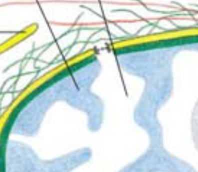
blue= heterochromatin
white= euchromatin
Wobble base
some tRNAs only require accurate base-pairing at first two positions of codon
tolerate mismatch (wobble base) at third position
Treadmilling
behavior of actin filaments
actin subunits added at + end and removed from - end at same rate
filament appears to move through cytoplasm with little change in length
Treadmilling & dynamic instability allow cells to do what?
rapidly remodel cytoskeleton
Intermediate filaments
not present in all cell types
in many cells, form extensive network in cytoplasm and provide mechanical strength
Nuclear lamins
type of intermediate filament
form meshwork under inner nuclear membrane & support nucleus + anchor chromatin
Intermediate filament soluble subunit
tetramer
Structure of intermediate filament
elongated molecules with central alpha-helical domain
no structural polarity
2 monomers —> parallel coiled-coil dimer
2 dimers associate antiparallel —> tetramer
8 tetramers associate laterally —> intermediate filament
Accessory proteins function
crosslink intermediate filaments and connect them to other cytoskeletal systems
Filaggrin
accessory protein
bundles keratin filaments
Plectin
accessory protein
links & bundles vimentin & other intermediate filaments
SUN-KASH complexes
connect cytoskeleton to the nucleus across nuclear envelope