Anatomy and Physiology Final Practical Appendix 2
 Call Kai
 Learn
 Practice Test
 Spaced Repetition
 Match
 Flashcards
 Knowt Play
1/89

There's no tags or description
Looks like no tags are added yet.
90 Terms
Dorsal Column, Lateral Column, Ventral Column, White matter
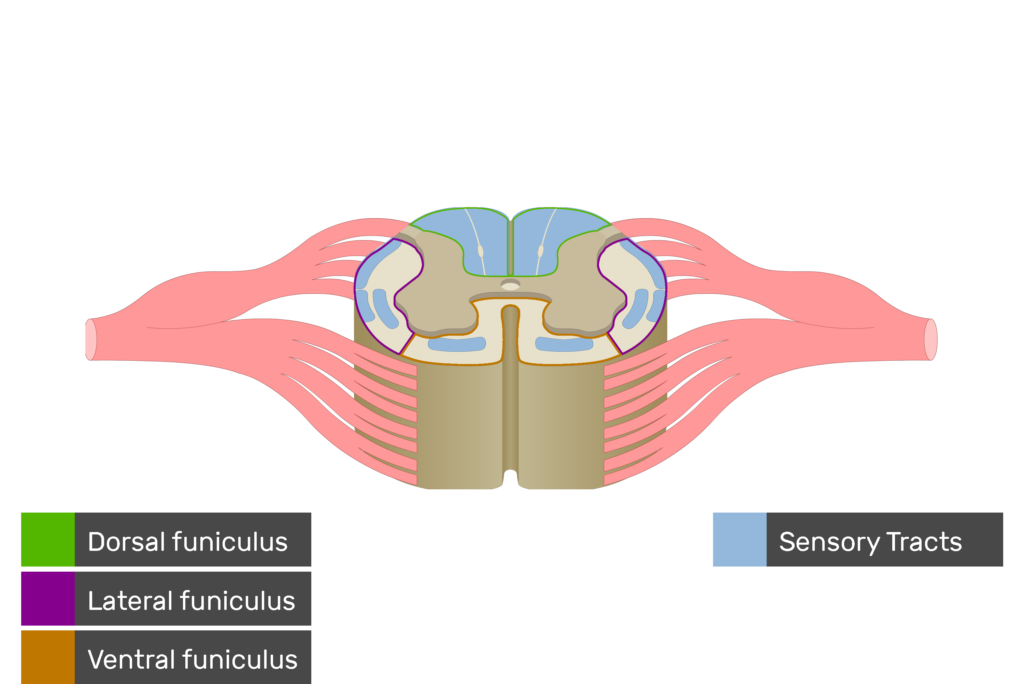
Anterior white commissure, white matter

Dorsal Grey Horn
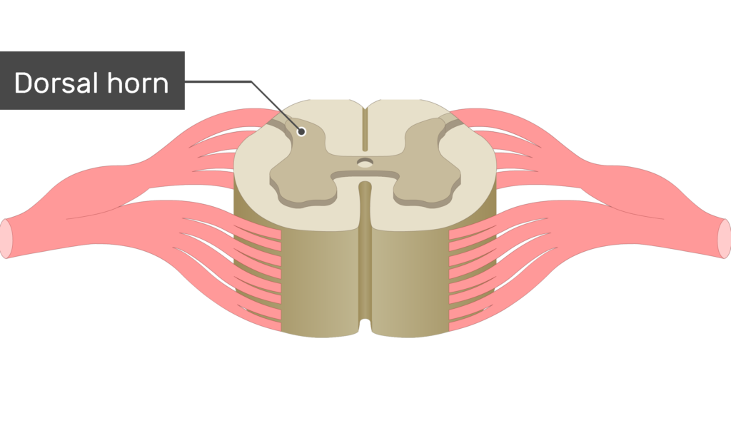
Lateral Gray Horn

Ventral Gray horn

Gray Commissure

Central Canal
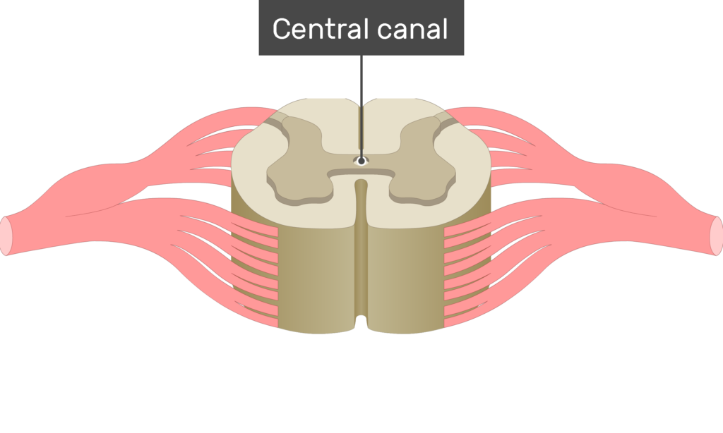
Posterior Spinal Vein

Dorsal Root

Spinal Ganglion

Ventral Root

Cerebellum

Pons

Medulla Oblongata
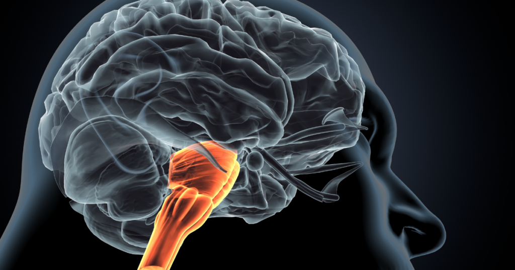
Medullary Cone

Cauda Equina

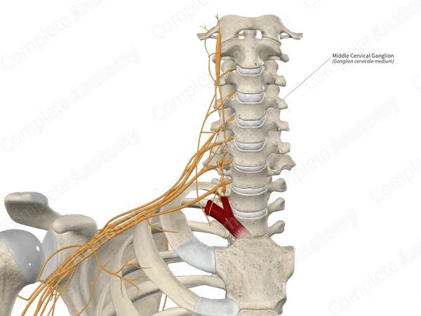
Cervical Ganglia
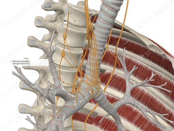
Thoracic Ganglia
Radial Nerve

Median Nerve

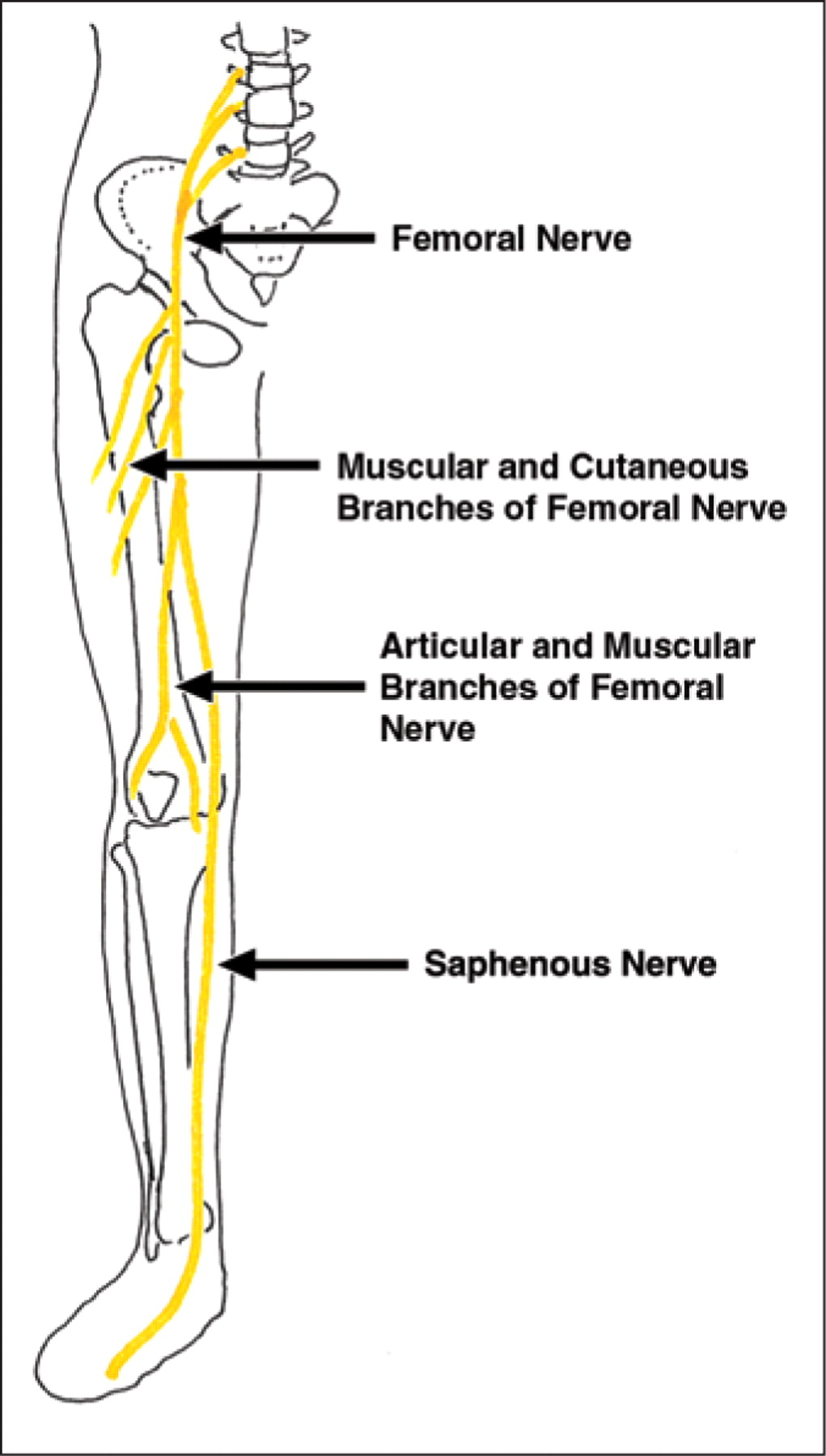
Femoral Nerve
Sciatic Nerve

Tibial Nerve

Cornea

Sclera

Choroid

Retina

Iris

Lens

Vitreous body

Optic Nerve II

Fovea Centralis

Vorticose Veins

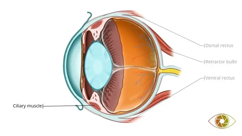
Ciliary Muscle
Central Retinal Artery and Vein

Auricle

External Acoustic Meatus

Tympanic Membrane

Tympanic Cavity
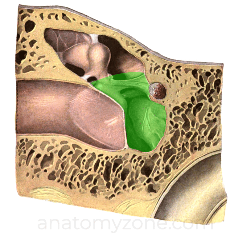
Malleus

Incus

Stapes

Oval Window

Round Window

Lateral Semicircular Canal

Anterior semicircular canal

Posterior semicircular canal

Cochlea

Vestibulocochlear Nerve

Motor Nerve Fiber
Cornea
Protective transparent outer layer of the eye that refracts (bends) incoming light toward the lens.
Sclera
Tough white outer coat of the eyeball that protects the eye and maintains its shape.
Choroid
Vascular middle layer of the eye that supplies blood to the outer retina and helps absorb stray light.
Retina
Innermost light-sensitive layer of the eye that converts light into nerve impulses.
Iris
Colored muscular structure that controls the size of the pupil and regulates the amount of light entering the eye.
Lens
Transparent structure behind the iris that focuses light onto the retina.
Vitreous body
Clear, gel-like substance filling the eyeball that helps maintain its shape and supports the retina.
Optic Nerve II
Cranial nerve II that transmits visual information from the retina to the brain.
Fovea Centralis
Small central area of the retina with the highest concentration of cones, responsible for sharp central vision.
Vorticose Veins
Veins of the choroid that drain blood from the middle layer of the eye.
Ciliary Muscle
Smooth muscle that changes the shape of the lens for focusing on near or far objects.
Central Retinal Artery and Vein
Blood vessels that supply oxygen to and drain blood from the retina.
Auricle
Outer visible part of the ear that collects sound waves and directs them into the ear canal.
External Acoustic Meatus
Canal that carries sound waves from the auricle to the tympanic membrane.
Tympanic Membrane
Thin membrane that vibrates when sound waves strike it, transferring vibrations to the ossicles.
Tympanic Cavity
Air-filled middle ear chamber that contains the ossicles and transmits sound vibrations.
Malleus
Hammer-shaped ossicle that receives vibrations from the tympanic membrane and passes them to the incus.
Incus
Anvil-shaped ossicle that transmits vibrations from the malleus to the stapes.
Stapes
Stirrup-shaped ossicle that transmits vibrations to the oval window of the inner ear.
Oval Window
Membrane-covered opening between the middle ear and inner ear that receives vibrations from the stapes.
Round Window
Membrane-covered opening that releases pressure waves in the cochlea, allowing fluid to move freely.
Lateral Semicircular Canal
Semicircular canal that detects head rotation in the horizontal plane to help maintain balance.
Anterior semicircular canal
Semicircular canal that detects head movement in the vertical plane to help maintain balance.
Posterior semicircular canal
Semicircular canal that detects head movement in the vertical plane, helping maintain balance and orientation.
Cochlea
Spiral-shaped inner ear structure that converts sound vibrations into nerve impulses.
Vestibulocochlear N
Cranial nerve VIII that carries sound and balance information from the inner ear to the brain.